Research Article 
 Creative Commons, CC-BY
Prevalence of additional canals in maxillary first molar in a Nepalese population: A Clinical Study
*Corresponding author: Rupam Tripathi, Department of Conservative Dentistry and Endodontics, UCMS, College of dental surgery, Bhairahawa, Nepal.
Received: January 08, 2019; Published: January 16, 2019
DOI: 10.34297/AJBSR.2019.01.000508
Abstract
Background and aims: There is a wide range of variations in the literature with respect to frequency of occurrence of the number of canals in each root, the number of roots and incidence of fusion. This study aims to investigate the prevalence of additional canals in permanent maxillary first molar in the Nepalese Population.
Methods: This was a descriptive study in which maxillary first permanent molars (n-201) of Nepalese population were examined using in vivo technique. Radio-graphs were taken. Access opening was done and gentle troughing of the pulpal floor was done, to look for additional canal.
Results: A total of 201 patients were treated. Of these, additional canals in me-siobuccal root was found in 34.1% of the cases and in distobuccal and palatal root, the additional canals were found in 0.49% of the cases.
Conclusions: Complete knowledge of the internal anatomy and anatomic varia-tion of root canal system is crucial for the success of the root canal treatment, as missed canals contribute to the failure of root canal treatment in a short dura-tion.
Keywords: Maxillary first molar, Palatal root, Root canal system, Variable anat-omy.
Introduction
Successful root canal therapy requires a thorough knowledge of root and root canal morphology. In spite of all procedural protocol if clinicians miss an addi-tional root canal it could pose a great challenge and lead to failure of root canal treatment. There is a wide range of variation in the literature with respect to frequency of occurrence of the number of canals in each root, the number of roots and inci-dence of fusion. Morphology of pulp systems varies greatly in different races and in different individuals within the same population [1].
Internal complexities of the root canal are genetically determined and have de-finitive importance in anthropology, so there is a need for the identification of root canal morphologies of different asian populations [2]. The anatomy of maxil-lary first permanent molar is complex which presents a constant challenge for the Endodontists. An awareness and understanding of the existence of additional canals and rare root canal morphology is essential [3].
The most common configuration described in the root canal anatomy of maxil-lary first molar is the presence of three roots with three canals, while the most frequent variation is the presence of second mesiobuccal canal (MB2).In maxil-lary first permanent molars, the broad buccolingual dimension of the mesiobuc-cal root and associated concavities on its mesial and distal surface is consistent with the majority of the mesiobuccal roots having two canals while there is usu-ally a single canal in each of the distobuccal and palatal roots [4,5]. The aim of this study was to show the prevalence of an additional canal in maxil-lary first permanent molars in a Nepalese population as there is no previous lit-erature regarding it.
Methods
This in vivo study was conducted in the Department of Conservative Dentistry and Endodontics between January 2014 and November 2015. Informed consent regarding objectives of the study was taken from all the patients. Ethical clear-ance was obtained from the college. Patient’s Maxillary first permanent molar having radiological and clinical evidence of pulpal pathology was included in the study. The included teeth were free of root resorption, had no calcifications or open apices. Preoperative radiographs were taken on different angulation i.e, straight on, mesio-oblique and distooblique to evaluate the number of roots and canals. No retreatment cases were included in the study. Selected cases had conventional root canal treatment done.
Local anesthesia (Ubistesin Forte/ 3M ESPE, Seefeld-Germany) was adminis-tered. An endodontic access opening was done with Endo Z bur (Dentsply, Tulsa Dental, and USA) under rubber dam isolation. The outline of the access cavity was modified to a rhomboidal shape to improve visibility of the second mesi-obuccal canal orifice. Gentle troughing of the pulpal floor palatally along the ori-fice of the mesiobuccal canal was done to identify the possibility of a second me-siobuccal canal or any other additional canal, with a half round bur. The contents of the pulp chamber were removed and sharp endodontic explorer and magnify-ing glass was used to explore the developmental grooves carefully to locate the orifices of the canals.
Copious amounts of 3% sodium hypochlorite (Prime Dental Products, Pvt, Ltd) solution irrigant was used. Pulp tissue was extirpated using barbed broaches (Nerve Broaches/ Alfred Becht- GmbH, Germany) or H-Files (Mani inc, Japan). A size 10 K file (Dentsply) was introduced into the canal to de-termine the canal patency and a working length radiograph was taken, using the paralleling technique. Apex locator (Root ZX, J. Morita, and Kyoto) was used to take a second working length as an adjunct to the radiographic method. Apical patency was confirmed with a small file (#15 or #20 NitiFlex) throughout the procedures after each larger file size. Preparation was completed using stepback of 1mm increments. Irrigants used were 2.5% NaOCl solution and normal saline. Each canal was dried using sterile paper points. Afterward, the canals were med-icated with a calcium hydroxide paste for 1 week, and then they were filled by the lateral compaction technique.
Results
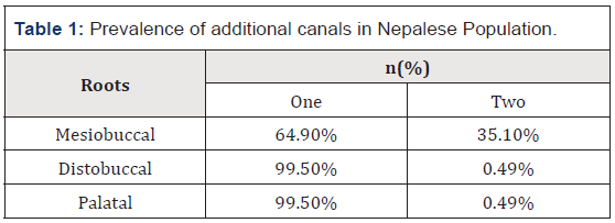
In the current study, the ratio of male to females was 1.5:1. The mean age of participants were 35.75±16.39. The additional canal in mesiobuccal root was found greater in females (59.18%) in comparison to males (40.82%) wheareas in distobuccal and palatal root, the additional canals were found in males. The in vivo study revealed that additional canals in mesiobuccal root was found in 34.1% of the cases and in distobuccal and palatal root, the additional canals were found in 0.49% of the cases (Table 1).
Discussion
Anatomical variations can occur in maxillary permanent first molars. The cur-rent study shows the prevalence of an additional canal in maxillary first perma-nent molar in a Nepalese population which was then compared with different asian populations. This study was conducted in Terai part of Nepal which consists of mostly Caucasian population. The present in vivo study revealed that most of the maxillary first permanent molars studied had three canals. The extra canals found in 35.1% of cases were exclusively in the mesiobuccal root in nepalese population whearas these additional canals were found to be greater in Chinese, Pakistani and Indian populations [6-8]. The additional canal in mesiobuccal root was found less in nepalese population than that of other asian populations, this maybe because present study was in vivo. There is the ease of manipulating the tooth outside the mouth in in-vitro technique than in-vivo technique. This could be also due to the fact that surgical loupes and operating microscope were not used in this study as modifications in endodontic access preparation. There are reports of high incidence of two canals in the mesio-buccal root of the maxillary molars. Wein et. al. [9] found that teeth with a fourth canal occurred more fre-quently than those with three canals (51.5% versus 48.5%). The incidence of MB2 is lower in present study.
In palatal root, additional canal was found in 0.49% of the cases which is less than that of Chinese and Indian populations and greater than that of Pakistani population. There are fewer reported cases of two root canals present in the palatal root of the maxillary molars. A review study on the anatomy of the maxil-lary first molar from a study done by Cleghorn et. al. [10] reported an incidence of the mesiopalatal canal (MP) at 56.8% and a prevalence of the distopalatal canal (DP) at 1.7%. Bratto Filho et. al. [11] reported that the frequency of extra roots and root canals in palatal roots are 2.05% (ex-vivo results), 0.62% (clinical results) and 4.55% (CBCT results). Stone and Stroner [12] examined more than 500 ex-tracted molars and found less than 2% incidence of multiple systems in which a single palatal root contains two separate orifices, canals and foramina.
The current study provides an insight on the prevalence of additional canals in maxillary permanent first molar among the Nepalese population which can be helpful in determining the successful outcome of root canal system. This study contributes to our knowledge and awareness of the complexity of the root canal morphology among the nepalese population. Although such cases occur infre-quently, the clinician should interpret the preoperative radiograph appropriately and not only look for two mesio-buccal canals, as additional canals can also be found in distobuccal and palatal roots. If these extra roots or extra canals are left untreated, they can harbour microorganisms and lead to failure of treatment.
Conclusions
Dentists should be aware of extra canals and roots when considering endodontic treatment of a maxillary first molar. Thus, it is essential to highlight the need to look for unusual morphology and additional roots and root canals for a good en-dodontic outcome.
References
- Ahmed HA, AbuBakr NH, Yahia NA, Ibrahim YE (2007) Root and canal morphology of permanent mandibular molars in a Sudanese population. Int Endod J 40:766–771.
- Neelakantan P, Subbarao C, Ahuja R, Chandragiri VS, Gutmann JL (2010) Cone-beamcomputed tomography studyof root and canal morphology of maxillary first and second molars in an Indian population. J Endod 36:1622–1627.
- Walton R, Torabinejad M (1996) Principles and practice of endodontics, (2nd edn), Phil-adelphia, WB Saunders Co, USA.
- Ash M, Nelson S (2003) Wheeler’s dental anatomy, physiology and occlusion, (8th edn), Saunders, Philadelphia, USA.
- Malagnino V, Gallotini L (1997) Some unusual clinical cases on root anatomy of permanent maxillary molar. J Endod 23:127–128.
- Zheng QH, Wang Y, Zhou XD, Wang Q, Zheng GN, et al. (2010) A conebeam computed tomography study of maxillary first permanent molar root and canal morphology in a Chinese population. J Endod 36(9):1480- 1484.
- Mothanna Alrahabi, Muhammad Sohail Zafar (2015) Evaluation of root canal mor-phology of maxillary molars using cone beam computed tomography. Pak J Med Sci 31(2): 426–430.
- Atool Chandra Bhuyan, Rubi Kataki, Pynshngain Phyllei, Gurdeep Singh Gill (2014) Root canal configuration of permanent maxillary first molar in Khasi popula-tion of Meghalaya: An in vitro study J Conserv Dent 17(4): 359–363.
- Weine FS, Healey HJ, Gerstein H, Evanson L (1969) Canal configuration in the mesi-obuccal root of the maxillary first molar and its endodontic significance. Oral Surg Oral Med Oral Pathol 28:419-425.
- Cleghorn BM, Christie WH, Dong C (2006) Root and root canal morphology of the human permanent maxillary first molar: a literature review. J Endod 32:813-821.
- Christie WH, Peikoff MD, Fogel HM (1991) Maxillary molars with two palatal roots: A retrospective clinical study. J Endod 17(2):80-84.
- Holderrieth S, Gernhardt CR (2009) Maxillary Molars with Morphologic Variations of the Palatal Root Canals: A Report of Four Cases. J Endod 35:1060-1065.


 We use cookies to ensure you get the best experience on our website.