Research article 
 Creative Commons, CC-BY
Real Asymptomatic Patients with COVID-19
*Corresponding author: Chen Zhihai, Center of Infectious Diseases, Beijing Ditan Hospital, Capital Medical University, Jingshun East Street, Chaoyang District, Beijing, China.
Received: October 23, 2020; Published: November 06, 2020
DOI: 10.34297/AJBSR.2020.10.001565
Abstract
Objective: To evaluate the clinical features, virological course, and follow-up of COVID-19 patients without symptoms who were admitted to Beijing Ditan hospital, and pays attention to the epidemiological significance of asymptomatic cases.
Methods: 10 cases in a cluster who diagnosed COVID-19 were enrolled in Beijing Ditan Hospital on February 27th in 2020. They had a close contact with the same COVID-19 patient. Clinical data were collected, including general status, clinical manifestations, laboratory test results, imaging characteristics, treatment regimens, outcomes, and follow-up.
Results: 7 were male and 3 were female, with an age range of 22 to 59 years and a median age of 49 years. Three patients were asymptomatic. The median age of the asymptomatic patients was 52, older than the symptomatic patients (52 vs 40, P = 0.37). 10 patients manifested as pneumonia. 7 cases of double lung and 3 cases of single lung. the asymptomatic patients had a significantly higher LDH than symptomatic patients (P = 0.03). The days of Negative for SARS-CoV2 RNA and Hospital stay were no difference (28.33±22 vs 22.88±12.04, P=0.7; 34±16.37 vs 28.57±12.12, P=0.57). At the 4-week follow-up after discharge, the lung lesions of 5 patients with symptoms were completely absorbed, the lung lesions of 2 patients with asymptoms were completely absorbed (71.4%vs66.7%, P=0.62).
Conclusion: The clinical course and prognosis in asymptomatic and symptomatic patients was similar, pay attention to the clinical management and epidemiological significance of asymptomatic patients.
Keywords: COVID-19, Asymptomatic, Symptomatic
Introduction
Coronavirus disease 2019 (COVID-19) is caused by SARS-CoV-2, a newly emergent coronavirus, that was first recognized in Wuhan, China, in December 2019. There have been around 26,994,442 reported cases of coronavirus disease 2019 (COVID-2019) and 880,994 reported deaths to date (07/09/2020). The symptoms are usually fever, cough, sore throat, breathlessness, fatigue, malaise, among others. The disease is mild in most people; in some (usually the elderly and those with comorbidities), it may progress to pneumonia, acute respiratory distress syndrome (ARDS) and multi organ dysfunction. Many people are asymptomatic [1]. An asymptomatic case is a person infected with SARS-CoV-2 who does not develop symptoms, but they can transmit the disease to others [2]. Although studies have described rates of asymptomatic infection, ranging from 4% to 32%, it is unclear whether these reports represent truly asymptomatic infection by individuals who never develop symptoms, transmission by individuals with very mild symptoms, or transmission by individuals who are asymptomatic at the time of transmission but subsequently develop symptoms [3]. A systematic review on this topic even suggested that true asymptomatic infection is probably uncommon [4]. In this study, we report 3 cases of COVID-19 pneumonia without any respiratory symptoms during hospitalization, and they came from a cluster of ten COVID-19 cases who had a definite history of exposure to the same COVID-19 patient. We aimed to compare clinical features, the virological courses, outcomes, and follow-up between asymptomatic and symptomatic patients.
Methods
Data Sources
Data were analyzed from 10 patients admitted to the infection department identified to be nucleic acid-positive for severe acute respiratory syndrome coronavirus 2 (SARS-CoV-2) in Beijing Ditan hospital on February 27, 2020. The inclusion procedures and criteria were in accordance with the Guidelines for COVID-19 Diagnosis and Treatment (Trial version 7) published by the National Health Commission of the People’s Republic of China. All had a definite history of exposure to the same COVID-19 patient. After admission, the patients would receive empirical antiviral treatment and supportive care followed by consecutive PCR testing and chest CT scan according to a standard protocol. Patients would be discharged from the hospital if they met all of the following criteria: nucleic acid test negative twice in a row; chest CT showed no aggravation; improved symptoms; and normal body temperature for three consecutive days. These patients had returned to the hospital for follow-up visits 2 and 4 weeks after discharge.
Data Collection
Basic information (age, gender, smoking history, and comorbidities) was collected for each patient. Clinical manifestations were recorded, along with disease condition changes. Laboratory test results were compiled, including standard blood counts (absolute white blood cells and lymphocytes), blood biochemistry (alanine transaminase, aspartate transaminase, creatine kinase, creatinine, fasting glucose, Electrolyte), coagulation function, procalcitonin, C-reactive protein, erythrocyte sedimentation rate, and myocardial enzyme spectrum. Additional data collected included medical imaging, treatment regimens.
Data Analysis
Continuous data are expressed as medians and ranges, and categorical data are presented as counts and percentages. The association between two categorical variables was tested with T- Test. All tests were 2-sided with P < 0.05 as significance threshold. The analysis was performed with SPSS 19.
Results
General Information
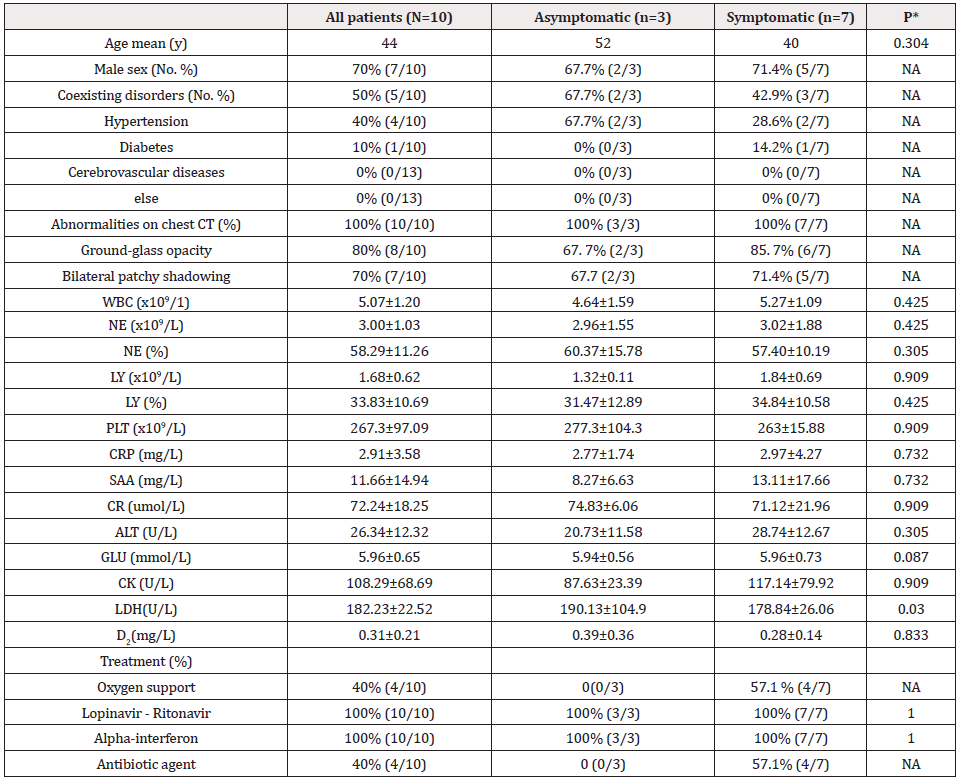
As shown in Table 1, Basic characteristics of the study population are summarized in Table 1. Of these patients, 7 were male and 3 were female, with an age range of 22 to 59 years and a median age of 49 years. Three patients were asymptomatic. The median age of the asymptomatic patients was 52, older than the symptomatic patients (52 vs 40, P = 0.37) Hypertension (4/10), diabetes (2/10) were the common coexisting conditions.
Radiologic, Laboratory Findings and Treatment
Of 10 patients who underwent chest computed tomography on admission, 100% manifested as pneumonia (and see asymptomatic Imaging Features, Figure 1). The most common patterns on chest computed tomography were ground-glass opacity. 7 cases of double lung and 3 cases of single lung. On admission, there was no statistical difference between the two groups in laboratory tests (except LDH). the asymptomatic patients had a significantly higher LDH than symptomatic patients (P = 0.03). All patients received antiviral therapy (Lopinavir–Ritonavir and Alpha-interferon) of the symptomatic cases, 4 received oxygen therapy and antibacterial therapy.
Clinical Course and Follow-Up
As shown in Table 2, the lung lesions of all patients began to absorb at the 12th day. the days of Negative for SARS-CoV2 RNA and Hospital stay were compared between the two groups, no difference was observed (28.33±19.22 vs 22.88±12.04,P=0.7; 34±16.37 vs 28.57±12.12,P=0.57). At the 4-week follow-up after discharge, the lung lesions of 5 patients with symptoms were completely absorbed, the lung lesions of 2 patients with asymptoms were completely absorbed (71.4% vs 66.7%, P=0.62) (Table 1&2) (Figure 1).

Table 1: Demographic, clinical, laboratory, and radiographic findings of patients on admission.
Abbreviations: WBC: White Blood Cell NE: Neutrophil LY: Lymphocyle PLT: Platelet CRP: C-Reactive protein CR: Creatinine ALT: Alanine Aminotransferase GLU: Glucose CK: Creatine Kinase LDH: Lactate Dehydrogenase D2: D-Dimer

Figure 1: CT images of 3 asymptomatic patients with COVID ‐ 19 on admission Note: A. Multiple patchy shadowing in lower lobe of left lung (case 1) B. Image shows multiple ground-glass opacities and minor consolidation in bilateral lungs (case2) C. Ground-glass opacity with consolidation in middle lobe of right lung (case3)
Conclusion
The clinical course and outcome in asymptomatic and symptomatic patients were similar, pay attention to the clinical management and epidemiological significance of asymptomatic patients.
Discussion
The clinical spectrum of COVID-19 can range from asymptomatic infection to mild upper respiratory tract illness to severe interstitial pneumonia with respiratory failure and even death [5]. It is estimated that non-severe patients with no symptoms or mild symptoms could represent 30%–60% of all infections [6] a large study on 72 314 Chinese patients reported that 1% were asymptomatic [7]. We currently do not know where 2019-nCoV falls on the scale of human-to-human transmissibility. Transmission of 2019-nCoV Infection from an Asymptomatic Contact in Germany [8]. Therefore, if asymptomatic patients are not identified in a timely manner and quarantined, they could become moving sources of infection and lead to massive transmission of disease [9]. A lack of severe disease manifestations affects our ability to contain the spread of the virus. Identification of chains of transmission and subsequent contact tracing are much more complicated if many infected people remain asymptomatic or mildly symptomatic. In this manner, a virus that poses a low health threat on the individual level can pose a high risk on the population level, with the potential to cause disruptions of global public health systems and economic losses. This possibility warrants the current aggressive response aimed at tracing and diagnosing every infected patient and thereby breaking the transmission chain of 2019-nCoV.
Asymptomatic patients currently are receiving more and more attention. On February 5, 2020, the "Diagnosis and Treatment Protocol for COVID-19 (Fifth ed., Trial)" was released by the National Health Commission of the People's Republic of China, and asymptomatic infection was first included as a source of infection [10]. Asymptomatic infection were laboratory-confirmed positive for the COVID-19 virus by testing the nucleic acid of the pharyngeal swab samples who did not show any subjective symptoms, including asymptomatic carriers and incubation period infection [11] the latter can develop symptoms (fever, cough, fatigue, etc.) during hospitalization. It is important to evaluate the virological course, clinical features, and outcomes of asymptomatic. In the study performed by Hu, et.al [12], described clinical features of 24 asymptomatic patients on their study, five cases (20.8%) developed symptoms (fever, cough, fatigue, etc.) during hospitalization. Twelve (50.0%) cases showed typical CT scan images of ground-glass chest and 5 (20.8%) presented stripe shadowing in the lungs. The remaining 7 (29.2%) cases had a normal CT image and had no symptoms during hospitalization and none of the 24 cases developed severe COVID-19 pneumonia. they also found that young cases (<15 years old) were prone to be asymptomatic. In the previous study, one asymptomatic child (aged 10 years) had radiological ground-glass lung opacities [13]. It is unclear why children are less susceptible to COVID-19. Potential explanations include that children have less robust immune responses (i.e. nocytokine storm), partial immunity from other viral exposures, and lower rates of exposure to SARS-CoV-2 [14]. In the present study, all patients had typical CT images, and 3 patients (mean aged 52) had no symptoms during hospitalization. There was no statistical difference between the two groups in laboratory tests (except LDH), clinical course and follow-up data. We cannot explain the difference about LDL, which may be due to the small sample size.
In conclusion, Individuals of all ages were involved in the COVID-19 asymptomatic infection. The actual prevalence of asymptomatic cases of COVID-19 could not be determined. There are limited data on the symptoms of individuals with mild COVID-19. Further research on the asymptomatic individuals of COVID-19 is essential for effective control of the pandemic spread of SARS-CoV-2.
This study has several limitations obviously, this study is limited by the small sample size. Large-scale multicenter studies are needed to verify our findings.
References
- Singhal T (2020) A Review of Coronavirus Disease-2019 (COVID-19). Indian J Pediatr 87(4): 281-286.
- Samsami M, Zebarjadi Bagherpour J, Nematihonar B, Tahmasbi H (2020) COVID-19 Pneumonia in Asymptomatic Trauma Patients; Report of 8 Cases. Arch Acad Emerg Med 8(1): e46.
- Tabata S, Imai K, Kawano S, Mayu Ikeda, Kodama T, et al. (2020) Clinical characteristics of COVID-19 in 104 people with SARS-CoV-2 infection on the Diamond Princess cruise ship: a retrospective analysis. Lancet Infect Dis 20(9): 1043-1050.
- Byambasuren O, Cardona M, Bell K, Clark J, McLaws M, et al. (2020) Estimating the extent of asymptomatic COVID-19 and its potential for community transmission: systematic review and meta-analysis. MedRxiv.
- Guan WJ, Ni ZY, Hu Y, Wen hua Liang, Chun quan, et al. (2020) Clinical Characteristics of Coronavirus Disease 2019 in China. N Engl J Med 382(18): 1708-1720.
- Mizumoto K, Kagaya K, Zarebski A, Chowell G (2020) Estimating the asymptomatic proportion of coronavirus disease 2019 (COVID-19) cases on board the Diamond Princess cruise ship, Yokohama, Japan, 2020. Euro Surveill 25(10): 2000180.
- Wu Z, McGoogan JM (2020) Characteristics of and Important Lessons From the Coronavirus Disease 2019 (COVID-19) Outbreak in China: Summary of a Report of 72 314 Cases From the Chinese Center for Disease Control and Prevention. JAMA 323(13): 1239-1242.
- Rothe C, Schunk M, Sothmann P, Gisela B, Guenter F et al. (2020) Transmission of 2019-nCoV Infection from an Asymptomatic Contact in Germany. N Engl J Med 382(10): 970-971.
- Zhou P, Yang XL, Wang XG, Ben HU, Lei Z, et al. (2020) A pneumonia outbreak associated with a new coronavirus of probable bat origin. Nature 579(7798): 270-273.
- (2020) National Health Commission of the People's Republic of China. Notice on the issuance and dissemination of the Diagnosis and Treatment Protocol for COVID-19 (Fifth Edition).
- (2020) COVID-19 Joint Prevention and Control Mechanism of the State Council. Management standards for asymptomatic COVID-19 cases (EB/OL].
- Hu Z, Song C, Xu C, Jin G, Chen Y, Xu X, et al. (2020) Clinical characteristics of 24 asymptomatic infections with COVID-19 screened among close contacts in Nanjing, China. Science China Life Sciences 63(5): 706-711.
- Chan JF, Yuan S, Kok KH, Wang To, Hin Chu, et al. (2020) A familial cluster of pneumonia associated with the 2019 novel coronavirus indicating person-to-person transmission: a study of a family cluster. Lancet 395(10233): 514-523.
- Wiersinga WJ, Rhodes A, Cheng AC, Peacock SJ, Prescott HC (2020) Pathophysiology, Transmission, Diagnosis, and Treatment of Coronavirus Disease 2019 (COVID-19): A Review. JAMA 324(8): 782-793.


 We use cookies to ensure you get the best experience on our website.