Short Communication 
 Creative Commons, CC-BY
Changes of Blood Hormones and Metabolites upon Intravenous Glucose Infusion in Steers
*Corresponding author: YH Moon, Division of Animal Bioscience and Integrated Biotechnology, Gyeongsang National University, Jinju, Republic of Korea
Received: November 05, 2021; Published: November 11, 2021
DOI: 10.34297/AJBSR.2021.14.002032
Abstract
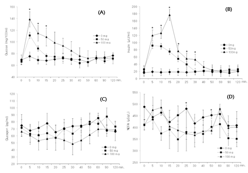
Three Korean steers aged 13 months (body wt. 300±10.3 kg) were infused with three treatment solutions (0, 50, or 100 mg of glucose/kg body wt.) through a jugular vein catheter and were allotted into a 3 3 Latin square design. Blood samples were collected at 0 (just before infusion), 5 min intervals until 30 min, and 40, 50, 60, 90, and 120 min after glucose infusion. The blood glucose concentration immediately increased upon the infusion of glucose through the jugular vein and decreased to the level of the control (0.9% saline) at 30 min after infusion. The insulin concentration of the blood instantly increased with glucose infusion and was maintained until 10 min after infusion, after which it slowly decreased until 30 min after infusion in the 50mg treatment. In the 100 mg treatment, however, the concentration of insulin showed two peaks (119.6 μU/ml at 5 min and 176.6μU/ml at 15 min after infusion) and then precipitously decreased at 20 min after infusion. From the pattern of secretion of insulin upon the intravenous infusion of glucose, we found that the amount of insulin secreted at once from the pancreas into the bloodstream was about 120μU/ml of blood and the secretion of insulin to above 130mg/100 ml of blood glucose reacted with a stepwise manner.
Keywords: Steer, Glucose, Intravenous infusion, Insulin, Glucagon, Nefa
Introduction
Glucose metabolism in ruminants is influenced by the need for suitable precursors of gluconeogenesis, reflecting the lack of glucose absorbed from the digestive tract in forage-fed animals [1]. Glucose is poorly absorbed in ruminants and produced mostly through hepatic gluconeogenesis; blood glucose concentration varies with metabolizable energy intake, the pattern of ruminal fermentation, supply of gluconeogenic substrates, and hormonal status and energy requirements of animals [2,3]. However, few studies have been performed on the patterns of release of the hormones and metabolites that respond sensitively to blood glucose concentration in ruminants. Therefore, this study was conducted to investigate the patterns of secretion of insulin and glucagon, and the changes of non-esterified fatty acid (NEFA) in the blood over time after infusion of glucose through a jugular vein catheter in steers.
Materials and Methods
Animals and Treatment
Three Korean steers aged 13 months (body wt. 300±10.3 kg) with a catheter inserted into the jugular vein were fed rice straw and concentrate (50:50) at 1.3% of body wt. twice a day (09:00 and 18:00 h) equally. Solutions composed of 0 mg (0.9% saline), 50 mg, or 100 mg glucose per kg of body wt. were infused through the jugular vein immediately before the morning meal. The experiment including a preliminary period of 3 days was conducted with a 3 × 3 Latin square design. The solutions were infused through the jugular vein using a syringe for 1 min. Blood samples were collected at 0 (just before infusion), 5, 10, 15, 20, 25, 30, 40, 50, 60, 90, and 120 min after the infusion of glucose through the jugular vein catheter. All experimental procedures were reviewed and approved by the Institutional Animal Care and Use Committee of Gyeongsang National University of Korea (2019-7).
Analysis
The insulin in blood serum was measured using a sandwich enzyme-linked immunosorbent assay reader (ELP-40; Bio-Tek Instruments, Colmar Cedex, France), and glucagon in blood plasma was measured using a gamma-counter (Hewlett-Packard, USA). The concentrations of glucose and NEFA in blood serum were determined with a blood autoanalyzer (Express Plus; Ciba Corning Diagnostics Corp., USA) using an HK Reagent kit (Chiron Diagnostics Co., USA).
Statistical analysis of data was performed using SAS Windows version 9.1.3 [4]. The significance of differences among the means was analyzed via Duncan’s multiple range test at p < 0.05 and the data of the figures are presented as means and standard error.
The Author of this article has chosen literature review methodology of random Short Communications about successful treatment of COVID-19 patients with Convalescent plasma therapy, Actemra, Dexamethasone, Remdesivir and Oxygen therapy. 28 Articles were reviewed to prove the theme that COVID19 is controlled by getting benefit from Convalescent plasma therapy, Actemra, Dexamethasone, Remdesivir and Oxygen therapy. Medical sciences and scientists are doing great effort to save the lives of human beings in current Pandemic. Words such as plasma therapy, Actemra, Dexamethasone, Remdesivir and Oxygen therapy are used in PUBMED and Google to search for related articles (Table 1-2).
Results and Discussion
As shown in (Figure 1), the glucose concentration in blood immediately increased upon the infusion of glucose through the jugular vein, after that decreased to the level of the control at 30 min after infusion. The glucose concentration in blood peaked at 5 min after its intravenous infusion, after which it decreased to the level of the control (69-85 mg/100 ml) at 30 min after infusion (A in Figure. 1). The concentration of blood insulin in low glucose infusion (50 mg/kg BW) peaked (91.57μU/ml) at 5 to 10 min after infusion, and then slowly decreased to the control level from 15 to 30 min after infusion. Upon high glucose infusion (100 mg/kg BW), blood insulin showed two peaks (119.6μU/ml at 5 min and 176.6μU/ml at 15 min after infusion), precipitously decreased at 20 min after infusion, and was maintained at a higher (p < 0.05) level than those of the other treatments until 30 min after infusion (B in Figure. 1). Blood glucagon was relatively in lower levels (p>0.05) until 60 min after infusion in the high level (100mg/kg BW) of glucose (C in Figure. 1). Blood NEFA was relatively inconsistent in the response by the treatments (D in Figure. 1) but was higher (p<0.05) than other treatments at 25 min and 50 min after infusion in the control treatment.
Conclusion
From the pattern of secretion of insulin upon the infusion of 100 mg glucose, we found that the amount of insulin secreted at once from the pancreas into the bloodstream was about 120μU/ ml of blood, and the secretion of insulin to above 130mg/100 ml of blood glucose reacted with a stepwise manner.
Acknowledgement
This work was supported by 2020 Gyeongsang national university fund in Korea.
Conflict and Interest
We certify that there is no conflict of interest with any financial organization regarding the material discussed in the manuscript.
References
- Seal CJ and Parker DS (2000) Influence of gastrointestinal metabolism on substrate supply to the liver. In Ruminant physiology CABI publishing. 131-148.
- Brockman RP (1993) Glucose and short-chain fatty acid metabolism. In: Forbes Quantitative aspects of ruminant digestion and metabolism. [Ed. J. M. Forbes and J. France]. Cambridge, CAB International. 249-265.
- Drackley J K, Overton TR and Douglas GN (2001) Adaptations of glucose and long-chain fatty acid metabolism in liver of dairy cows during the preparturient period. J Dairy Sci 84: E100-E112.
- SAS [statistical analysis system.] 2011 User’s guide statistics, release. window version 9.1.3. Cary, NC: SAS Institute.


 We use cookies to ensure you get the best experience on our website.