Research Article 
 Creative Commons, CC-BY
A Comparative Study on the Microstructural Change of Bony Endplate During Spinal Degeneration in Rats
*Corresponding author: Zongping Luo, Orthopedic Institute, Department of Orthopedics, The First Affiliated Hospital of SooChow University, 708 Renmin Rd, SuZhou, Jiangsu, 215007, PR China.
Received: February 10, 2022; Published: March 07, 2022
DOI: 10.34297/AJBSR.2022.15.002151
Abstract
Background and Objective: Bony endplate is a kind of sponge-like bone porous tissue, with a capillary network inside the heterogeneous foramina to provide the tissue structure foundation for the nutrition metabolism of the intervertebral disc. However, changes in bony endplate structure and foramina during spinal degeneration are poorly understood. This study aims to study the microstructural change of bony endplate and foramina during spinal degeneration in rats.
Methods: Degeneration was made in the lumbar spine and coccygeal vertebra, respectively, of rats. Structural changes in bony endplate and foramina during spinal degeneration were analyzed by scanning electron microscopy and atomic force microscopy.
Results: In lumbar spine degeneration, there was no significant change in the degenerative bony endplate, and osteophytes appeared in some of the foramina. Sagittal section results showed that there was no significant change in the number and primary morphology of the foramina. Atomic force microscope test showed that modulus of elasticity has no obvious change between the degenerated bony endplate and the normal bony endplate. In the coccygeal vertebra degeneration model, with the time of degeneration, the foramina on the surface of the bony endplate grew more and more osteophyte, also with the reduction in the number of the foramina and the disappear of normal “8” or “吕” shape. From the sagittal position, the cross foramina in the bony endplate began to close or even disappear, and the number of bony channels decreased significantly. Atomic force microscope showed that bony endplate of the 8-week degenerated coccygeal vertebra was tougher than normal bony endplate.
Conclusion: Mild degeneration of the spine will not lead to changes in the bony endplate, while severe degeneration will lead to changes in the bony endplate, such as decreasing number of the foramina and osteophytes formation.
Keywords: Bony endplate, Spinal degeneration, Sponge-like, Porous structure
Abbreviations: IVD - Intervertebral Disc
Introduction
The intervertebral disc (IVD) degeneration is a common spinal disorder that has been implicated in its pathogenesis and pathological changes [1]. Some pathological structural changes in IVD, such as tears in the anulus fibrosus has been observed in spinal degeneration patients [2,3]. Especially, structural changes of cartilaginous bony endplate that lie between the rigid vertebral bodies of the spine are one of the pathological degenerative changes of IVD, which are a major cause of back pain even neurological deficits in human [4]. In a healthy state, vertebral bony endplates have a sponge-like porous structure that provides the tissue structure foundation for the nutrition metabolism of the intervertebral disc. In the adult with the spinal degenerative disorder, IVD degeneration significantly correlated with intervertebral disc-endplate degeneration [5,6], and the altered cell nutrition in the IVD caused by bony endplate structural dysfunction is considered the main cause for disk degeneration [7]. However, very little data exists on acute structural change of vertebral bony endplate at the onset and progression of IVD degeneration. In the previous study, we observed the unique three-dimensional structure combined with interior foramina on the surface using scanning electron microscopy [8]. In this study, we will observe the change process of bony endplate during spinal degeneration by detailed visualization and microstructural characterization using scanning electron microscopy and atomic force microscopy.
Materials and Methods
Animals
Fifty-four male Sprague Dawley rats were housed in a 12:12 h bright light-dark (1,000 lux) cycle cage and were allowed to acclimatize for 7 days before the commencement of treatments. Animal experiments protocols have been approved by the Animal Care and Animal Research Ethics Committee of Orthopaedic Institute, The First Affiliated Hospital of Soo Chow University.
Models’ Establishment
Experiment I
Thirty-two rats were randomly divided into two groups: a normal group and a model group (n=16 in each group). For lumbar spine degeneration establishment, rats were anesthetized with chloral hydrate and exposed to the lumbar vertebrae. The spinous process and its associated ligaments were removed to destabilize the lumbar spine. The wound was sutured and treated with a local injection of penicillin avoiding infection. Normal rats exposed only the lumbar vertebrae and did not remove other tissues. The two groups of rats were raised in the same feeding environment, and two rats were randomly selected and killed every other month. The lumbar vertebrae of rats were taken, and the changes of the tissues were observed with the scanning electron microscope. At the end of 6 months, no accidental death was found in either group. In the remaining 4 rats of each group, 2 were examined by scanning electron microscopy and 2 by atomic force microscopy.
Experiment II
After 6 months of treatment of lumbar process de-spinous instability, the degeneration of lumbar bony endplate was not obvious, so experiment II was further designed to promote the degeneration of the spine through coccygeal vertebra compression. Twenty-two rats were randomly divided into two groups: a normal group and a model group (n=11 of each group). For coccygeal vertebra degeneration establishment, rats were anesthetized with chloral hydrate. At the central point of the coccygeal vertebra of the model group of rats from the L7-L10 vertebra, a fine needle was injected horizontally, and a pre-made mold was used to pressurize it (the nail spacing was 12mm under normal conditions, and the nail spacing was changed to 11mm after pressurization).
A small dose of penicillin was injected around the nail track to prevent infection. The normal group of rats did not do any treatment. Both groups were kept in the same feeding environment, and two rats were randomly selected and killed at weeks 2, 4, and 8. The coccygeal vertebra of rats was taken, and the changes of the tissues were observed with the scanning electron microscope. At the end of 8 weeks, one rat died in the model group and none in the normal group. In the remaining 4 rats of each group, 2 were examined by scanning electron microscopy and 2 by atomic force microscopy.
Specimen processing
According to L1 - L6, the lumbar spine was cut into 6 portions with the integrity of each vertebral bodies and the bony endplate with the intervertebral disc and articular process joints were cut up to remove the spinal cord and respectively put in the six with cover tube (without the use of glutaraldehyde soaking pretreatment, because of protein denaturation, unable to take the next step processing). Each tube has two vertebral bodies. The coccygeal vertebra was divided into 4 parts according to C7-C10 (only the distal half of C7 was taken, and the proximal half of C10 was taken, because only the two parts of the C7 and C10 coccyx were compressed to produce degeneration), and they were put into 4 tubes with lidded (also without glutaraldehyde immersion and other pretreatments), with 2 vertebral bodies in each tube. A volume of 20ml of the mixed solution of collagenase type I and collagenase type II (1:1 of both with a concentration of 1%) was added to each test tube and put into a 37℃ incubator. The mixed solution was changed every 2 days. After 6 days, the specimens soaked with collagenase were removed, and all the soft tissues and cartilage on the surface were cleaned, and the integrity of the bony endplate was preserved. After repeated washing with normal saline, gradient dehydration with 70%, 80%, and 90% alcohol was carried out and then air-drying in a cool and ventilated place for 24 hours. After the above treatment, the osseous endplate has been separated from the vertebral body (part of the bony endplate has been completely separated from the vertebral body). If not, the bony endplate can be completely detached from the vertebral body with only a slight external force. If the special condition that the bony endplate of the coccygeal vertebra after 8 weeks of partial pressure has been closely combined with the vertebral body and cannot be separated from the vertebral body at all, the degenerative bony endplate can only be sawed off with the part of the vertebral body.
Scanning electron microscopy detection
After the specimen treatment, four bony endplates (two upper and lower bony endplates for each vertebral body) could be obtained from the samples in each test tube, and 20ml Formic acid formalin mixed solution was added to each test tube for decalcification. Three days later, use the “the double cutters and slice method” along the endplate by sagittal cut. The complete section including annulus fibrosus and nucleus pulposus area of the bony endplate and complete not damaged was directly pasted in the conductive adhesive paste of the electron microscope samples on the stage, put in a cool ventilated place dry naturally for 24 hours, and sprayed gold following by using a scanning electron microscope observation.
Atomic force microscopy
After specimen processing, the highest degree degeneration and the good integrity of L4 and C8 bony endplate of the model rat and the L4 and C8 bony endplate of the normal rat was compared using the atomic force microscopy. Because of bony endplate surface is uneven, so it picks a point to test rather than use the scanning function of atomic force microscopy.
Statistical analysis
Statistical analysis of all data was performed using IBM SPSS Statistics 26 software (Armonk, NY, USA).
Results
Changes In the Surface Characteristics of The Bony Endplate In Lumbar Spine Degeneration Rat
In the surface of the section of the bony endplate, the microstructure after 6-month degeneration has no obvious change and there is the only osteophyte that occurred in some foramina (Figure 1). The foramina kept its primary structure of the “8” or “ 吕” (Figure 1). These data suggesting the lumbar spine was mild degeneration.
Changes In the Foramina of The Bony Endplate in Lumbar Spine Degeneration Rat
According to the observation of the sagittal section, there was no obvious change in the number and shape of the foramina (Figure 2) suggesting that the mild lumbar spine degeneration had no obvious interior foramina degeneration.
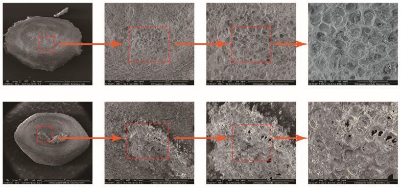
Figure 1: Microstructural characterization of lumbar spine bony endplate by scanning electron microscopy. The upper is the bony endplate after instability for 6 months and the lower is the normal bony endplate. Zoom in from left to right.

Figure 2: Microstructural characterization of the lumbar spine bony endplate in the sagittal section by scanning electron microscopy. The upper is the bony endplate after instability for 6 months and the lower is the normal bony endplate. Zoom in from left to right.
Changes In the Surface Characteristics of The Bony Endplate in Coccygea Vertebral Degeneration Rat
Compared with normal bony endplate, 4-week depressioninduced the mild degeneration and 8-week induced severe degeneration (Figure 3). With the time of degeneration, foramina on the surface of bony endplate grew more and more osteophyte, which made the loss of smooth form (Figure 3). Some foramina were closed, so the total number of foramina decreased. Except, the foramina lost its normal “8” or “吕” shape (Figure 3).
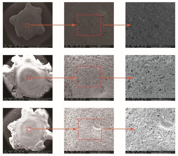
Figure 3: Microstructural characterization of coccygeal vertebra bony endplate by scanning electron microscopy. The upper is normal bony endplate, the middle is the bony endplate after 4-week degeneration and the lower is the bony endplate after 8-week degeneration. Zoom in from left to right.
Changes In the Foramina of The Bony Endplate in Coccygeal Vertebra Degeneration Rat
According to the observation of the sagittal section, on the 8-week degeneration, the interior foramina of bony endplate began to close and even disappear. The number of bony channels was decreased (Figure 4). However, there was no obvious change in shape, osteophyte formation, and the number of foramina in the backing of bony endplate (Figure 5).
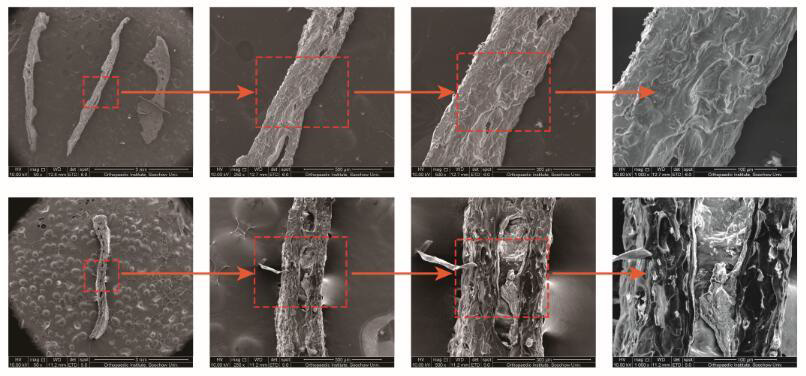
Figure 4: Microstructural characterization of the coccygeal vertebral bony endplate in the sagittal section by scanning electron microscopy. The upper is the bony endplate after 8-week degeneration and the lower is the normal bony endplate. Zoom in from left to right.
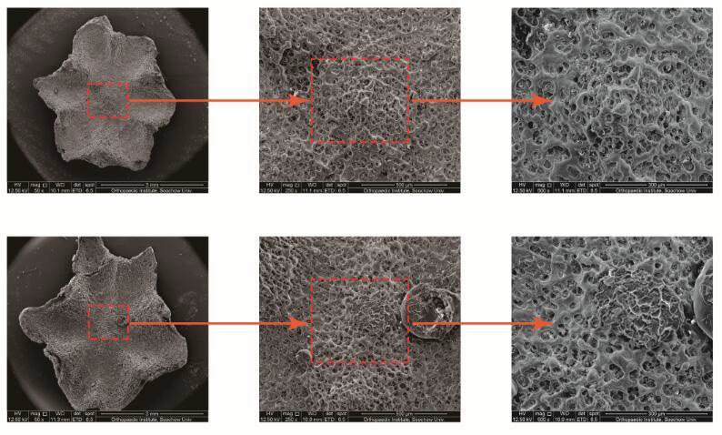
Figure 5: Microstructural characterization of coccygeal vertebra bony endplate in the reverse side after 8-week degeneration by scanning electron microscopy. The lower is normal control. Zoom in from left to right.
The Elasticity Modulus of The Bony Endplate of The Degenerated Spine by Atomic Mechanics Microscopy
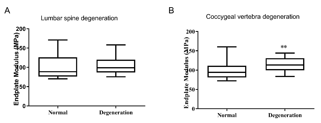
In rat with lumbar spine degeneration, the elasticity modulus of bony endplate was 104.7±5.07MPa,which had no significant difference from normal rat (103.5±7.59 MPa) (P>0.05). The bony endplate of the rat with 8-week coccygeal vertebra degeneration had a 114.8±3.85 elasticity modulus, which significantly higher than normal bony endplate (99.53±5.19) (P<0.05).
Discussion
The cartilaginous bony endplate transmits complex and multiaxial loads between the disc and vertebra to ensure proper range-of-motion. Evidence is attempting to define the involvement of bony endplate in the spinal degeneration due to its critical role in supplying intervertebral disk nutrition [7,9]. The present study compared the change in microstructural characterization of the cartilaginous bony endplate in both mild and severe spinal degeneration and to understand the change process of bony endplate during spinal degeneration. Observations based on rat lumbar spine degeneration and coccygeal vertebra degeneration showed the significant structural change in bony endplate and foramina in severe spinal degeneration of coccygeal vertebra.
Due to an avascular structure, cells in the center of the disc of an adult must survive from their nearest blood supply. The approximately 40% porous in the central region of the bony endplate is such foundational support for the vascular network [10] to enable the nutrient and metabolite exchange for the disc adjacent to the bony endplate. Recently researchers found that changes to endplate morphology and composition impair its permeability associate with disc degeneration [11]. In our experiment I, in the model of spinal degeneration induced by the unstable lumbar spine in rats, there was no obvious structural change in the microstructure of the lumbar bony endplate, and the most foramina kept their primary structure of “8” or “吕” shape, suggesting no obvious degeneration occurs. However, osteophytes can be seen in some of the foramina. It indicates that the degeneration of the lumbar spine endplate in rats is mild, which may be because the rats walk on all fours and carry less load on the spine. Even if the structure around the lumbar spine is seriously damaged, no obvious degeneration will occur in a short period in the rat. In our experiment II, in the coccygeal vertebra degeneration rat, the foramina lose its normal “8” or “吕” shape and begin to close, suggesting bony endplate of rat coccygeal vertebra has severe degeneration. Endplate biomechanical function is determined by multiple structural factors, such as thickness, porosity, and curvature, and it is the guarantee of the normal function of the bony endplate [12-15].
We evaluated the function of the bony endplate by examining the elastic modulus of the mild and severe degenerated endplate and found that in experiment I, there was no significant change in the elastic modulus of the bony endplate after degeneration, and in experiment II, the elastic modulus of the bone endplate after degeneration was significantly changed compared with the normal one, indicating that mild degeneration would not cause fundamental changes in the bone quality of the bony endplate, and if the spine experienced more serious degeneration, the bone quality of the bone endplate would undergo fundamental changes. According to the results of the atomic force microscope in the above two experiments, mild degeneration of the spine will not lead to changes in the bone quality of the bony endplate, while more severe degeneration will lead to changes in the bone quality of the bony endplate.
The absence of significant degeneration in the bony endplate in experiment I does not mean that the internal vascular network is normal. However, the integrity of its internal foramina suggests that there may still be a relatively complete capillary network. In experiment II, the bony endplate after 8 weeks of pressure showed obvious changes, and the internal foramina had been seriously deformed or even closed and disappeared, which indicated that the internal capillary network showed obvious changes, and even many vascular networks had disappeared. Further, during the degeneration, no significant changes were observed in the foramina at the back of the bony endplate (the side facing the vertebral body). In combination with the results of previous experiments, it can be concluded that during spinal degeneration, the capillary network inside the bony endplate will undergo significant degeneration, but the capillary network inside the bony endplate penetrating from the side of the vertebral body does not undergo significant change.
In this study, elastic modulus, and microstructure in the experiment I have no obvious change while significantly changed in experiment II. We can conclude that in the process of spinal degeneration, the first degeneration occurs in the capillary network, which has an impact on the nutrition and metabolism of the surrounding structure and finally leads to a series of changes in the bony endplate, including osteophytes, foramina closure, and changes in the bone quality. Still, there are shortcomings in this study due to experimental restrict need further to be investigated: what is the changing process of the capillary network inside the heterogeneous foramina during bony endplate degeneration? Whether the capillary network of the back of bony endplate changes although this site has no significant degeneration?
Conclusion
In summary, based on previous experiments, this study studied the degeneration process of bony endplates in the process of spinal degeneration. The bony endplate is also an important factor in the process of spinal degeneration and to understand the change of the bony endplate makes better comprehension of the degeneration of the spine. However, there are still many deficiencies in this experiment. In the next experiment, we will try to make up for the deficiencies in this experiment and learn more about the changes in bony endplates in the process of spinal degeneration.
Acknowledgements
Not applicable.
Conflict of interest
The authors declare that they have no competing interests.
References
- Wiet MG, Piscioneri A, Khan SN, Ballinger MN, Hoyland JA, et al. (2017) Mast Cell-Intervertebral disc cell interactions regulate inflammation, catabolism and angiogenesis in Discogenic Back Pain. Sci Rep 7(1): 12492.
- Stich S, Jagielski M, Fleischmann A, Meier C, Bussmann P, et al. (2020) Degeneration of Lumbar Intervertebral Discs: Characterization of Anulus Fibrosus Tissue and Cells of Different Degeneration Grades. Int J Mol Sci 21(6): 2165.
- Inoue N, Espinoza Orias AA (2011) Biomechanics of intervertebral disk degeneration. Orthop Clin North Am 42(4) 487-499.
- Senck S, Trieb K, Kastner J, Hofstaetter SG, Lugmayr H, et al. (2020) Visualization of intervertebral disc degeneration in a cadaveric human lumbar spine using microcomputed tomography. Journal of Anatomy 236(2): 243-251.
- Ding WY, Wu HL, Shen Y, Zhang W, Li BJ, et al. (2011) Correlation between intervertebral disc-endplate degeneration and bony structural parameter in adult degenerative scoliosis and its significance. Chinese Journal of Surgery 49(12): 1123-1127.
- Dolan P, Luo J, Pollintine P, Landham PR, Stefanakis M, et al. (2013) Intervertebral disc decompression following endplate damage: implications for disc degeneration depend on spinal level and age. Spine (Phila Pa 1976) 38(17): 1473-1481.
- Ruiz Wills C, Foata B, Gonzalez Ballester MA, Karppinen J, Noailly J (2018) Theoretical Explorations Generate New Hypotheses About the Role of the Cartilage Endplate in Early Intervertebral Disk Degeneration. Front Physiol 9: 1210.
- Fu B, Jiang H, Che Y, Yang H, Luo Z (2020) Microanatomy of the lumbar vertebral bony endplate of rats using scanning electron microscopy. Orthop Traumatol Surg Res 106(4): 731-734.
- Urban JP, Roberts S (2003) Degeneration of the intervertebral disc. Arthritis Res Ther 5(3): 120-30.
- Rodriguez AG, Slichter CK, Acosta FL, Rodriguez AE, Burghardt AJ, et al. (2011) Human disc nucleus properties and vertebral endplate permeability. Spine (Phila Pa 1976) 36(7): 512-520.
- Fields AJ, Ballatori A, Liebenberg EC, Lotz JC (2018) Contribution of the endplates to disc degeneration. Curr Mol Biol Rep 4(4): 151-160.
- Hulme PA, Boyd SK, Ferguson SJ (2007) Regional variation in vertebral bone morphology and its contribution to vertebral fracture strength. Bone 41(6): 946-57.
- Langrana NA, Kale SP, Edwards WT, Lee CK, Kopacz AJ (2006) Measurement and analyses of the effects of adjacent end plate curvatures on vertebral stresses. Spine J 6(3): 267-278.
- Zhao FD, Pollintine P, Hole BD, Adams MA, Dolan P (2009) Vertebral fractures usually affect the cranial endplate because it is thinner and supported by less-dense trabecular bone. Bone 44(2): 372-379.
- Dudli S, Enns BE, Pauchard Y, Rommeler A, Fields AJ, et al. (2018) Larger vertebral endplate concavities cause higher failure load and work at failure under high-rate impact loading of rabbit spinal explants. J Mech Behav Biomed Mater 80: 104-110.



 We use cookies to ensure you get the best experience on our website.