
Research Article 
 Creative Commons, CC-BY
Mesenchymal Stem Cell Therapeutic Potential in Regenerative Medicine and Its Future Direction
*Corresponding author: AK Azad, Department of Pharmacy, Daffodil International University, Bangladesh.
Received: February 21, 2022; Published: February 28, 2022
DOI: 10.34297/AJBSR.2022.15.002142
Abstract
MSCs (mesenchymal stem cells) are stromal cells that can self-renew and differentiate into mesenchymal and non-mesenchymal lineages. These cells’ inherent characteristics make them an appealing possibility for clinical use. MSCs are of particular interest since they can be quickly grown in vitro and obtained from a tiny aspirate of bone marrow or adipose tissues. Furthermore, they are an even more appealing candidate for regenerative medicine because of their ability to control immune responses. Allogeneic transplantation of these cells is possible without significant danger of immunological rejection. MSCs release various immunomodulatory chemicals that provide a regenerative milieu for wounded tissues or organs, reducing damage and promoting self-regulated tissue regeneration. Autologous/allogeneic MSCs supplied through the bloodstream can boost the number of MSCs recruited to tissue trauma sites and promote healing. MSCs are currently being studied for cell and gene therapy applications in treating a variety of severe human diseases and genetic disorders. This paper reviews the current clinical and nonclinical evidence regarding MSCs’ use in tissue regeneration and their potential therapeutic involvement in various illnesses.
Keywords: MSCs, Immunology, Osteogenesis and Chondrogenesis, T-lymphocyte
Introduction
In embryonic development, stem cells are immature tissue precursor cells that can self-renew and specialize into various cell types [1,2]. MSCs, commonly known as multipotent stromal cell differentiation, is conscience cells that can be detected in nearly all postnatal organs and tissues [3,4]. MSCs are also known as multipotent mesenchymal stromal cells. In recent years, MSCs have been increasingly popular for research because they can be easily retrieved from a small vasodilate of bone marrow or adipose tissue and grown to therapeutic sizes in a laboratory setting, eliminating the requirement for animal models in most cases. Aside from these advantages, MSCs offer many other benefits, including the ability to be preserved for an extended time while losing considerable potency and the absence of harmful effects following allogeneic MSC transplantation [5]. When Frieden stein and colleagues revealed the first method for identifying MSCs (also known as “stromal cells”) from whole bone marrow aspirates based on differential adhesion qualities in 1976, it was considered a breakthrough. Several characteristics of these cells include adhesion, clonogenicity, nonphagocytic, and fibroblastic, with the potential to produce colony-forming units-fibroblastic (CFU-F). [6] Molecular descriptions of bone marrow stromal cells in the late 1980s [7,8] by Owen and Frieden stein. To describe the subtype of marrow stromal cells involved in metagenesis, bone marrow stromal cells were further identified and designated as mesenchymal stem cells [9,10] after they were discovered in the bone marrow. Shortly after these discoveries were made, researchers began investigating the therapeutic potential of MSCs [11], and there have been no reports of ill consequences from MSC transplantation since then. In this work, we aimed to integrate recent MSC research achievements and their medicinal consequences into a single comprehensive report.
MSCs of an Immunophenotype
The identification of MSCs by the use of particular markers has proven to be a difficult task. Instead of a single surface marker, a panel of surface markers is used to describe Human MSCs (hMSCs), which can be produced from either freshly harvested tissues or cryopreserved samples. MSCs must express the surface markers CD105 (SH2), CD73 (SH3/4), and CD90, and they must be negative for the surface markers CD34, CD45, CD14, CD79, or CD19, and HLA-DR, according to the International Society for Cellular Therapy recommendations [9]. Additionally, hMSCs are negative for a number of additional antigens, including CD4, CD8, CD11a, CD14, CD15, CD16, CD25, CD31, CD33, CD49b, CD49d, CD49f, CD50, CD62E, CD62L, CD62P, CD80, CD86, CD106 (vascular cell adhesion molecule [VCAM]-1), cadherin V, and glycophorin A. Human mesenchymal stem cells [hMSCs] are positive for the following antigens: CD10, CD13, CK-8, CK-18, nestin, and von Willebrand factor [5,12,13]. On the other hand, hMSCs are negative for the following antigens: CD10, CD13, CK-8, CK-18, nestin, and von Willebrand factor [5,12,13]; CD44, CD49e (a5-integrin); CD Surface marker expression varies depending on the tissue-derived MSCs come from, for example, only adipose tissue-derived MSCs express high amounts of CD34 [14], and only bone marrow-derived MSCs, but not placenta-derived MSCs, express CD271 [15]. In another article [16], the phenotypic expression of surface markers is discussed in greater detail.
MSC’s Differentiation Potential
MSCs must also be capable of sticking to plastic and differentiating into in vitro osteoblasts, adipocytes, and chondroblasts [9], other than surface indicators. Differentiation involving transcription factors is governed by genetic processes. Some regulatory genes can control the difference between a given pathway phenotype and stimulate the Differentiation of progenitor cells to a specific lineage. In addition to the growth hormones and induction chemicals, the biomaterial scaffolded microenvironment can also supply MSCs with adequate circumstances of proliferation and difference [17]. Although MSCs may distinguish between in vitro several tissues, the resulting cell population does not perfectly imitate the targeted tissues in terms of their biochemical and biomechanical properties [18].
Mesodermic Distinguishing
In theory, mesodermal distinguishing is easily achieved for MSC because it is of the same embryonic origin. The mesodermal Differentiation (osteogenic, adipogenic, and chondrogenic) is relatively well explored in the literature. Dexamethasone (Dex), β-glycerophosphate (β-GP), and ascorbic acid phosphate (aP) mixtures were commonly employed for osteogenic differentiation induction [18,19]. Osteogenic MSC development is a complex process that is closely controlled by a wide variety of transcription and signaling pathways [20]. Run-related transcription factor 2 (Runx2) and caveolin-1 are regarded to be a crucial osteogenic differentiation regulator, which is precisely regulated by various active and repressive activators [19-21]. Bone morphogenetic proteins (BMPs) have been demonstrated to improve osteogenic MSC differences, specifically BMP-2, BMP-6, and BMP-9. The BMP9- induced osteogenic Differentiation [22] is implicated in Smads, p38, and Extracellular signal-regulated Kinase-1/2 (ERK1/2).
The vascular endothelial growth factor (VEGF) and fundamental fibroblast growth factor (bFGF) synergistically stimulate the osteogenic development of rat bone marrow-derived mesenchymal stem cells at extremely low concentrations. Wnt signaling was also implied in the osteogenic development of MSCs, apart from core-binding factor alpha-1/osteoblast-specific factor2 (cbfa1/ osf2) [23,24] Recently a study by Alm et al. has shown that 100 nM of temporary dexamethasone treatment minimizes inter-and intraindividual differences in osteoblast development of human bone marrow MSCs [25]. A scaffold or matrix designed to provide differentiating clues might be an alternative method. The sustained attachment and proliferation of MSCs supported by silicatesubstituted calcium phosphate (Si-CaP) was osteogenesis [26]. Dex and isobutyl-methylxanthine (IBMX) and indomethacin (IM) were used to differentiate the adipogenesis and seen by staining the lipid droplets in cells with the solution Oil Red O. [27] Adipogenesis was associated with peroxisome proliferator-activated receptors β2 (PPAR-α2), CCAAT/enhancer-binding protein (C/EBP) and retinoic C receptor [17].
The Phosphatidylinositol 3-kinase (PI3 K) activated by Epac is responsible for activating protein kinase B (PKB)/cAMP response element-binding protein (CREB) signals and upregulating expression, which activate transcription of adipogenic genes in turn, while Rho/Focal Adhesion (FAC)/mitogen-activated protein kinase (MEK/ERK/Runx2 signals) are responsible for osteogenesis, The transforming growth factor (TGF)-β1 and TGF-β2 are found to be involved in chondrogenesis differentiation [28]. Cartilagebased Differentiation of MSCs are characterized by upregulations of the particular cartilage genes, collagen type II, IX, aggrecans, and collagen and proteoglycan biosynthesis. The results showed putative functions of Wnt/β-catenin in the Differentiation between osteogenesis and chondrogenesis in mesenchymal cells [18]. A recent analysis revealed that miR-449a regulates the chondrogenesis of human MSCs by targeting the enhancer-binding factor of the lymphoid directly [29]. Elevated β-catenin stimulates Runx2, which causes Differentiation of osteoblast, whereas decreased β-catenin induces chondrogenesis [30], the opposite effect on the gene expression. Fibroblastic Factor-2 (FGF-2), which may lead to an early differentiation, may improve the kinetics of MSC chondrogenesis by a priming mechanism [31].
Differentiation of the Ectoderm
DMSO, butylated hydroxy anisole (BHA,” β-mercaptothion, KCL, forskolin, and hydrocortisone may be stimulated to in vitro neural development of MSCs [17]. In addition, neuronal Differentiation is observed in Notch-1 and protein kinase A (PKA) paths [32]. The downregulation of caveolin-1 in the presence of other stimulatory elements increases neuronal development of MSCs by altering the Notch signaling pathway [33].
Differentiation from the Endoderm
Hepatocyte growth factor and oncostatin M were utilized to induce cuboid cell diagnostic indicators (α-fetoprotein, 6-phosphate glucose, tyrosine aminotransferase, and CK-18) and albumin synthesis in vitro for liver differentiating [34]. Recent research has revealed strategies for the development of pancreatic β-cell x-rays with satisfactory results from adult stem cells. These cells had a certain shape, high insulin-1 mRNA expression, and insulin and nest synthesis [35,36]. Tissue-driven tissue-morphic mesenchymal stem cells may also be differentiated into endodermic islet cells (Sox17, Foxa2, and GATA4), then pancreatic (Pancreatic, and duodenal homeobox 1[Pdx1], Ngn2, Aerostatic Differentiation, Box 4 [PAX4] and Glut-2), and finally into pancreatic hormone expression (Insulin, glucagon, and somatostatin) (Insulin, glucagon, and somatostatin) and then pancreas [37].
Homing and Migration
The MSC’s physical niche and migration signals give essential information on their role and tissue interactions. Researchers paid more attention to Bone Marrow derived MSCs to disclose evidence about their therapeutic action. In vivo, it is challenging to locate the niche of MSCs. In addition, it is exceedingly difficult to detect dynamic movements of MSC due to the lack of any unique MSC marker and challenges in testing the marrow cavity. Most investigators utilize genetic markers like Y-chromosome, but these methods do not dissolve the dynamics of cell and time responses in female or fluorescent protein reporter genes [5]. Non-invasive in vivo imaging via bioluminescence imaging (BLI) can be a potential option. The fundamental advantage of BLI is that just 100 cells can be detected in vivo, even at very low levels of signals [38,39]. Significant progress has been made in this sector, but the migration of MSCs to the tissue niche is still illusory. In radiation-induced multi-organ failure, ischemic brain injury, myocardial infarction, and acute renal failure, the migration of MSCs to injuring tissues has yet been described [40].
Still, the mechanisms that regulate the migration of MSCs to injured tissues remain unknown. Human MSCs express various combinations of the CCR1, CCR4, CCR7, CCR9, CCR10, CXCR1, CXCR3, CXCR4, CXCR5, and CX3CR1 chemokine receptors [41]. The chemokines that affect the trafficking of MSCs are yet unclear; as of now, 39 chemokines with varied activities to influence the trafficking of hematopoietic cells, particularly leukocytes, have been identified [41]. Of these chemokines, the factor-1 (SDF1) generated from the stromal cell is reasonably well investigated for the migration of MSCs. The CXCR4 receptor of the SDF-1-induced cell migration is widely expressed in cells of the immune system and the central nervous system (CNS). SDF-1 has been identified in vivo with the rat model of myocardial infarction as a significant mediator in stromal progenitor migration to wounded tissue [42,43]. Hiasa et al., 2004 reported that the over-expression of human ischemic muscle SDF-1 caused endothelial progenitor cells to mobilize and improved myocardial repair.
Studies have also shown that SDF-1 levels have been elevated in infarct tissue after myocardial infarction, and this rise is correlated to the amount of MSCs that live in the heart [42,43]. On the other hand, Ip et al. research, 2007 Suggested that for their myocardial migration, MSCs utilize Intein β1, not CXCR4[44]. In addition, the MSCs can be improved in the regeneration of the skeletal tissue with growth factor delivery, as coupled MSCs and erythropoietin infusion have shown better outcomes in the treatment of limb ischemia [45]. Lysophosphatidic bioactive lipid acid (LPA) plays a major role in the migration of human lung resident MSCs through a signaling route that includes beta-catenin activation mediated by LPA1[46]. The anti-inflammatory milieu is more adaptable than the proinflammatory environment to the therapeutic hMSCs [47]. Another crucial stage in the tissue migration of circulating cells is crossing the endothelial barrier.
MSCs adhere to the endothelial cells is, similar to leucocytes, a vital step and a limited collection of molecules, like selectin-P, integrin β1, and VCAM-1 and appear to perform critical functions in this association [48]. A pre-treating of endothelial cells with some pro-apoptotic agents (angiogenesis) and inflammatory cytokines, as well as growth factor (e.g., interleukin (IL)-8, neurotrophin-3, TG-β, IL-1β, TNF-α, platelet-derived growth factor (EGF)) and growth factors (e.g., interleukin, TGF-β), is possible in vivo. Further investigations on the molecular mechanism behind migration and homing will give a stimulus for the therapeutic application of MSCs.
Action Mechanism / Action Mode
The method via which MSCs exert their antiproliferative activity has yet to be clarified, while numerous mechanisms and molecules that are likely to operate in concert and/or alternate ways depending on the environment to which MSCs are exposed have been suggested. Several investigations have demonstrated that MSCs are able to replace injured in vivo tissues [49,50]. Multiple techniques for tissue engineering have also been documented in the areas of undifferentiated or differentiated MSCs with or without biomaterial assistance [49,50]. MSCs demonstrated promise to replace different tissues such as cartilage, skeleton, tendon, vasculature, liver, kidneys, and nerves [51]. It remains, however, unknown how many originally given MSCs establish residence in the tissue and maintain a proper terminally differentiated phenotype because considerable amounts of transplanted population are apoptotic or move towards the lungs and liver during the early stage. A study with Li et al. and Askari et al. on stroke and heart injury suggests that the presence of transitory MSCs seems adequate to cause therapeutic effect [52,53]. In conjunction with these findings, resident MSCs also strive to suppress transitory and permanent immune surveillance systems and to establish an optimum curative environment by securing factors and modifying the local microenvironment [51].
T-lymphocyte activation and proliferation tests in vitro since 2002 have been employed in various investigations to detect immunomodulatory effects of human, murine, and baboon MSCs [54-56]. These investigations have shown that the MSCs can decrease the proliferation and activation of lymphocytes in response to allogeneic antigens. In addition, MSCs can trigger the formation of CD8+ T (Treg) regulatory cells as a means of suppressing lymphocyte allogenic responses [56] and prohibiting monocyte and CD34+ progenitor differentiation to antigen-presenting dendritic cells [57]. T lymphocytes activated by MSC are stopped in the G1 phase as a result of the downregulation of cyclin D2[58]. MSCs are also able to block the proliferation of NK cells induced by IL-2 or IL-15 [59,60]. MSCs have also been demonstrated to influence the proliferation, activation, IgG secretion, Differentiation, generation of antibodies, and chemotactic conduct [51]. A severe grade IV acute graft-versus-host diseases (GvHD) treatment with expanded allogeneic MSCs has successfully resolved in vivo immunomodulatory property of MSCs [61].
Moreover, MSCs decreased expression of major Class II (MHCII), Dendritic cell (DC) histocompatibility complex expression [51]. Interesting, antigen-specific T-cell proliferation in rheumatoid arthritis was further suppressed by allogeneic MSCs, which were differentiated into chondrogenic phenotypes [62], while GMS cells evaded immune rejection and caused the ectopic production of bones in vivo [56]. More than one other research has shown, however, that MSC immunomodulatory effects are not universal and unconditional and that the phenotypic of the MSCs is temporary and environment-dependent [63]. One of the most important therapeutic properties of MSCs is cytokine secretion [64]. The secretion of MSCs does not include molecules such as TGF-β, IL- 10, IL-6, cyclooxygenase-1 (COX1), and COX-2 that are responsible for the secretion of prostaglandin E2 (PEG2). DC development was partially suppressed by IL-6, and tissue inflammation decreased by IL-10, TGF-β1, and IL-6 [57,65] ary cultures of T-cells was contributed exclusively to the supernatants derived from the cocultures of stromal and activated T cells [58,66,67]. The combination of the mediating immunosuppression of MSCs does not only occur from direct inhibitory action but also involves the recruitment of other regulatory effects. Details of immunological modulation are discussed elsewhere [68,69].
MSCs in Different Diseases
Diabetes Mellitus
Diabetes mellitus (DM) is characterized by hyperglycemia caused by deficiencies in insulin secretion, action, or both. Diabetes type I, also known as juvenile-onset diabetes, is characterized by beta-cell death, which is usually caused by an autoimmune T cellmediated process and results in an absolute lack of insulin in the body, which is essential for glucose metabolism. Type 2 diabetes, often known as adult-onset diabetes, is characterized by Insulin’s failure to efficiently metabolize glucose [70]. Despite the fact that Type 1 and Type 2 diabetes have separate pathogenic pathways, they share symptoms such as glucose intolerance, hyperglycemia, and hyperlipidemia. Diabetes is also a factor. Other illnesses, such as adult blindness and kidney disease, failure, leg and foot amputation, pregnancy difficulties as well as a cardiac attack [70].
Insulin therapy as it is now is neither. capable of perfectly simulating endogenously produced Insulin is neither released nor safe, as it frequently causes hypoglycemia. [70] coma, As a result, strategies that promote either the expansion or of the body’s existing beta-cells or the supply of stem cells Insulin-producing cells produced from cells would be useful in the future. choices for treatment MSCs, as previously stated, are capable of They can differentiate into a variety of cell types, making them potentially useful. a major source for the therapy of human debilitation Diabetes, for example [71]. MSC differentiation in insulin-producing cells in vitro It is commonly known that stem cells (IPCs) exist. The process of bone differentiation Multistep differentiation procedures is used to generate bone marrow-derived MSCs. A variety of protocols are included in the protocols. In high glucose conditions, nicotinamide, activin A, and -cellulin medium. Differentiated cells exhibit a morphology at the end of the culture. high morphology resembling pancreatic islet-like cells Expression of PDX-1, Insulin, and glucagon genes, and glucose-insulin production are reliant [72].
Similar outcomes were obtained. when umbilical cord blood MSCs were employed as a treatment, IPCs can be obtained from a variety of sources. Insulin was released by the obtained isletlike clusters. and C peptide in vitro in response to normal glucose concentration [73]. The formation of new islets from pancreatic tissue It has also been discovered that epithelial cells can be cultured in vitro [74]. These in vitro tests, Alpha and delta cells were found in the islets, and they responded favorably. to an in vitro glucose challenge, and after being implanted in nonobese patients, NOD mice reversed insulin-dependent diabetes. [74] Combination transfection of three transcriptional factors Differentiation is caused by factors such as PDX-1, NeuroD1, and MafA. transformation of bone marrow MSCs into insulin-producing cells [75]. In MSCs obtained from bone marrow were transformed in another investigation. by inhibiting two genes in vitro into insulin-producing cells, silencing transcription factor/neuronal repressor element-1 Restriction silencing factor (Rest/Nrsf) and sonic hedgehog are both transcription factors. (Shh) as well as by overexpressing Pdx1. Both genes and proteins were expressed by the reprogrammed bone marrow-derived MSCs. Islet cell-specific [76].
Despite the fact that it is quite preliminary, the most promising outcomes for diabetes cell-based therapy Timper et al. presented the findings when they demonstrated the IPCs could be generated from adipose-derived MSCs [77]. MSCs’ immunomodulatory activity is thought to be similarly significant for diabetic treatment, particularly in I have type 1 diabetes. Esquire preclinical. ‘s study suggested suggesting mesenchymal stem cells have no antidiabetic impact not only in terms of their transdifferentiation potential but also in terms of their competence to influence immunological response and pancreatic function [78] Microenvironment They hypothesized that in the pancreatic in mice with Type I diabetes treated with MSCs, a cytokine The profile changed from proinflammatory to anti-inflammatory. observed. MSC transplantation had little effect on pancreatic cell numbers. apoptosis, but the local expression was restored. [78] Trophic factor A study by Ho et al, on the other hand, suggested that the long-term therapeutic benefit of MSCs was related to the Engraftment and differentiation of MSCs in insulinproducing cells, as well as immunomodulation characteristics [79].
Despite the fact that the mechanism of action was not specified, Wharton’s jelly derived MSCs show promise in phase I clinical investigation. long-term positive effect on newly diagnosed Type I diabetes patients. In comparison to DM Type I, there has been less research. MSC transplantation in type 2 diabetes (T2D) has been studied, but Initial preclinical and pilot clinical trials yielded promising findings. Metabolic regulation was improved by MSC inoculums. in T2D experimental models [80-83] (Figure 1). MSCs are used in a variety of ways. was also used in numerous diabetesrelated complications, including cardiomyopathy, nephropathy, polyneuropathy, and neuropathy. Diabetes-related wounds [71]. Chronic hyperglycemia is to blame. for myocardial remodeling and is a key component in the evolution of diabetic cardiomyopathy (DCM) characterized by cardiomyocyte hypertrophy and apoptosis, as well as changes in the quality and content of the increased collagen as a result of extracellular matrix (ECM) Matrix Metalloproteinase (MMP) 2 and 9 deposition Activities play an important part in the pathogenesis of cardiomyopathy; decreased MMP-2 activity leads to increased collagen accumulation, whilst increased MMP-9 activity leads to decreased collagen accumulation. Endothelial cell death is elevated, and capillary permeability is reduced. density, as well as inadequate myocardial perfusion [84,85].
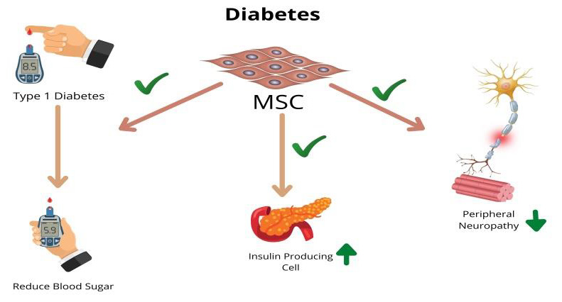
Figure 1.
In rats, Intravenous injection of bone marrow-derived MSCs improved myogenesis and angiogenesis in a DCM model [86]. MMP-2 levels were shown to rise after MSC transplantation in this investigation. activity and a decrease in MMP-9 activity, which results in increases myocardial arteriolar density and lowers collagen volume, leading to cardiac remodeling attenuation as well as better myocardial function [86]. In a mouse model, MSCs were administered in a methodical manner, which resulted in an improvement of kidney function and glomerular structure regeneration as MSCs have the ability to regenerate a diabetic’s necrotic segment. [87,88] Kidneys Because MSCs are unable to multiply in the kidney, [89], an alternative scenario for kidney improvement MSCs’ ability to scavenge cytotoxic agents could be a function. molecules, or to encourage neovascularization [71]. Diabetic The most prevalent consequence is polyneuropathy (DPN). a type of diabetes characterized by nerve fiber destruction DPN is distinguished by the degeneration of neural cells. as well as reduced nerve blood flow (NBF). one-month following, the MSCs were discovered to be making bFGF by intramuscular injection. and VEGF, which resulted in an increase in the capillary ratio. muscle fibers, which was followed by an improvement in hyperalgesia and a related brain fiber functional enhancement [90].
Despite research indicating that MSCs have the ability to In vitro differentiation into neural cells, it was not detected. during in vivo diabetic rat model experiments [90]. research on Systematic and local administration was demonstrated in rats and mice. MSCs generated from bone marrow promote diabetic wound healing wounds. MSCs infusion resulted in an increase in various parameters. Growth factors are critical for wound healing success. These substances induced cell adhesion at the site of damage. as well as encouraged the cell to secrete more chemokines, resulting in neovascularization and the establishment of infiltration of inflammation primarily composed of mononuclear cells and devoid of tissue [91] necrosis. The results of promising exploratory and preclinical investigations have led to phase I and phase II clinical trials. which are eagerly anticipated. The results of this research will be decisive. the future of cell-based therapy for the most heinous diseases mankind’s degenerative illness.
Cardiac Diseases
Ischemic heart disease is the primary cause of death in developed countries, and it is associated with severe morbidity. The use of MSCs in cardiac repair has been extensively researched in preclinical and clinical investigations. Following an acute myocardial infarction (MI), the heart has a reduced ability for self-renewal and undergoes remodeling, resulting in decreased left ventricular function [92]. Intensive research on MSCs as a future cell-based treatment method for heart repair has been conducted over the last decade, and many of these studies have been transferred into clinical trials [92]. Somatic reprogramming, trans differentiation, paracrine signaling, and direct electrophysiological coupling have all been proposed as pathways for MSC-mediated cardiac improvement [93]. Several in vivo rodent and swine investigations have shown that MSCs can engraft and differentiate within the heart. Shake et al. and Toma et al. reported that transplanted MSCs engrafted into damaged myocardium and produced cardiomyocyte markers such as-actin, desmin, tropomyosin, and myosin heavy chain [94,95]. Quevedo et al. observed the ability of allogeneic MSCs to engraft and develop into cardiomyocytes, smooth muscle cells, and endothelium in a pig model of chronic ischemic cardiomyopathy [96].
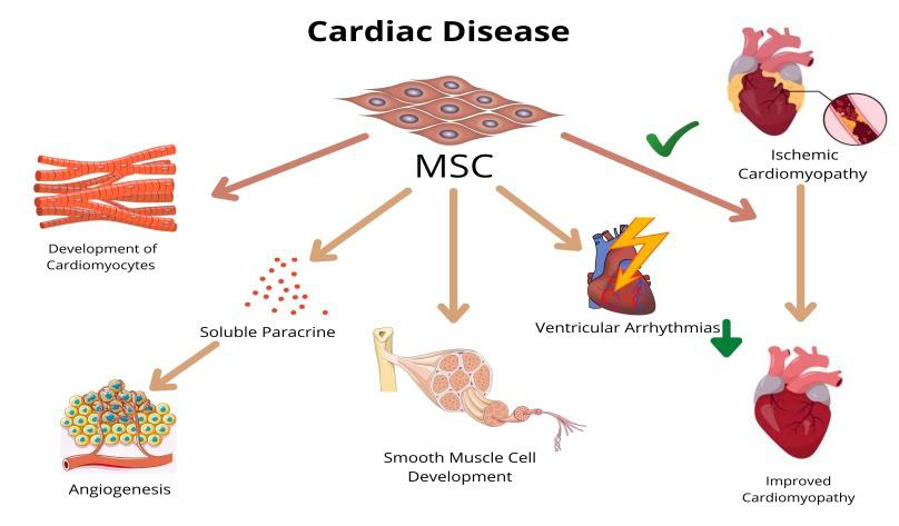
Figure 2.
Several other investigations [97-99] found that MSCs develop into cardiomyocytes in vivo. In contrast to previous findings of engraftment, Dixon et al. demonstrated that male mesenchymal precursor cells implanted into post-MI sheep failed to engraft [100]. Functional recovery following MSC transplantation has been thoroughly demonstrated and recognized. MSC transplantation reduced infarct size enhanced left ventricular ejection fraction (LVEF) and boosted vascular density and myocardial perfusion in most animal models of MI. Clinical trials for MI and ischemic cardiomyopathy have begun based on comprehensive preclinical studies and established safety. Intracoronary infusion of bone marrow derived MSCs in patients with sub-acute MI corrected perfusion deficits three months later and left ventriculography revealed better ejection fraction (EF) and left ventricular chamber size [101]. Similarly, intravenous injection of MSCs in acute MI showed a reduction in ventricular arrhythmias and better pulmonary function, with patients having a 6% gain in EF at three months [102] (Figure 2).
Trans endocardial intramyocardial injection of MSCs in ischemic cardiomyopathy revealed reversal remodeling and enhanced regional contractility of treated scar three months after injection, which lasted for 12 months. End-diastolic volume (EDV) and end-systolic volume (ESV) improved as well [103]. A smallscale clinical trial was conducted on patients with chronic MI who were treated with a collagen scaffold previously seeded with bone marrow mononuclear cells to improve engraftment of transplanted MSCs, but only marginal ventricular wall remodeling and improved diastolic function were detected [104,105]. Regardless of multiple studies on MSC transplantation, insight into the mechanisms of inpatient and animal models the issues underlying the effect of MSC transplantation remain unresolved. Vague. A recent study revealed that IL-6 is important. Janus kinase secretion and activation of signal transducers and transcription activators (JAK-STAT) in cardiac repair by MSC transplantation [106].
A recent study revealed showed paracrine signaling boosted the survival of Akt-induced calcium changes in ventricular myocytes signaling, which resulted in transplanted MSCs having an antiapoptotic impact [107]. The occurrence of MSC engraftment and heart differentiation is low in comparison to the After-cell transplantation; there was a significant functional recovery. This implies that engraftment and Differentiation may occur not be the primary effect MSCs, as previously said, are known to release soluble paracrine substances that have been identified proposed to play a role in endogenous cardio myogenesis as well as angiogenesis. The mechanism, however, by which These factors’ interactions have yet to be investigated.
Liver Disorders
Liver transplant is the most favored treatment option for liver illnesses; however, donor organ shortages are the primary reason that whole organ or hepatocyte transplants are not performed on a regular basis. As a result, generating hepatocyte-like cells from MSCs has become a viable alternative to isolating primary hepatocytes. MSCs have been shown to adapt functional properties of differentiated hepatocytes and successfully engraft into mouse liver under certain growth circumstances [108]. Xenografting of allogeneic MSCs into allyl alcohol- (AA-) treated rat liver resulted in hepatocyte-like cells with positive immunostaining for albumin, CK-19, CK-18, and asialoglycoprotein receptor [109]. In a rat model, MSCs aid in the recovery from chemically induced liver injury and also aid in the reduction of hepatic fibrosis [110]. A similar outcome was seen in a rat model of hepatic cirrhosis [111]. MSCs were found to be diffusely engrafted in the liver parenchyma, with CK19 positive and albumin generating hepatocytes.
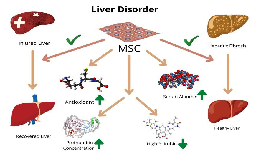
Figure 3.
Despite the low engraftment rate, MSCs demonstrated therapeutic effects such as repairing injured hepatocytes, restoring intracellular glycogen, and resolving fibrosis. Similarly, bone marrow-derived MSCs protected rats from experimental liver fibrosis [112-114]. MSCs and MSC-derived hepatocytes engrafted in mice livers developed into functional hepatocytes and cured liver failure when implanted intrasplenic ally or intravenously [115]. Contrary to this finding, Burra et al. proposed that systemic treatment of umbilical cord MSCs promotes the resolution of a severe liver injury without Differentiation or modification [116]. MSC transplantation not only improved liver function caused by degenerative disease, but also significantly improved liver damage caused by Schistosoma japonicum. MSCs transplantation, in combination with the standard medication praziquantel, increased the life duration of infected mice by reducing egg granuloma diameter and serum TGF-1 and hyaluronic acid concentrations [117] (Figure 3).
The cytoprotective mechanism of MSCs is still a mystery. A recent study suggested that the cytoprotective effect was related to bone marrow derived MSCs promoting antioxidant response [118]. According to a recent study [119], MSCs are attracted to the wounded liver in a beta1-integrin and CD44 dependent way. Researchers discovered that the technique of stem cell transplantation affects the result in preclinical experiments. In a swine model of acute liver failure, portal vein transplantation provided the best results, not only supporting liver regeneration but also extending host survival [120]. In the rat liver fibrosis model, however, intravenous injection transplantation has been demonstrated to produce the best effects and protect the liver from fibrosis via IL-10 expression [121]. Humans are also investigating the potential of MSCs in liver healing. Both differentiated [115] and undifferentiated MSCs transplantation improved liver function in phase I and II clinical trials for hepatic cirrhosis [122-124].
Follow-up at 3 and 6 months after transplant demonstrated a partial improvement in liver function tests, with an increase in prothrombin concentration and serum albumin levels, a decrease in high bilirubin, and a lower Model for End-Stage Liver Disease score (MELD) [122]. In patients with decompensated liver cirrhosis, umbilical cord-MSC transplantation resulted in a considerable reduction in ascites volume. During one-year followup investigations, umbilical cord -MSC therapy also dramatically improved liver function, as evidenced by an increase in serum albumin levels, a decrease in total serum bilirubin levels, and a decrease in MELD scores [125]. Another detailed clinical observation for liver failure suggested that autologous bonemarrow MSCs transplanted patients showed marked improvement in the levels of alanine aminotransferase, albumin, total bilirubin, prothrombin time, and MELD from 2- 3 weeks after transplantation, but long-term follow-up did not show any significant difference between the control and transplanted groups [126]. Although preclinical and clinical trials have yielded encouraging results, further research is needed to transfer these findings into standard treatment. Scientists want to improve the therapeutic efficacy of MSCs by pretreatment with different chemicals [127] and evaluating genetically engineered MSCs [128].
Kidney disease
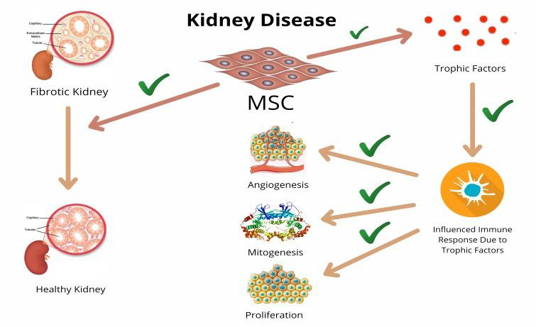
Figure 4.
There have been numerous cases of MSCs repopulating injured kidneys, with varying degrees of significance. In cyclosporineimmunocompromised rats, intraparenchymal injection of bone marrow-derived MSCs decreases kidney fibrosis following ischemiareperfusion [129]. Exogenous injection of MSCs to mice with acute renal damage was found to improve both morphological and functional renal healing by MSC trans-differentiation into tubular epithelium [130]. Engraftment was seen in just 2–2.5 percent of the implanted MSCs [130,131]. MSCs may be a beneficial technique for preserving podocyte viability and reducing glomerular inflammation and sclerosis due to their affinity for injured kidneys and capacity to establish a local prosurvival environment [132]. Furthermore, a study with female mice that received male bone marrow for tubular damage discovered that approximately 4% of tubular cells were positive for the Y-chromosome, implying that a small but significant number of engrafted bone-marrow-derived cells engaged in kidney regeneration [133] (Figure 4).
In contrast, another study found that arterial injection of MSCs reduced necrosis, improved kidney function, and boosted mesangial cell proliferation and expression of -smooth muscle actin (-SMA), but no incorporation of MSCs into kidney structures [134]. These studies show that direct engraftment of exogenously supplied and transdifferentiating MSCs is not the primary mechanism by which MSCs improve renal healing [134]. There is growing evidence that MSCs can induce kidney repair via paracrine and/or endocrine processes, in which they produce trophic growth factors that influence the immune response and thereby facilitate repair [134]. MSCs’ ability to inhibit the release of proinflammatory cytokines and secrete a variety of trophic growth factors that promote angiogenesis, mitogenesis, and proliferation while decreasing apoptosis may collectively mediate the protective and regenerative effects in laboratory rodent kidneys [134,135]. Recent research of targeted delivery of bone marrow derived MSCs called this idea into question. In their investigation, the researchers demonstrated not just homing of bone marrow derived MSCs, but also kidney healing in a rat model of acute renal injury [136]. In a preliminary clinical investigation of chronic renal disease, two intravenous transplants of 1 million MSCs/kg body demonstrated a significant change in serum creatinine and creatinine clearance levels before and after MSC injection at 1, 3, and 6 months [137].
Bone Disease
Because of the scarcity of autologous bone grafts and the unsuitability of allografts, there has been considerable interest in using MSCs to promote repair. Murine model studies yielded highly encouraging findings, particularly for bone regeneration and metabolic bone diseases [138]. MSCs have been used successfully for bone regeneration since their first use in 1951 [138]. Compared to the control group, a ceramic scaffold packed with enlarged MSCs dramatically improved bone formation in the femurs of athymic rats [139] (Figure 5). Using in vitro grown MSCs loaded on porous hydroxyapatite scaffolds, researchers could repair bone nonunion and diaphyseal anomalies while also achieving successful implant integration [140,141]. Angiographic assessment of implants after seven years revealed vascularization of the grafted zone, which is critical for the graft’s longevity and future stability. Another study found that differentiated bone-marrow-derived stem cells can assist patients in achieving the goal length of the femora and tibiae in patients undergoing distraction osteogenesis [142].
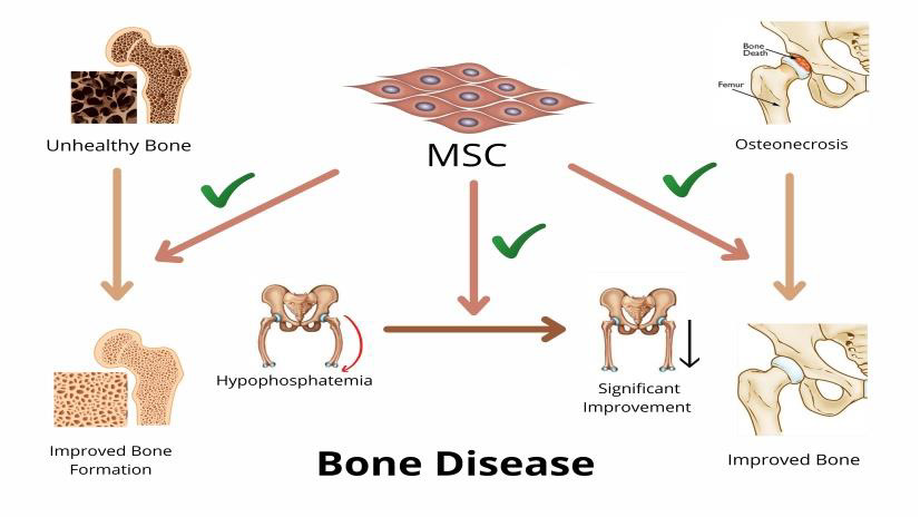
Figure 5.
MSCs have been employed successfully in the treatment of steroid-induced femoral head osteonecrosis [143]. Scaffolds seeded with bone marrow derived MSCs have also been employed in spinal fusion, but further research with appropriate controls is needed before we can draw any firm conclusions [144]. In bone repair, SDF- 1 and its receptor CXCR4 have been demonstrated to operate as a possible homing signal for MSCs [145]. According to the findings of another study, the volume and concentration of autologous MSCs implanted influenced bone regeneration, meaning that at least 1000 MSCs per cm3 are required to achieve union [146]. When mesenchymal stem cells, platelet-rich plasma, and synthetic bone replacement were used together, it was discovered that the combination was more effective at inducing new bone synthesis (osteogenesis) than either platelet-rich plasma or synthetic bone replacement used alone [145].
In Osteogenesis imperfecta (OI), bone fragility and other symptoms of connective tissue malfunction are present in addition to the normal bone formation process. In a study in which MSCs from wild-type mice were implanted into transgenic mice with a phenotype of weak bones similar to OI, the MSCs were found to operate as a source of continuous cell renewal in a variety of nonhematopoietic organs [147]. Adult bone marrow donor cells from transgenic mice engrafted into hematopoietic and nonhematopoietic tissues generated up to 20% of all type I collagen in the host bone and prevented perinatal mortality in mice with dominant OI [148]. Allogeneic bone marrow transplantation in three children with OI resulted in osteoblast engraftment of about 2.0 percent donor cells, histologic alterations indicative of new bone production, and a rise in total body bone mineral content [149]. However, the trial only had a 6-month clinical follow-up and did not directly compare results to controls. In three children (out of five) with severe OI, the same group demonstrated linear growth, total body bone mineral content, and fracture rate [150].
Growth rates reduced and eventually plateaued as time passed following transplant, although bone mineral content increased [150]. Horwitz et al. treated six children with OI with a systemic infusion of MSCs for bone repair in another research. When compared to similar unaffected youngsters, five children demonstrated an acceleration in bone growth [151]. Direct application of MSCs to the fracture, on the other hand, is thought to be more practicable [138]. Le Blanc et al. injected allogeneic HLA mismatched MSCs into a 32-week-old fetus and demonstrated the transplanted cells’ participation in bone turnover using a Y-chromosome-specific probe [152]. Hypophosphatasia, a heritable metabolic condition, was helped by an allogeneic transplant of the patient’s osteoblasts [153]. The results demonstrated that when patient osteoblasts were replaced with donor cells, there was a significant improvement in bone without any alterations in the biochemical hallmark of hypophosphatasia, which was validated clinically and radiologically [153].
Autoimmune Diseases
MSCs’ ability to modulate the actions of numerous immune effector cells may be involved in the etiology of autoimmune illnesses, making them a useful tool for autoimmune disease treatment [154]. GvHD, which is otherwise untreatable and deadly, is one of the priority target diseases. The first report on the successful use of MSCs for the treatment of severe steroid refractory acute GvHD was published in 2004, and it used e Vivo expanded haploidentical human MSCs [61]. MSCs from haploidentical donors were used in 18 cases in phase I and phase II clinical trials, while MSCs from HLA mismatched donors were used in 69 cases. Of the 55 severe acute GvHD steroid-resistant patients, 30 had a full response, while nine did not improve [155] [ Figure 6].
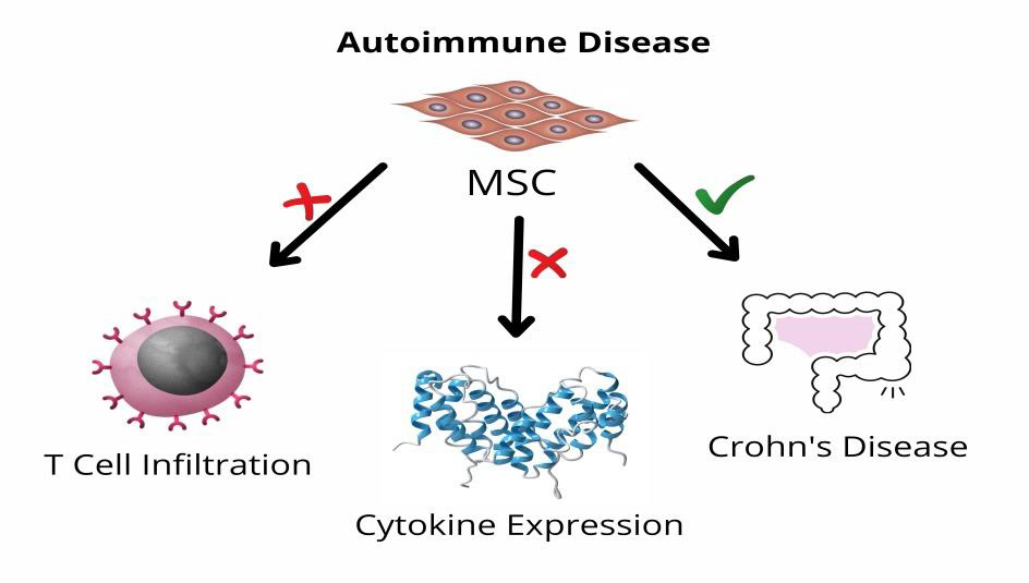
Figure 6.
In Phase II therapeutic trial with allogeneic MSCs, 94% of the 31 patients with acute GvHD showed initial reaction to MSCs; 77% showed full answer; 17% showed a partial response without infusion or ectopic formation of tissue [156]. Interestingly, Mills et al. reported in 2009 mixed results from a large size phase III clinical trial involving 192 acute GvHD patients and 260 steroid resistant GvHD patients. According to the findings of this investigation, the success of MSC treatment may be dependent on the class of tissue affected by GvHD patients. MSCs may provide therapeutic effects for Crohn’s disease [157], but large-scale clinical trials are needed to acquire clear results. Dalal et al. [158] recently evaluated the role of MSCs in Crohn’s disease, which is not explored in-depth in this study. T cells and macrophages are involved in experimental autoimmune encephalomyelitis (EAE), a CNS autoimmune illness. The current standard of care for EAE is to target T cells in order to promote immunosuppression or tolerance. Furthermore, numerous studies have demonstrated the therapeutic potential of human and mouse MSCs for EAE treatment by demonstrating improved clinical progress, stimulation for tissue repair, decrease in demyelination, and T cell and macrophage infiltration of the CNS [159-164].
Though there is limited evidence for MSC engraftment in the CNS over time [165,166], MSCs in the periphery appear to enhance tissue healing and prevent autoimmune disturbance in the CNS. Yamout et al. treated ten patients with advanced Multiple Sclerosis (MS) with ex vivo grown bone-marrow-derived MSCs, with 50% showing beneficial effects, indicating that MSCs are safe and practical for use in the treatment of MS patients [167,168]. Amyotrophic lateral sclerosis (ALS) is an autoimmune illness that causes loss of upper and lower motor neurons in the cerebral cortex, brainstem, and spinal cord, resulting in death within five years of onset [169]. Patients with MS and ALS were treated with intravenous MSC infusion in a phase I/II study, which resulted in an increase in the proportion of CD4+ CD45+ Treg cells in the patient’s peripheral blood [170]. Despite promising preclinical data, definitive evidence of MSCs’ therapeutic effect on neurodegenerative diseases is missing [171,172]. Furthermore, human clonal MSCs have been shown to improve pancreatic function in rat models of moderate and severe acute pancreatitis (AP) by inhibiting T cell infiltration, lowering inflammatory mediator or cytokine expression, and boosting Foxp3 regulatory T cells [64].
Future Direction
The available data until now does not clearly demonstrate Differentiation and engraftment but supports its immunomodulation properties anonymously. We know the chemicals used by MSCs to modulate immune-effector cells, some of which might be involved in autoimmune pathogenesis. The underlying mechanisms underlying these compounds continue to be obscure. In addition, MSCs can not only use immunomodulate immune cells but can also evade immunological rejection. However, this feature depends on the MSCs’ microenvironment. Recent data indicate that the inflammatory milieu with autoimmune disorders can modify the polarization of MSC into an immunosuppressive phenotype [63]. Interestingly enough, in one case, MSCs have been protected from diseases, and the other with the same condition have worsened the clinical parameters [173,174]. The variation in the time parameter when MSCs are infused following illness induction may lead to a range of inflammatory environments around MSCs, in both circumstances, which might affect the function of MSCs. For such conflicts, it can be suggested that, thus, a micro-approximation check before the treatment can assist in determining how the patient reacts to MSC therapy for a specific condition. This information can have a tremendous impact on the clinical usage of MSCs.
Further investigations should therefore be targeted at understanding the processes underlying MSC immunomodulation to employ MSCs for therapeutic purposes. The clinical usefulness of MSCs is not restricted to treating Autoimmune disorders; MSCs were also evaluated as cell carriers for gene therapy and the improvement of hematopoietic stem cell grafting in tissue regeneration. More recently, a concept of engineered MSCs for cancer therapy was proposed [175-178]. The use of MSCs as an isolated cancer treatment is disputed; nevertheless, some intelligent researches have shown that MSCs developed with certain anticancer genes were used successfully to clock cancer cells and thereby reduce the progression of the tumor [179-183]. These MSC-containing anticancer genes can locate on a particular tumor site, no matter whether tumor kind or invasivity and provide anticancer chemicals [175-178].
Therefore, well-designed future research is required in the actual clinical environment outside the laboratory. Fortunately, in many cases, the clinical trials described so far have not shown any seriously detrimental consequences of MSCs on the illness status. The use of MSCs in therapeutic applications can therefore be regarded safe. However, further data is needed to establish the long-term safety, MSC immunogenicity, appropriate source, and a number of cells to be injected into no immunocompromised animals. Although consistency in specific areas of MSC therapies is not now present, the potential immunomodulatory characteristics of MSCs are outstanding to become the basis of future medicines.
References
- T Graf (2002) Differentiation plasticity of hematopoietic cells. Blood 99(9): 3089-3101.
- FM Watt, BLM Hogan (2000) Out of eden: stem cells and their niches. Science 287(5457): 1427-1430.
- CD Porada,ED Zanjani, G Almeida Porad (2006) Adult mesenchymal stem cells: a pluripotent population with multiple applications. Curr Stem Cell Res Ther 1(3): 365-369.
- Lda Silva Meirelles, PC Chagastelles, NB Nardi (2006) Mesenchymal stem cells reside in virtually all post-natal organs and tissues. J Cell Sci 119(11): 2204-2213.
- B Parekkadan, JM Milwid (2010) Mesenchymal stem cells as therapeutics. Annu Rev Biomed Eng 12: 87-117.
- AJ Friedenstein, UF Gorskaja, NN Kulagina (1976) Fibroblast precursors in normal and irradiated mouse hematopoietic organs. Exp Hematol 4(5): 267-274.
- M Owen, AJ Friedenstein (1988) Stromal stem cells: marrowderived osteogenic precursors. Ciba Found Symp 136: 42-60.
- M Owen (1988) Marrow stromal stem cells. Journal of Cell Science10: 63-76.
- M Dominici, K Le Blanc, I Mueller, I Slaper Cortenbach, Fc Marini, et al. (2006) Minimal criteria for defining multipotent mesenchymal stromal cells. The international society for cellular therapy position statement. Cytotherapy 8(4): 315-317.
- AI Caplan (1991) Mesenchymal stem cells. Journal of Orthopaedic Research 9(5): 641-650.
- HM Lazarus, SE Haynesworth, SL Gerson, NS Rosenthal, AI Caplan (1995) Ex vivo expansion and subsequent infusion of human bone marrow-derived stromal progenitor cells (mesenchymal progenitor cells): implications for therapeutic use. Bone Marrow Transplant 16(4): 557-564.
- H Motaln, C Schichor, TT Lah (2010) Human mesenchymal stem cells and their use in cell-based therapies. Cancer 116(11): 2519-2530.
- G Chamberlain, J Fox, B Ashton, J Middleton (2007) Concise review: mesenchymal stem cells: their phenotype, differentiation capacity, immunological features, and potential for homing. Stem Cells 25(11): 2739-2749.
- V Planat Benard, JS Silvestre, B Cousin, Mireille André, Maryse Nibbelink, et al. (2004) Plasticity of human adipose lineage cells toward endothelial cells: physiological and therapeutic perspectives. Circulation 109(5): 656-663.
- VL Battula, S Treml, PM Bareiss, Friederike Gieseke, Helene Roelofs, et al. (2009) Isolation of functionally distinct mesenchymal stem cell subsets using antibodies against CD56, CD271, and mesenchymal stem cell antigen-1. Haematologica 94(2): 173-184.
- K Sivasubramaniyan, D Lehnen, R Ghazanfari, Malgorzata Sobiesiak, Abhishek Harichandan, et al. (2012) Phenotypic and functional heterogeneity of human bone marrow- and amnion-derived MSC subsets. Ann N Y Acad Sci 1266: 94-106.
- DC Ding, WC Shyu, SZ Lin (2011) Mesenchymal stem cells. Cell Transplantation 20(1): 5-14.
- NS Hwang, C Zhang, YS Hwang, S Varghese (2009) Mesenchymal stem cell differentiation and roles in regenerative medicine. Wiley Interdiscip Rev Syst Biol Med 1(1): 97-106.
- CM Teven, X Liu, N Hu, Ni Tang, Stephanie H Kim, et al. (2011) Epigenetic regulation of mesenchymal stem cells: a focus on osteogenic and adipogenic differentiation. Stem Cells Int 2011: 201371.
- ZL Deng, KA Sharff, N Tang, Wen-Xin Song, Jinyong Luo, et al. (2008) Regulation of osteogenic differentiation during skeletal development. Front Biosci 13(6): 2001-2021.
- N Baker, G Zhang, Y You, RS Tuan (2012) Caveolin-1 regulates proliferation and osteogenic differentiation of human mesenchymal stem cells. Journal of Cellular Biochemistry 113(12): 3773-3787.
- DJ Xu, YZ Zhao, J Wang, JW He, YG Weng, et al. (2012) Smads p38 and ERK1/2 are involved in BMP9-induced osteogenic differentiation of C3H10T1/2 mesenchymal stem cells. BMB Reports 45(4): 247-252.
- H Zheng, Z Guo, Q Ma, H Jia, G Dang (2004) Cbfa1/osf2 transduced bone marrow stromal cells facilitate bone formation in vitro and in vivo. Calcif Tissue Int 74(2): 194-203.
- T Gaur, CJ Lengner, H Hovhannisyan, Ramesh A Bhat, Peter V N Bodine, et al. (2005) Canonical WNT signaling promotes osteogenesis by directly stimulating Runx2 gene expression. J Biol Chem 280(39): 33132-33140.
- JJ Alm, TJ Heino, TA Hentunen, HK Vaananen, HT Aro (2012) Transient 100 nM dexamethasone treatment reduces inter- and intraindividual variations in osteoblastic differentiation of bone marrow-derived human mesenchymal stem cells. Tissue Eng Part C Methods 18(9): 658-666.
- K Cameron, P Travers, C Chander, T Buckland, C Campion, et al. (2013) Directed osteogenic differentiation of human mesenchymal stem/precursor cells on silicate substituted calcium phosphate. J Biomed Mater Res A 101(1): 13-22.
- Z Tang, D Shi, B Jia, Jiarong Chen, Chen Zong, et al. (2012) Exchange protein activated by cyclic adenosine monophosphate regulates the switch between adipogenesis and osteogenesis of human mesenchymal stem cells through increasing the activation of phosphatidylinositol 3-kinase. Int J Biochem Cell Biol 44(7): 1106-1120.
- CYC Huang, KL Hagar, LE Frost, Y Sun, HS Cheung (2004) Effects of cyclic compressive loading on chondrogenesis of rabbit bone-marrow derived mesenchymal stem cells. Stem Cells 22(3): 313-323.
- S Paik, HS Jung, S Lee, DS Yoon, MS Park, et al. (2012) miR-449a regulates the chondrogenesis of human mesenchymal stem cells through direct targeting of lymphoid enhancer-binding factor-1. Stem Cells Dev 21(18): 3298-3308.
- TF Day, X Guo, L Garrett Beal, Y Yang (2005) Wnt/𝛽-catenin signaling in mesenchymal progenitors controls osteoblast and chondrocyte differentiation during vertebrate skeletogenesis. Developmental Cell 8(5): 739-750.
- T Cheng, C Yang, N Weber, HT Kim, AC Kuo (2012) Fibroblast growth factor 2 enhances the kinetics of mesenchymal stem cell chondrogenesis. Biochem Biophys Res Commun 426(4): 544-550.
- J Yanjie, S Jiping, Z Yan, Z Xiaofeng, Z Boai, et al. (2007) Effects of Notch-1 signalling pathway on differentiation of marrow mesenchymal stem cells into neurons in vitro. Neuro Report 18(14): 1443-1447.
- S Wang, Q Kan, Y Sun, Rui Han, Guangyu Zhang, et al. (2013) Caveolin-1 regulates neural differentiation of rat bone mesenchymal stem cells into neuron by modulating notch signaling. Int J Dev Neurosci 31(1): 30-35.
- KD Lee, TKC Kuo, J Whang Peng, Yu-Fen Chung, Ching-Tai Lin, et al. (2004) In vitro hepatic differentiation of human mesenchymal stem cells. Hepatology 40(6): 1275-1284.
- LB Chen, XB Jiang, L Yang (2004) Differentiation of rat marrow mesenchymal stem cells into pancreatic islet 𝛽-cells. World J Gastroenterol 10(20): 3016-3020.
- DR Bhandari, KW Seo, KH Roh, JW Jung, SK Kang, et al. (2010) REX-1 expression and p38 MAPK activation status can determine proliferation/differentiation fates in human mesenchymal stem Cells. PLoS ONE 5(5): e10493.
- V Chandra, Swetha G, S Phadnis, PD Nair, RR Bhonde (2009) Generation of pancreatic hormone-expressing islet-like cell aggregates from murine adipose tissue-derived stem cells. Stem Cells 27(8): 1941-1953.
- P Ray, A De, JJ Min, RY Tsien, SS Gambhir (2004) Imaging tri-fusion multimodality reporter gene expression in living subjects. Cancer Res 64(4): 1323-1330.
- TR Brazelton, HM Blau (2005) Optimizing techniques for tracking transplanted stem cells in vivo. Stem Cells 23(9): 1251-1265.
- WR Otto, NA Wright (2011) Mesenchymal stem cells: from experiment to clinic. Fibrogenesis & Tissue Repair 4(20).
- F Granero Molto, JA Weis, L Longobardi, A Spagnoli (2008) Role of mesenchymal stem cells in regenerative medicine: application to bone and cartilage repair. Expert Opin Biol Ther 8(3): 255-268.
- KI Hiasa, M Ishibashi, K Ohtani, Shujiro Inoue, Qingwei Zhao, et al. (2004) Gene transfer of stromal cell-derived factor-1𝛼 enhances ischemic vasculogenesis and angiogenesis via vascular endothelial growth factor/endothelial nitric oxide synthase-related pathway: nextgeneration chemokine therapy for therapeutic neovascularization. Circulation 109(20): 2454-2461.
- J Ma, J Ge, S Zhang, Aijun Sun, Jianying Shen, et al. (2005) Time course of myocardial stromal cell-derived factor 1 expression and beneficial effects of intravenously administered bone marrow stem cells in rats with experimental myocardial infarction. Basic Res Cardiol 100(3): 217-223.
- JE Ip, Y Wu, J Huang, L Zhang, RE Pratt, et al. (2007) Mesenchymal stem cells use integrin 𝛽1 not CXC chemokine receptor 4 for myocardial migration and engraftment Mol Biol Cell 18(8): 2873-2882.
- X Hou, X Wu, J Ma, X Lv, X Jin (2010) Erythropoietin augments the efficacy of therapeutic angiogenesis induced by allogenic bone marrow stromal cells in a rat model of limb ischemia. Mol Biol Rep 37(3): 1467-1475.
- L Badri, VN Lama (2012) Lysophosphatidic acid induces migration of human lung-resident mesenchymal stem cells through the 𝛽-catenin pathway. Stem Cells 30(9): 2010-2019.
- DO Freytes, JW Kang, I Marcos Campos, G VunjakNovakovic (2013) Macrophages modulate the viability and growth of human mesenchymal stem cells. J Cell Biochem 114(1): 220-229.
- B Ruster, SGottig, RJ Ludwig, Roxana Bistrian, Stefanie Müller, et al. (2006) Mesenchymal stem cells display coordinated rolling and adhesion behavior on endothelial cells. Blood 108(12): 3938-3944.
- A Mirza, JM Hyvelin, GY Rochefort, Patrick Lermusiaux, Daniel Antier, et al. (2008) Undifferentiated mesenchymal stem cells seeded on a vascular prosthesis contribute to the restoration of a physiologic vascular wall. Journal of Vascular Surgery 47(6): 1313-1321.
- Z Sheng, X Fu, S Cai, Yonghong Lei, Tongzhu Sun, et al. (2009) Regeneration of functional sweat gland-like structures by transplanted differentiated bone marrow mesenchymal stem cells. Wound Repair Regen 17(3): 427-435.
- CEP Aronin, RS Tuan (2010) Therapeutic potential of the immunomodulatory activities of adult mesenchymal stem cells. Birth Defects Res C Embryo Today 90(1): 67-74.
- Y Li, J Chen, XG Chen, L Wang, SC Gautam, et al. (2002) Human marrow stromal cell therapy for stroke in rat: neurotrophins and functional recovery. Neurology 59(4): 514-523.
- AT Askari, S Unzek, ZB Popovic, Corey K Goldman, Farhad Forudi, et al. (2003) Effect of stromalcell-derived factor 1 on stem-cell homing and tissue regeneration in ischaemic cardiomyopathy. Lancet 362(9385): 697-703.
- MD Nicola, C Carlo Stella, M Magni, Marco Milanesi, Paolo D Longoni, et al. (2002) Human bone marrow stromal cells suppress T-lymphocyte proliferation induced by cellular or nonspecific mitogenic stimuli. Blood 99(10): 3838-3843.
- A Bartholomew, C Sturgeon, M Siatskas, Karen Ferrer, Kevin McIntosh, et al. (2002) Mesenchymal stem cells suppress lymphocyte proliferation in vitro and prolong skin graft survival in vivo. Exp Hematol 30(1): 42-48.
- F Djouad, P Plence, C Bony, Philippe Tropel, Florence Apparailly, et al. (2003) Immunosuppressive effect of mesenchymal stem cells favors tumor growth in allogeneic animals. Blood 102(10): 3837-3844.
- F Djouad, LM Charbonnier, C Bouffi, Pascale Louis Plence, Claire Bony, et al. (2007) Mesenchymal stem cells inhibit the differentiation of dendritic cells through an interleukin-6-dependent mechanism. Stem Cells 25(8): 2025-2032.
- S Glennie, I Soeiro, PJ Dyson, EWF Lam, F Dazzi (2005) Bone marrow mesenchymal stem cells induce division arrest anergy of activated T cells. Blood 105(7): 2821-2827.
- PA Sotiropoulou, SA Perez, AD Gritzapis, CN Baxevanis, M Papamichail (2006) Interactions between human mesenchymal stem cells and natural killer cells. Stem Cells 24(1): 74-85.
- GM Spaggiari, A Capobianco, S Becchetti, MC Mingari, L Moretta (2006) Mesenchymal stem cell-natural killer cell interactions: evidence that activated NK cells are capable of killing MSCs, whereas MSCs can inhibit IL-2-induced NK-cell proliferation. Blood 107(4): 1484-1490.
- K Le Blanc, I Rasmusson, B Sundberg, Cecilia Götherström, Moustapha Hassan, et al. (2004) Treatment of severe acute graft-versus-host disease with third party haploidentical mesenchymal stem cells. Lancet 363(9419): 1439-1441.
- ZH Zheng, XY Li, J Ding, JF Jia, P Zhu (2008) Allogeneic mesenchymal stem cell and mesenchymal stem celldifferentiated chondrocyte suppress the responses of type II collagen-reactive T cells in rheumatoid arthritis. Rheumatology 47(1): 22-30.
- F Dazzi, M Krampera (2011) Mesenchymal stem cells and autoimmune diseases. Best Practice and Research Clinical Haematology 24(1): 49-57.
- T Yi, SU Song (2012) Immunomodulatory properties of mesenchymal stem cells and their therapeutic applications. Arch Pharm Res 35(2): 213-221.
- YO Joo, KK Mee, SS Mi, Hyun Ju Lee, Jung Hwa Ko, et al. (2008) The anti-inflammatory and anti-angiogenic role of mesenchymal stem cells in corneal wound healing following chemical injury. Stem Cells 26(4): 1047-1055.
- ME Groh, B Maitra, E Szekely, ON Koc (2005) Human mesenchymal stem cells require monocyte-mediated activation to suppress alloreactive T cells. Exp Hemat 33(8): 928-934.
- S Jones, N Horwood, A Cope, F Dazzi (2007) The antiproliferative effect of mesenchymal stem cells is a fundamental property shared by all stromal cells. J Immunol 179(5): 2824-2831.
- MP De Miguel, S Fuentes Julian, A Blazquez Martinez, CY Pascual, MA Aller, et al. (2012) Immunosuppressive properties of mesenchymal stem cells: advances and applications. Curr Mol Med 12(5): 574-591.
- W Li, G Ren, Y Huang, J Su, Y Han, et al. (2012) Mesenchymal stem cells: a double-edged sword in regulating immune responses. Cell Death Differ 19(9): 1505-1513.
- PK Mishra, SR Singh, IG Joshua, SC Tyagi (2010) Stem cells as a therapeutic target for diabetes. Front Biosci 15(2): 461-477.
- V Volarevic, N Arsenijevic, ML Lukic, M Stojkovic (2011) Concise review: mesenchymal stem cell treatment of the complications of diabetes mellitus. Stem Cells 29(1): 5-10.
- Y Sun, L Chen, XG Hou, Wei kai Hou, Jian jun Dong, et al. (2007) Differentiation of bone marrow-derived mesenchymal stem cells from diabetic patients into insulin-producing cells in vitro. Chin Med J 120(9): 771-776.
- KC Chao, KF Chao, YS Fu, SH Liu (2008) Islet-like clusters derived from mesenchymal stem cells in Wharton’s jelly of the human umbilical cord for transplantation to control type 1 diabetes. PLoS One 3(1): e1451.
- JA Cancelas, DA Williams (2006) Stem cell mobilization by 𝛽2-agonists. Nat Med 12(3): 278-279.
- QS Guo, MY Zhu, L Wang, Xiang Jun Fan, Yu Hua Lu, et al. (2012) Combined transfection of the three transcriptional factors, PDX-1, NeuroD1, and MafA, causes differentiation of bone marrow mesenchymal stem cells into insulin-producing cells. Exp Diabetes Res 2012: 672013.
- HT Li, F X Jiang, P Shi, Tao Zhang, Xiao-Yu Liu, et al. (2012) In vitro reprogramming of rat bone marrow-derived mesenchymal stem cells into insulin-producing cells by genetically manipulating negative and positive regulators. Biochem Biophy Res Commun 420(4): 793-798.
- K Timper, D Seboek, M Eberhardt, Philippe Linscheid, Mirjam Christ Crain, et al. (2006) Human adipose tissue-derived mesenchymal stem cells differentiate into insulin, somatostatin, and glucagon expressing cells. Biochem Biophys Res Commun 341(4): 1135-1140.
- F Ezquer, M Ezquer, D Contador, M Ricca, V Simon, et al. (2012) The antidiabetic effect of mesenchymal stem cells is unrelated to their transdifferentiation potential but to their capability to restore Th1/Th2 balance and to modify the pancreatic microenvironment. Stem Cells 30(8): 1664-1674.
- JH Ho, TC Tseng, WH Ma, Wei Kee Ong, Yu Fan Chen, et al. (2012) Multiple intravenous transplantations of mesenchymal stem cells effectively restore long-term blood glucose homeostasis by hepatic engraftment and beta-cell differentiation in streptozocin-induced diabetic mice. Cell Transplant 21(1): 217-234.
- Y Si, Y Zhao, H Hao, Jiejie Liu, Yelei Guo, et al. (2012) Infusion of mesenchymal stem cells ameliorates hyperglycemia in type 2 diabetic rats: identification of a novel role in improving insulin sensitivity. Diabetes 61(6): 1616-1625.
- R Jiang, Z Han, G Zhuo, Xiaodan Qu, Xue Li, et al. (2011) Transplantation of placentaderived mesenchymal stem cells in type 2 diabetes: a pilot study. Front Med 5(1): 94-100.
- M Yu, W Zhou, Y Song, Fengbin Yu, Dehua Li, et al. (2011) Development of mesenchymal stem cell-implant complexes by cultured cells sheet enhances osseointegration in type 2 diabetic rat model. Bone 49(3): 387-394.
- A Bhansali, V Upreti, N Khandelwal, N Marwaha, Vivek Gupta, et al. (2009) Efficacy of autologous bone marrow-derived stem cell transplantation in patients with type 2 diabetes mellitus. Stem Cells Dev 18(10): 1407-1416.
- TM Camp, SC Tyagi, RM Senior, MR Hayden, SC Tyagi (2003) Gelatinase B(MMP-9) an apoptotic factor in diabetic transgenic mice. Diabetologia 46(10): 1438-1445.
- YS Yoon, S Uchida, O Masuo, Manfred Cejna, Jong Seon Park, et al. (2005) Progressive attenuation of myocardial vascular endothelial growth factor expression is a seminal event in diabetic cardiomyopathy: restoration of microvascular homeostasis and recovery of cardiac function in diabetic cardiomyopathy after replenishment of local vascular endothelial growth factor. Circulation 111(16): 2073-2085.
- N Zhang, J Li, R Luo, J Jiang, JA Wang (2008) Bone marrow mesenchymal stem cells induce angiogenesis and attenuate the remodeling of diabetic cardiomyopathy. Exp Clin Endocrinol Diabetes 116(2): 104-111.
- MB Herrera, B Bussolati, S Bruno, V Fonsato, GM Romanazzi, et al. (2004) Mesenchymal stem cells contribute to the renal repair of acute tubular epithelial injury. Int J Mol Med 14(6): 1035-1041.
- F Ezquer, M Ezquer, V Simon, Fabian Pardo, Alejandro Yañez, et al. (2009) Endovenous administration of bone marrow-derived multipotent mesenchymal stromal cells prevents renal failure in diabetic mice. Biol Blood Marrow Transplant 15(11): 1354-1365.
- RH Lee, MJ Seo, RL Reger, Jeffrey L Spees, Andrey A Pulin, et al. (2006) Multipotent stromal cells from human marrow home to and promote repair of pancreatic islets and renal glomeruli in diabetic NOD/scid mice. Proc Natl Acad Sci U S A 103(46): 17438-17443.
- T Shibata, K Naruse, H Kamiya, Mika Kozakae, Masaki Kondo, et al. (2008) Transplantation of bone marrow-derived mesenchymal stem cells improves diabetic polyneuropathy in rats. Diabetes 57(11): 3099-3107.
- DS Kwon, X Gao, YB Liu, Deborah S Dulchavsky, Andrew L Danyluk, et al. (2008) Treatment with bone marrow-derived stromal cells accelerates wound healing in diabetic rats. Int Wound J 5(3): 453-463.
- AR Williams, JM Hare (2011) Mesenchymal stem cells: biology, pathophysiology, translational findings, and therapeutic implications for cardiac disease. Circ Res 109(8): 923-940.
- TJ Cashman, V Gouon Evans, KD Costa (2013) Mesenchymal stem cells for cardiac therapy: practical challenges and potential mechanisms. Stem Cell Rev Rep 9(3): 254-265.
- JG Shake, PJ Gruber, WA Baumgartner, Guylaine Senechal, Jennifer Meyers, et al. (2002) Mesenchymal stem cell implantation in a swine myocardial infarct model: engraftment and functional effects. Ann Thorac Surg 73(6): 1919-1925.
- C Toma, MF Pittenger, KS Cahill, BJ Byrne, PD Kessler (2002) Human mesenchymal stem cells differentiate to a cardiomyocyte phenotype in the adult murine heart. Circulation 105(1): 93-98.
- HC Quevedo, KE Hatzistergos, BN Oskouei, Gary S Feigenbaum, Jose E Rodriguez, et al. (2009) Allogeneic mesenchymal stem cells restore cardiac function in chronic ischemic cardiomyopathy via trilineage differentiating capacity. Proc Natl Acad Sci U S A 106(33): 14022-14027.
- RR Makkar, MJ Price, M Lill, Malka Frantzen, Kaname Takizawa, et al. (2005) Intramyocardial injection of allogenic bone marrow-derived mesenchymal stem cells without immunosuppression preserves cardiac function in a porcine model of myocardial infarction. J Cardiovasc Pharmacol Ther 10(4): 225-233.
- GV Silva, S Litovsky, JAR Assad, Andre LS Sousa, Bradley J Martin, et al. (2005) Mesenchymal stem cells differentiate into an endothelial phenotype, enhance vascular density, and improve heart function in a canine chronic ischemia model. Circulation 111(2): 150-156.
- YJ Yang, HY Qian, J Huang, Jian Jun Li, Run Lin Gao, et al. (2009) Combined therapy with simvastatin and bone marrow-derived mesenchymal stem cells increases benefits in infarcted swine hearts. Arterioscler Thromb Vasc Biol 29(12): 2076-2082.
- JA Dixon, RC Gorman, RE Stroud, Shenikqua Bouges, Hamamoto Hirotsugu, et al. (2009) Mesenchymal cell transplantation and myocardial remodeling after myocardial infarction. Circulation 120(1): S220-S229.
- SL Chen, WW Fang, F Ye, Yu-Hao Liu, Jun Qian, et al. (2004) Effect on left ventricular function of intracoronary transplantation of autologous bone marrow mesenchymal stem cell in patients with acute myocardial infarction. Am J Cardiol 94(1): 92-95.
- JM Hare, JH Traverse, TD Henry, Nabil Dib, Robert K Strumpf, et al. (2009) A randomized, double-blind, placebo-controlled, dose-escalation study of intravenous adult human mesenchymal stem cells (prochymal) after acute myocardial infarction. J Am Coll Cardiolo 54(24): 2277-2286.
- AR Williams, B Trachtenberg, DL Velazquez, Ian McNiece, Peter Altman, et al. (2011) Intramyocardial stem cell injection in patients with ischemic cardiomyopathy: functional recovery and reverse remodeling. Circ Res 108(7): 792-796.
- JC Chachques, JC Trainini, N Lago, Osvaldo H Masoli, Jose L Barisani, et al. (2007) Myocardial assistance by grafting a new bioartificial upgraded myocardium (MAGNUM clinical trial): one year follow-up. Cell Transplantation 16(9): 927-934.
- JC Chachques, JC Trainini, N Lago, M Cortes Morichetti, O Schussler, et al. (2008) Myocardial assistance by grafting a new bioartificial upgraded myocardium (MAGNUM trial): clinical feasibility study. Ann Thorac Surg 85(3): 901-908.
- A Shabbir, D Zisa, H Lin, Michalis Mastri, Gregory Roloff, et al. (2010) Activation of host tissue trophic factors through JAK-STAT3 signaling: a mechanism of mesenchymal stem cell-mediated cardiac repair. Am J Physio Heart Circ Physiol 299(5): H1428-H1438.
- J DeSantiago, DJ Bare, I Semenov, RD Minshall, DL Geenen, et al. (2012) Excitationcontraction coupling in ventricular myocytes is enhanced by paracrine signaling from mesenchymal stem cells. J Mol Cell Cardiol 52(6): 1249-1256.
- P Stock, S Bruckner, S Ebensing, M Hempel, MM Dollinger, et al. (2010) The generation of hepatocytes from mesenchymal stem cells and engraftment into murine liver. Nature Protocols 5(4): 617-627.
- Y Sato, H Araki, J Kato, Kiminori Nakamura, Yutaka Kawano, et al. (2005) Human mesenchymal stem cells xenografted directly to rat liver are differentiated into human hepatocytes without fusion. Blood 106(2): 756-763.
- YJ Chang, JW Liu, PC Lin, Li Yi Sun, Chih Wen Peng, et al. (2009) Mesenchymal stem cells facilitate recovery from chemically induced liver damage and decrease liver fibrosis. Life Sci 85(13-14): 517- 525.
- S Hwang, HN Hong, HS Kim, Se Ra Park, You Jin Won, et al. (2011) Hepatogenic differentiation of mesenchymal stem cells in a rat model of thioacetamide-induced liver cirrhosis. Cell Biol Int 36(3): 279-288.
- DC Zhao, JX Lei, R Chen, Wei Hua Yu, Xiu Ming Zhang, et al. (2005) Bone marrow-derived mesenchymal stem cells protect against experimental liver fibrosis in rats. World J Gastroenterol 11(22): 3431-3440.
- MT Abdel Aziz, HM Atta, S Mahfouz, HH Fouad, NK Roshdy, et al. (2007) Therapeutic potential of bone marrow-derived mesenchymal stem cells on experimental liver fibrosis. Clin Biochem 40(12): 893-899.
- PC Tsai, TW Fu, YMA Chen, Tsui Ling Ko, Tien Hua Chen, et al. (2009) The therapeutic potential of human umbilical mesenchymal stem cells from Wharton’s jelly in the treatment of rat liver fibrosis. Liver Transpl 15(5): 484-495.
- TK Kuo, SP Hung, CH Chuang, Chien Tsun Chen, Yu Ru V Shih, et al. (2008) Stem cell therapy for liver disease: parameters governing the success of using bone marrow mesenchymal stem cells. Gastroenterology 134(7): 2111-2121.
- P Burra, D Arcidiacono, D Bizzaro, Tatiana Chioato, Rosa Di Liddo, et al. (2012) Systemic administration of a novel human umbilical cord mesenchymal stem cells population accelerates the resolution of acute liver injury. BMC Gastroenterol 12(88).
- H Xu, H Qian, W Zhu, Xu Zhang, Yongmin Yan, et al. (2012) Mesenchymal stem cells relieve fibrosis of Schistosoma japonicum-induced mouse liver injury. Exp Biol Med (Maywood) 237(5): 585-592.
- KA Cho, SY Woo, JY Seoh, HS Han, KH Ryu (2012) Mesenchymal stem cells restore CCl4-induced liver injury by an antioxidative process. Cell Biol Int 36(12): 1267-1274.
- V Aldridge, A Garg, N Davies, David C Bartlett, Janine Youster, et al. (2012) Human mesenchymal stem cells are recruited to injured liver in a 𝛽1-integrin and CD44 dependent manner. Hepatology 56(3): 1063-1073.
- H Cao, J Yang, J Yu, Qiaoling Pan, Jianzhou Li, et al. (2012) Therapeutic potential of transplanted placental mesenchymal stem cells in treating Chinese miniature pigs with acute liver failure. BMC Med 10: 56.
- W Zhao, JJ Li, DY Cao, Xiao Li, Lin Ying, et al. (2012) Intravenous injection of mesenchymal stem cells is effective in treating liver fibrosis. World J Gastroenterol 18(10): 1048-1058.
- M El Ansary, I Abdel Aziz, S Mogawer, Samah Abdel Hamid, Olfat Hammam, et al. (2012) Phase II Trial: undifferentiated versus differentiated autologous mesenchymal stem cells transplantation in Egyptian patients with HCV induced liver cirrhosis. Stem Cell Rrv Rep 8(3): 972-981.
- P Kharaziha, PM Hellstrom, B Noorinayer, Farivar Farzaneh, Katayoun Aghajani, et al. (2009) Improvement of liver function in liver cirrhosis patients after autologous mesenchymal stem cell injection: a phase I-II clinical trial. Eur J Gastroenterol Hepatol 21(10): 1199-1205.
- M Mohamadnejad, K Alimoghaddam, M Mohyeddin Bonab, Mohamad Bagheri, Maryam Bashtar, et al. (2007) Phase 1 trial of autologous bone marrow mesenchymal stem cell transplantation in patients with decompensated liver cirrhosis. Arch Iran Med 10(4): 459-466.
- Z Zhang, H Lin, M Shi, Ruonan Xu, Junliang Fu, et al. (2012) Human umbilical cord mesenchymal stem cells improve liver function and ascites in decompensated liver cirrhosis patients. J Gastroenterol Hepatol 27(2): 112-120.
- L Peng, DY Xie, BL Lin, Jing Liu, Hai peng Zhu, et al. (2011) Autologous bone marrow mesenchymal stem cell transplantation in liver failure patients caused by hepatitis B: short-term and long-term outcomes. Hepatology 54(3): 820-828.
- G Ali, S Mohsin, M Khan, Ghazanfar Ali Nasir, Sulaiman Shams, et al. (2012) Nitric oxide augments mesenchymal stem cell ability to repair liver fibrosis. J Transl Med 10:75.
- JW Cho, CY Lee, Y Ko (2012) Therapeutic potential of mesenchymal stem cells overexpressing human forkhead box A2 gene in the regeneration of damaged liver tissues. J Gastroenterol Hepatol 27(8): 1362-1370.
- C Alfarano, C Roubeix, R Chaaya, C Ceccaldi, D Calise, et al. (2012) Intraparenchymal injection of bone marrow mesenchymal stem cells reduces kidney fibrosis after ischemia-reperfusion in cyclosporineimmunosuppressed rats. Cell transplant 21(9): 2009-2019.
- M Morigi, B Imberti, C Zoja, Daniela Corna, Susanna Tomasoni, et al. (2004) Mesenchymal stem cells are renotropic, helping to repair the kidney and improve function in acute renal failure. J Am Soc Nephrol 15(7): 1794-1804.
- MB Herrera, B Bussolati, S Bruno, L Morando, G Mauriello Romanazzi, et al. (2007) Exogenous mesenchymal stem cells localize to the kidney by means of CD44 following acute tubular injury. Kidney Int 72(4): 430-441.
- C Zoja, PB Garcia, C Rota, Sara Conti, Elena Gagliardini, et al. (2012) Mesenchymal stem cell therapy promotes renal repair by limiting glomerular podocyte and progenitor cell dysfunction in adriamycin-induced nephropathy. Am J Physio Renal Physiol 303(9): F1370- F1381.
- TH Yen, MR Alison, HT Cook, R Jeffery, W R Otto, et al. (2007) The cellular origin and proliferative status of regenerating renal parenchyma after mercuric chloride damage and erythropoietin treatment. Cell Proliferation 40(2): 143-156.
- AF Wise, SD Ricardo (2012) Mesenchymal stem cells in kidney inflammation and repair. Nephrology 17(1): 1-10.
- BD Humphreys, JV Bonventre (2008) Mesenchymal stem cells in acute kidney injury. Annual Review of Medicine 59: 311-325.
- HL Tang, ZG Wang, Q Li, Hai Tao Ran, Yuan Yi Zheng, et al. (2012) Targeted delivery of bone mesenchymal stem cells by ultrasound destruction of microbubbles promotes kidney recovery in acute kidney injury. Ultrasound Med Biol 38(4): 661-669.
- M El Ansary, G Saadi, SM Abd El Hamid (2012) Mesenchymal stem cells are a rescue approach for recovery of deteriorating kidney function. Nephrology 17(7): 650-657.
- M Griffin, SA Iqbal, A Bayat (2011) Exploring the application of mesenchymal stem cells in bone repair and regeneration. J Bone Joint Surg Br 93(4): 427-434.
- SP Bruder, AA Kurth, M Shea, WC Hayes, N Jaiswal, et al. (1998) Bone regeneration by implantation of purified, culture-expanded human mesenchymal stem cells J Orthop Res 16(2): 155-162.
- R Quarto, M Mastrogiacomo, R Cancedda, S M Kutepov, V Mukhachev, et al. (2001) Repair of large bone defects with the use of autologous bone marrow stromal cells. N Engl J Med 344(5): 385-386.
- M Marcacci, E Kon, V Moukhachev, Andrei Lavroukov, Sergej Kutepov, et al. (2007) Stem cells associated with macroporous bioceramics for long bone repair: 6- To 7-year outcome of a pilot clinical study. Tissue Eng 13(5): 947-955.
- H Kitoh, T Kitakoji, H Tsuchiya, Hirohito Mitsuyama, Hiroshi Nakamura, et al. (2004) Transplantation of marrow-derived mesenchymal stem cells and platelet-rich plasma during distraction osteogenesis-a preliminary result of three cases. Bone 35(4): 892-898.
- K Kawate, H Yajima, H Ohgushi, Noriko Kotobuki, Kazuya Sugimoto, et al. (2006) Tissue-engineered approach for the treatment of steroid-induced osteonecrosis of the femoral head: transplantation of autologous mesenchymal stem cells cultured with 𝛽-tricalcium phosphate ceramics and free vascularized fibula. Artif Organs 30(12): 960-962.
- Y Gan, K Dai, P Zhang, T Tang, Z Zhu, et al. (2008) The clinical use of enriched bone marrow stem cells combined with porous 𝛽-tricalcium phosphate in posterior spinal fusion. Biomaterials 29(29): 3973-3982.
- T Kitaori, H Ito, EM Schwarz, Ryosuke Tsutsumi, Hiroyuki Yoshitomi, et al. (2009) Stromal cell-derived factor 1/CXCR4 signaling is critical for the recruitment of mesenchymal stem cells to the fracture site during skeletal repair in a mouse model. Arthritis Rheum 60(3): 813-823.
- P Hernigou, A Poignard, F Beaujean, H Rouard (2005) Percutaneous autologous bone-marrow grafting for nonunions: influence of the number and concentration of progenitor cells. J Bone Joint Surg Am 87(7): 1430-1437.
- RF Pereira, MD O’Hara, AV Laptev, KW Halford, MD Pollard, et al. (1998) Marrow stromal cells as a source of progenitor cells for nonhematopoietic tissues in transgenic mice with a phenotype of osteogenesis imperfecta. Proc Natl Acad Sci U S A 95(3): 1142-1147.
- C Panaroni, R Gioia, A Lupi, Roberta Besio, Steven A Goldstein, et al. (2009) In utero transplantation of adult bone marrow decreases perinatal lethality and rescues the bone phenotype in the knockin murine model for classical, dominant osteogenesis imperfect. Blood 114(2): 459-468.
- EM Horwitz, DJ Prockop, LA Fitzpatrick, WW Koo, PL Gordon, et al. (1999) Transplantability and therapeutic effects of bone marrow-derived mesenchymal cells in children with osteogenesis imperfecta. Nat Med 5(3): 309-313.
- EM Horwitz, DJ Prockop, PL Gordon, WW Koo, LA Fitzpatrick, et al. (2001) Clinical responses to bone marrow transplantation in children with severe osteogenesis imperfecta. Blood 97(5): 1227-1231.
- EM Horwitz, PL Gordon, WKK Koo, Jeffrey C Marx, Michael D Neel, et al. (2022) Isolated allogeneic bone marrow-derived mesenchymal cells engraft and stimulate growth in children with osteogenesis imperfecta: implications for cell therapy of bone. Proc Natl Acad Sci U S A 99(13): 8932-8937.
- K Le Blanc, C Gotherstrom, O Ringden, Moustapha Hassan, Robert McMahon, et al. (2005) Fetal mesenchymal stem-cell engraftment in bone after in utero transplantation in a patient with severe osteogenesis imperfecta. Transplantation 79(11): 1607-1614.
- MP Whyte, J Kurtzberg, WH McAlister, Steven Mumm, Michelle N Podgornik, et al. (2003) Marrow cell transplantation for infantile hypophosphatasia. J Bone Miner Res 18(4): 624-636.
- A Uccelli, L Moretta, V Pistoia (2008) Mesenchymal stem cells in health and disease. Nat Rev Immunol 8(9): 726-736.
- K Le Blanc, F Frassoni, L Ball, Franco Locatelli, Helene Roelofs, et al. (2008) Mesenchymal stem cells for treatment of steroid-resistant, severe, acute graft-versushost disease: a phase II study. The Lancet 371(9624): 1579-1586.
- P Kebriaei, L Isola, E Bahceci, Kent Holland, Scott Rowley, et al. (2009) Adult human mesenchymal stem cells added to corticosteroid therapy for the treatment of acute graft-versus-host disease. Biol Blood Marrow Transplant 15(7): 804-811.
- M Duijvestein, AC Vos, H Roelofs, Manon E Wildenberg, Barbara B Wendrich, et al. (2010) Autologous bone marrow-derived mesenchymal stromal cell treatment for refractory luminal Crohn’s disease: results of a phase I study. Gut 59(12): 1662-1669.
- J Dalal, K Gandy, J Domen (2012) Role of mesenchymal stem cell therapy in Crohn’s disease. Pediatr Res 71: 445-451.
- L Bai, DP Lennon, V Eaton, Kari Maier, Arnold I Caplan, et al. (2009) Human bone marrowderived mesenchymal stem cells induce Th2-polarized immune response and promote endogenous repair in animal models of multiple sclerosis. Glia 57(11): 1192-1203.
- G Constantin, S Marconi, B Rossi, Stefano Angiari, Laura Calderan, et al. (2009) Adipose-derived mesenchymal stem cells ameliorate chronic experimental autoimmune encephalomyelitis. Stem Cells 27(10): 2624- 2635.
- E Gerdoni, B Gallo, S Casazza, Silvia Musio, Ivan Bonanni, et al. (2007) Mesenchymal stem cells effectively modulate pathogenic immune response in experimental autoimmune encephalomyelitis. Ann Neurol 61(3): 219-227.
- D Gordon, G Pavlovska, CP Glover, JB Uney, D Wraith, et al. (2008) Human mesenchymal stem cells abrogate experimental allergic encephalomyelitis after intraperitoneal injection, and with sparse CNS infiltration. Neurosci Lett 448(1): 71-73.
- M Rafei, PM Campeau, A Aguilar Mahecha, Marguerite Buchanan, Patrick Williams, et al. (2009) Mesenchymal stromal cells ameliorate experimental autoimmune encephalomyelitis by inhibiting CD4 Th17 T cells in a CC chemokine ligand 2-dependent manner. J Immunol 182 (10): 5994-6002.
- J Zhang, Y Li, J Chen, Yisheng Cui, Mei Lu et al. (2005) Human bone marrow stromal cell treatment improves neurological functional recovery in EAE mice. Exp Neurol 195(1): 16-26.
- I Kassis, N Grigoriadis, B Gowda Kurkalli, Rachel Mizrachi Kol, Tamir Ben Hur, et al. (2008) Neuroprotection and immunomodulation with mesenchymal stem cells in chronic experimental autoimmune encephalomyelitis. Arch Neurol 65(6): 753-761.
- J Zhang, Y Li, M Lu, Yisheng Cui, Jieli Chen, et al. (2006) Bone marrow stromal cells reduce axonal loss in experimental autoimmune encephalomyelitis mice. J Neurosci Res 84(3): 587- 595.
- MM Bonab, MA Sahraian, A Aghsaie, Sanaz Ahmadi Karvigh, Seyed Massoud Hosseinian, et al. (2012) Autologous mesenchymal stem cell therapy in progressive multiple sclerosis: an open label study. Curr stem cell res Ther 7(6): 407-414.
- B Yamout, R Hourani, H Salti, Wissam Barada, Taghrid El Hajj, et al. (2010) Bone marrow mesenchymal stem cell transplantation in patients with multiple sclerosis: a pilot study. J Neuroimmunol 227(1-2): 185-189.
- LP Rowland, NA Shneider (2001) Amyotrophic lateral sclerosis. The New England Journal of Medicine 344(22): 1688-1700.
- D Karussis, I Kassis, BGS Kurkalli, S Slavin (2008) Immunomodulation and neuroprotection with mesenchymal bone marrow stem cells (MSCs): a proposed treatment for multiple sclerosis and other neuroimmunological/neurodegenerative diseases. J Neurol Sci 265(1-2): 131-135.
- MS Freedman, A Bar Or, HL Atkins, Dimitrios Karussis, Francesco Frassoni, et al. (2010) The therapeutic potential of mesenchymal stem cell transplantation as a treatment for multiple sclerosis: consensus report of the international MSCT study group. Mult Scler 16(4): 503-510.
- L Mazzini, I Ferrero, V Luparello, D Rustichelli, M Gunetti, et al. (2010) Mesenchymal stem cell transplantation in amyotrophic lateral sclerosis: a phase I clinical trial. Exp Neurol 223(1): 229-237.
- A Augello, R Tasso, SM Negrini, R Cancedda, G Pennesi (2007) Cell therapy using allogeneic bone marrow mesenchymal stem cells prevents tissue damage in collagen-induced arthritis. Arthritis Rheum 56(4): 1175-1186.
- F Djouad, V Fritz, F Apparailly, Pascale Louis Plence, Claire Bony, et al. (2005) Reversal of the immunosuppressive properties of mesenchymal stem cells by tumor necrosis factor 𝛼 in collagen-induced arthritis. Arthritis Rheum 52(5): 1595-1603.
- LJ Dai, MR Moniri, ZR Zeng, JX Zhou, J Rayat, etcl. (2011) Potential implications of mesenchymal stem cells in cancer therapy. Cancer Lett 305(1): 8-20.
- Q Bao, Y Zhao, H Niess, Claudius Conrad, Bettina Schwarz, et al. (2012) Mesenchymal stem cell-based tumor-targeted gene therapy in gastrointestinal cancer. Stem Cells Dev 21(13): 2355-2363.
- D Bexell, S Scheding, J Bengzon (2010) Toward brain tumor gene therapy using multipotent mesenchymal stromal cell vectors. Mol Ther 18(6): 1067-1075.
- YL Hu, YH Fu, Y Tabata, JQ Gao (2010) Mesenchymal stem cells: a promising targeted-delivery vehicle in cancer gene therapy. Journal of Controlled Release 147(2): 154-162.
- SA Choi, SK Hwang, KC Wang, Byung Kyu Cho, Ji Hoon Phi, et al. (2011) Therapeutic efficacy and safety of TRAIL-producing human adipose tissuederived mesenchymal stem cells against experimental brainstem glioma. Neuro Oncol 13(1): 61-69.
- MR Loebinger, A Eddaoudi, D Davies, SM Janes (2009) Mesenchymal stem cell delivery of TRAIL can eliminate metastatic cancer. Cancer Res 69(10): 4134-4142.
- LS Sasportas, R Kasmieh, H Wakimoto, Shawn Hingtgen, Jeroen AJM van de Water, et al. (2009) Assessment of therapeutic efficacy and fate of engineered human mesenchymal stem cells for cancer therapy. Proc Natl Acad Sci U S A 106(12): 4822-4827.
- M Studeny, FC Marini, RE Champlin, C Zompetta, IJ Fidler, et al. (2002) Bone marrow-derived mesenchymal stem cells as vehicles for interferon-𝛽 delivery into tumors. Cancer Res 62(13): 3603-3608.
- H Xin, M Kanehira, H Mizuguchi, Takao Hayakawa, Toshiaki Kikuchi, et al. (2007) Targeted delivery of CX3CL1 to multiple lung tumors by mesenchymal stem cells. Stem Cells 25(7): 1618-1626.




 We use cookies to ensure you get the best experience on our website.