Review Article 
 Creative Commons, CC-BY
The Role of Mutations on Genes OPN1LW, OPN1MW, OPN1SW in Color Vision Deficiency Syndrome
*Corresponding author: Shahin Asadi, Medical Genetics-Harvard University. Director of the Division of Medical Genetics and Molecular Optogenetic Research, USA.
Received: February 10, 2022; Published: March 01, 2022
DOI: 10.34297/AJBSR.2022.15.002144
Abstract
Color vision deficiency syndrome, sometimes known as color blindness, is a genetic disorder that affects color perception. Red-green eyesight is the most common form of this syndrome. Affected people have a hard time recognizing some shades of red, yellow and green. Blue-yellow (also called Tritan defects), which rarely occurs, causes difficulty distinguishing between blue and green shades and makes it difficult to distinguish dark blue from black. The most severe and less common type of color vision deficiency syndrome is the blue monochrome cone, which causes severe visual acuity and severe reduction in color vision. Color vision deficiency syndrome is caused by mutations in the OPN1LW gene and the OPN1MW gene, which are located on the long arm of the sex X chromosome as Xq28, and the OPN1SW gene, which is located on the long arm of chromosome 7 as 7q32.1.
Keywords: Color vision deficiency syndrome, OPN1LW, OPN1MW, OPN1SW genes
Generalities of Color Vision Deficiency Syndrome
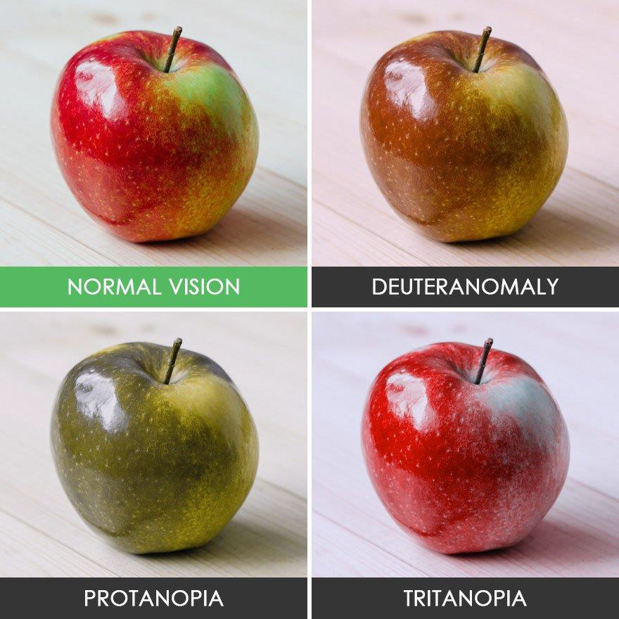
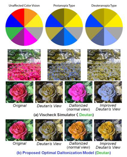
Color vision deficiency syndrome, sometimes known as color blindness, is a genetic disorder that affects color perception. Redgreen eyesight is the most common form of this syndrome. Affected people have a hard time recognizing some shades of red, yellow and green. Blue-yellow (also called Tritan defects), which rarely occurs, causes difficulty distinguishing between blue and green shades and makes it difficult to distinguish dark blue from black. These two types of color vision impair color perception, but do not affect visual acuity [1] (Figure 1).
Clinical Signs and Symptoms of Color Vision Deficiency Syndrome
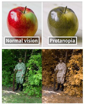
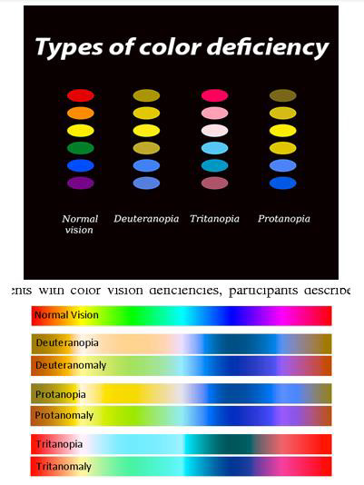
The most severe and less common type of color vision deficiency syndrome is the blue monochrome cone, which causes severe visual acuity and severe reduction in color vision. People with this form of the syndrome have more vision problems, which can include increased sensitivity to light (photophobia), rapid and involuntary movements of the pupil (nystagmus), and myopia. Blue-cone monochromacy is sometimes thought of as a form of achromatopsia, a disorder characterized by a partial or complete lack of color vision along with other vision problems [1] (Figure 2).
Etiology of Color Vision Deficiency Syndrome
Color vision deficiency syndrome is caused by mutations in the OPN1LW gene and the OPN1MW gene, which are located on the long arm of the sex X chromosome as Xq28, and the OPN1SW gene, which is located on the long arm of chromosome 7 as 7q32.1. The proteins produced by these genes play an important role in color vision. They are found in the retina, which is the light-sensitive tissue behind the eye. The retina consists of two types of lightreceiving cells, rods and cones, that transmit visual signals from the eye to the brain. The rods play a role in the eyes in low light. The cones provide visibility in bright light, including colored vision [1,2].
There are three types of cones, each containing a specific pigment (a photopigment called opsin) that is more sensitive to specific wavelengths. The brain combines the input of all three types of cones to produce natural colored vision [1,2] (Figure 3).

Figure 3: Schematic view of the sex X chromosome where the OPN1LW and OPN1MW genes are located in the long arm of this chromosome as Xq28 [1].
The OPN1LW, OPN1MW, and OPN1SW genes provide instructions for the synthesis of three opsin pigments in cones. The opsin synthesized from the OPN1LW gene is more sensitive to light in the yellow / orange part of the visible spectrum (long wavelength of light), and the cones of this pigment are known as long-wavelength sensitive or L-cones. The opsin synthesized from the OPN1MW gene is more sensitive to light in the middle of the visible spectrum (yellow / green light), and cones with this pigment are known as mid-wavelength sensitizers, or M cones. The opsin synthesized from the OPN1SW gene is more sensitive to light in the blue / purple part of the payroll spectrum (short wavelengths of light), and the cones of this pigment are known as short-wavelength sensitive or S-cones [1,2] (Figure 4).

Figure 4: Schematic view of chromosome 7 where the OPN1SW gene is located in the long arm of this chromosome as 7q32.1 [1].
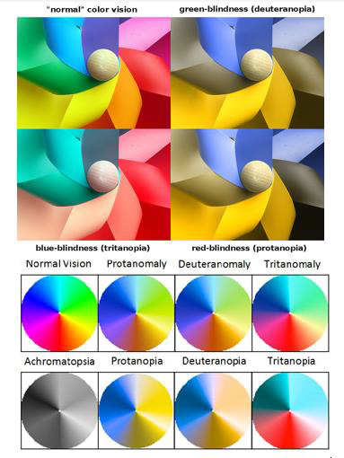
Genetic changes associated with the OPN1LW or OPN1MW gene cause a lack of red and green color vision. These changes lead to the absence of L or M cones and the production of abnormal opsin pigments in these cones, which affect the perception and recognition of red and green. Blue and yellow vision defects are caused by mutations in the OPN1SW gene. These mutations lead to premature destruction of S cones or the production of defective S cones. Dysfunction of the S cones alters the perception of blue, making it difficult or impossible to distinguish between blue and green shades and making it difficult to distinguish dark blue from black [1,3] (Figure 5).
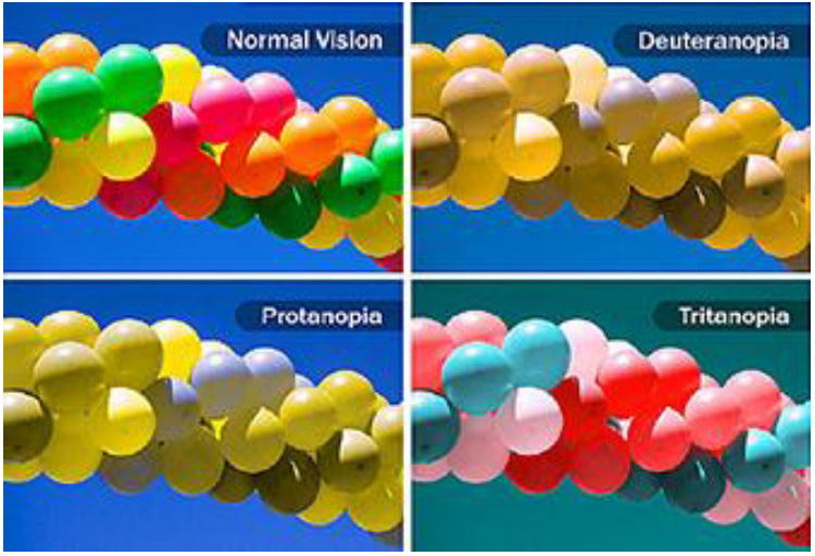
Figure 5: Another view of normal colored vision eyes versus eyes with color vision deficiency syndrome [1].
The blue monochrome cone occurs when genetic mutations in the OPN1LW and OPN1MW genes of both the L and M cones prevent normal function. In people with this condition, only the S cone has a function, which leads to a sharp decrease in visual acuity and poor color vision. Loss of L and M cones also causes other vision problems in people with blue monochrome cones [1,3] (Figure 6).
Some color vision problems are not caused by mutations in these genes. These conditions are known as non-hereditary color vision deficiency. They can be caused by other eye disorders such as retinal diseases, the nerve that carries visual information from the eye to the brain (the optic nerve), or areas of the brain that carry visual information processing. Non-hereditary color vision impairment can be a side effect of certain medications, such as chloroquine (used to treat malaria), or exposure to certain chemicals, such as organic solvents [1,4] (Figure 7).
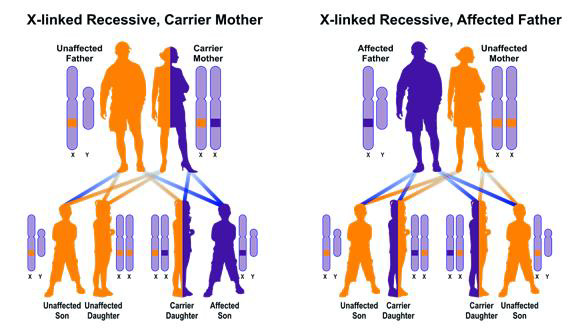
Red / green color vision deficiency syndrome and blue monochromatic cones follow a recessive X-linked hereditary pattern. The OPN1LW and OPN1MW genes are located on the X sex chromosome, which is one of two sex chromosomes. In men (who have only one X chromosome), one genetic change in each cell is enough to cause this condition. Men are much more likely than women to have the disease because of recessive X-linked disorders, because in women (who have two X chromosomes), genetic changes on both copies of the chromosome are needed to cause the disorder. The inherited characteristics of X are that fathers cannot pass on X-related traits to their sons [1,4] (Figure 8).
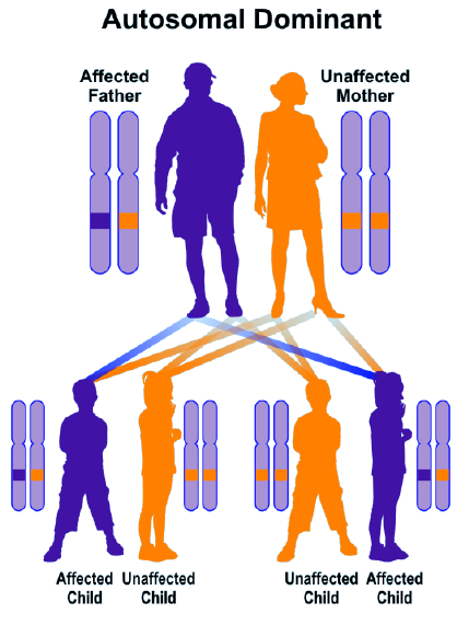
Blue / yellow color vision defects follow a predominantly autosomal inherited pattern. Therefore, a copy of the OPN1SW mutant gene (either parent) is required to cause the disorder, and the chance of having a child with the autosomal dominant disorder is 50% for each possible pregnancy [1,5] (Figure 9).
Frequency of Color Vision Deficiency Syndrome
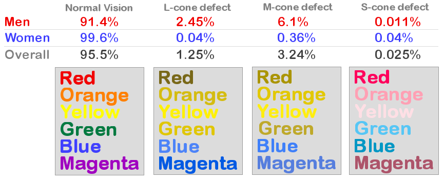
Red / green color vision deficiency is the most common form of color vision deficiency. This situation affects men much more than women. The disorder occurs in about 1 in 12 men and 1 in 200 women in Nordic populations. Red / green color vision defects have been less studied in almost all other populations [1,6]. Blue / yellow visual impairment affects men and women equally. It occurs in less than 1 in 10,000 people worldwide [1,6].
Monochrome blue cones are less common than other forms of color vision impairment, affecting approximately 1 in 100,000 people worldwide. Like red / green color vision defects, the monochrome blue cone affects men much more than women [1,6] (Figure 10).
Diagnosis of Color Vision Deficiency Syndrome
Colored vision deficiency syndrome is diagnosed based on clinical and physical findings of patients and some pathological and ophthalmological tests. The most accurate way to diagnose this syndrome is a molecular genetic test for the OPN1LW, OPN1MW, OPN1SW genes to check for possible mutations [1,6] (Figure 11).
Treatment Options for Color Vision Deficiency Syndrome
The treatment strategy and management of color vision deficiency syndrome is symptomatic and supportive. Treatment may be performed with the efforts and coordination of a team of specialists, including an ophthalmologist, eye surgeon, and optometrist. So far, no effective treatment for this syndrome has been identified and all clinical measures are taken to alleviate the suffering of the patients. Genetic counseling is also essential for all parents who want a healthy baby [1,6] (Figure 12,13).
Discussion and Conclusion
The most severe and less common type of color vision deficiency syndrome is the blue monochrome cone, which causes severe visual acuity and severe reduction in color vision. People with this form of the syndrome have more vision problems, which can include increased sensitivity to light (photophobia), rapid and involuntary movements of the pupil (nystagmus), and myopia. Genetic changes associated with the OPN1LW or OPN1MW gene cause a lack of red and green color vision. These changes lead to the absence of L or M cones and the production of abnormal opsin pigments in these cones, which affect the perception and recognition of red and green. Treatment may be performed with the efforts and coordination of a team of specialists, including an ophthalmologist, eye surgeon, and optometrist [1,6].
Conflict Of Interest
No conflict of interest.
References
- Asadi S (2022) Pathology in Medical Genetics Book, Vol 15, Amidi Publications Iran.
- Neitz J, Neitz M (2011) The genetics of normal and defective color vision. Vision Res 51(7): 633-651.
- Gardner JC, Michaelides M, Holder GE, Kanuga N, Webb TR, et al. (2009) Blue cone monochromacy: causative mutations and associated phenotypes. Mol Vis 15: 876-884.
- Deeb SS (2005) The molecular basis of variation in human color vision. Clin Genet 67(5): 369-377.
- Michaelides M, Hunt DM, Moore AT (2004) The cone dysfunction syndromes. Br J Ophthalmol 88(2): 291-297.
- Deeb SS (2004) Molecular genetics of color-vision deficiencies. Vis Neurosci 21(3): 191-196.




 We use cookies to ensure you get the best experience on our website.