Case Report 
 Creative Commons, CC-BY
Bilateral Herpes Zoster Ophthalmicus: A Painful Dermatomal Rash of Both Eyes in A 4 Years-Old Child
*Corresponding author:Stefan Bittmann, Ped Mind Institute (PMI), Department of Pediatrics, Hindenburgring , Gronau, Germany
Received:March 21, 2023; Published:March 30, 2023
DOI: 10.34297/AJBSR.2023.18.002463
Abstract
Zoster ophthalmicus is caused by reactivation of varicella zoster viruses, which belong to the group of herpes viruses. The symptoms and complaints, such as a dermatomal rash in the forehead area and painful inflammation of all tissues in the anterior and, less commonly, posterior ocular structures, can be very severe. Diagnosis is based on the characteristic findings in the anterior ocular structures associated with zoster dermatitis of the 1st trigeminal branch (V1). We present an interesting case of a 4 years-old boy with bilateral herpes zoster ophthalmicus with a bridging connection between both eyes.
Introduction Varicella zoster virus belongs to the family of herpes viruses (Herpesviridae). It consists of an icosahedron-shaped capsid in which a double-stranded DNA is embedded. The nucleocapsid is surrounded by a double membrane with pseudospikes. Varicella viruses have low genetic variability, and their only host is humans. After the initial infection has cleared, the viruses retract into the dorsal roots of the spinal and cranial ganglia, where they cannot be eliminated by the immune system. They thus persist in the body for life. When reactivated, they migrate along sensory nerves to the skin and lead to the characteristic picture of dermatomal painful zoster with erythema and of grouped papulovesicular, later pustular skin lesions. Herpes zoster occurs primarily in the elderly and/or immunocompromised. The risk of disease generally increases with age. The lifetime prevalence is 25 to 50%. As a result of increasing life expectancy and the associated growing number of elderly people in Germany, an increase in the incidence of herpes zoster and associated complications such as Post-Zoster Neuralgia (PZN) can be assumed. This trend continues to be exacerbated by the likewise increasing number of immunocompromised and organ transplant patients, such as tumor and AIDS patients. Approximately 99% of people over the age of 40 in Germany have experienced VZV infection. Approximately 20 to 30% will develop herpes zoster during their lifetime. According to estimates, the annual incidence of people suffering from zoster in Germany is around 400,000. Women fall ill significantly more often than men. Transmission of VZV is usually aerogenic through virus-containing droplets that enter the ambient air during coughing and are absorbed through the respiratory tract. In chickenpox, infectivity is very high. Infection can occur within a radius of several meters. In contrast, the infectivity of herpes zoster is rather low, as transmission only occurs via the secretion from the skin vesicles. The risk of infection can therefore be reduced by covering the skin lesions. However, it persists in principle until all efflorescence’s are completely encrusted. Moreover, herpes zoster is infectious only for people who have not previously contracted chickenpox. In the case of infection with the VZV, chickenpox initially develops in children as well as in adults, and herpes zoster develops only as a secondary manifestation. Diaplacental transmission of VZV during varicella disease in pregnant women is possible and may result in fetal varicella syndrome. The incubation period is usually 14 to 16 days, possibly as long as 28 days after passive immunization. Herpes zoster symptoms can vary widely. For example, the disease may be mild and cause only itching. On the other hand, shingles is often accompanied by considerable distress, and even light touch can cause considerable pain. The disease usually presents initially with nonspecific symptoms such as malaise, headache, pain in the limbs, and paresthesia’s, often followed by a phase of itchy exanthema and fever. Characteristic vesicular skin lesions also occur in herpes zoster. Their localization depends on the supply area of the affected nerves. In many cases, the skin vesicles initially develop in the trunk area and may spread from there to other parts of the body, including the hairy scalp and mucous membranes. Usually, only one dermatome is affected (zoster segmentalis); however, overlapping dermatome involvement is also possible. However, crossing the midline of the body is a rarity (zoster duplex). Very rarely, several skin segments are also affected asymmetrically on both sides of the body. Pain, sensory disturbance and itching often occur several days before the skin symptoms. The pain symptomatology in the prodromal phase often leads to a wide range of misdiagnoses, which can be misinterpreted as myocardial infarction, cholecystitis, toothache, etc., depending on the localization. Pain quality is often described as burning, stabbing, and pulsating. Local lymphadenopathy is possible. Pretreatment with anticoagulants or corticosteroids may sometimes result in skin hemorrhage. As a result of the acute symptoms as well as the sometimes chronic symptoms, herpes zoster can lead to a considerable and lasting impairment of the quality of life.
Keywords: Herpes zoster ophthalmicus-child-bridging
Case Report
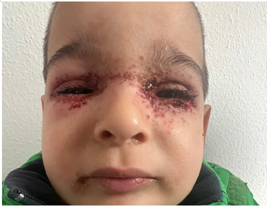
A previously healthy 4 years old boy was admitted to our emergency department because of an acute rash around both eyes. The patient had low-grade fever of around 38°C for two days. The history of infections in the surrounding area was unremarkable. All vaccinations had been performed according to the German vaccination schedule. Clinically, the boy presented in good general condition with a rash around both eyes with a bridging between both eyes (Figure1). O2 saturation was 100%. Many ruptured blisters and papules were noted around both eyes (Figure 1); otherwise, no other cutaneous abnormalities were apparent at this time. No neck stiffness was found. The rest of the physical examination including the remaining neuro status was unremarkable.

Figure 1:A 4 years-old boy presented with a bilateral herpes zoster ophthalmicus of both eyes. A herpetiform rash about both eyes with a bridging connection was present. There was no fever. Visual acuity and deeper ocular areas were not affected. Facial paralysis was not present. The smear for VZV-PCR was positive.
Discussion
In herpes zoster, the skin lesions are usually very painful and often first develop in the girdle region [1-24]. The cause is an infection with the varicella zoster virus [1-24]. This can cause two clinical pictures: Chickenpox as a primary infection and Shingles as a secondary manifestation due to reactivation of the viruses persisting in nerve cells [1-24]. The incidence of zoster is high and increasing, especially in the elderly and in immunocompromised individuals. The disease can heal spontaneously. On the other hand, it can also take a severe, potentially life-threatening course. Complications such as post-zoster neuralgia can also occur, which is associated with severe pain and a considerable loss of quality of life. The aim of treatment is to minimize the risk of complications and also to achieve rapid relief of the acute symptoms. Antiviral therapy is of particular importance in this context. If possible, this should be initiated within 72 hours of the appearance of the skin symptoms or within 48 hours of the manifestation of the characteristic skin vesicles [1,6,9,14]. Four different agents are available, which differ in their antiviral potency and also in their mode of administration. Two vaccines are now approved in Germany for the prevention of herpes zoster. Varicella-Zoster Virus (VZV) can manifest itself in two different clinical pictures: Varicella (chickenpox) in exogenous initial infection, which mostly occurs in childhood, and Herpes Zoster (shingles; zoster, ancient Greek for girdle) in endogenous reactivation. Herpes zoster occurs more frequently in older people beyond the fifth decade of life [1-24]. Contrary to what the name shingles suggests, the disease by no means manifests itself only in the shingles area of the body. On the contrary, severe courses with involvement of the eyes and ears and also of the internal organs may occur. The latency of VZV infection is ensured by an effective immune defense. If sufficient control can no longer be ensured as a result of a weakened immune system (e.g. in the context of natural aging processes or HIV infection), reactivation of virus replication may occur. Herpes zoster is often accompanied by severe pain and may cause persistent complications such as post-herpetic neuralgia, which emphasizes the importance of prompt diagnosis and effective therapy [7,9,12,22]. VZV is a member of the herpesvirus family (Herpesviridae). It consists of an icosahedron-shaped capsid in which a double-stranded DNA is embedded. The nucleocapsid is surrounded by a double membrane with pseudospikes. Varicella viruses have low genetic variability, and their only host is humans. After the initial infection has cleared, the viruses retract into the dorsal roots of the spinal and cranial ganglia, where they cannot be eliminated by the immune system. They thus persist in the body for life. When reactivated, they migrate along sensory nerves to the skin and lead to the characteristic picture of dermatomal painful zoster with erythema and of grouped papulovesicular, later pustular skin lesions. Herpes zoster occurs primarily in the elderly and/ or immunocompromised. The risk of disease generally increases with age. The lifetime prevalence is 25 to 50% [1-24]. As a result of increasing life expectancy and the associated growing number of elderly people in Germany, an increase in the incidence of herpes zoster and associated complications such as Post-Zoster Neuralgia (PZN) can be assumed. This trend continues to be exacerbated by the likewise increasing number of immunocompromised and organ transplant patients, such as tumor and AIDS patients. Approximately 99% of people over the age of 40 in Germany have experienced a VZV infection. Approximately 20 to 30% will develop herpes zoster during their lifetime [1-24]. According to estimates, the annual incidence of people suffering from zoster in Germany is around 400,000. Females fall ill significantly more often than males [1,8,9,11,20].
The diagnosis of herpes zoster is usually made clinically on the basis of the symptoms and primarily by inspection of the skin, including attention to the localization of the efflorescence’s [1-24]. The purely clinical diagnosis has a specificity of about 60 to 90 %, depending on the severity and localization. In the case of a typical clinical picture of herpes zoster, laboratory confirmation can usually be dispensed with. However, atypical manifestations are also possible (for example, in persons with immunodeficiency), so that specific laboratory diagnostics are indicated in individual cases. This should also be done in cases of CNS involvement, pneumonia, infections during pregnancy, and neonates. Differential diagnosis must consider herpes simplex virus infections (HSV1 especially in the head/neck area, HSV2 especially in the lumbosacral area) and zosteriform dermatological diseases [1-24]. Molecular detection of VZV DNA from swabs is now considered the gold standard for laboratory diagnosis of VZV infection. Modern real-time PCR methods have nearly 100% sensitivity and specificity when performed correctly. No fluid-filled vesicles are necessary for PCR detection. Viral DNA can usually be reliably detected even in the maculopapular or healing stage. If CNS infection is suspected, VZV PCR must be performed from cerebrospinal fluid. If zoster ophthalmicus is suspected, VZV DNA can be detected in aqueous humor or, in some cases, from an ocular smear. If systemic dissemination is suspected, serum or plasma is obtained for VZV PCR (quantitative PCR is recommended in these cases). Direct antigen detection is much less sensitive and specific than PCR. Serologic antibody detection is not suitable for acute diagnosis of zoster efflorescence’s. However, antibody diagnostics may prove useful for differential diagnosis in cases of seronegativity to differentiate zoster-like neurologic symptoms from herpes zoster. Due to its low sensitivity and the higher technical effort, viral culture is only of value in special cases (e.g. testing of drug sensitivity). The differential diagnosis should particularly consider the possibility of herpes simplex infection, hemorrhagic and bullous erysipelas, and bullous dermatoses such as bullous pemphigoid and pemphigus vulgaris, contact dermatitis, and also the possibility of insect bites. Treatment of herpes zoster should generally be initiated as early as possible. The goal of antiviral treatment of zoster in immunocompetent patients is to shorten the acute phase of the disease, as measured by reduction of fever, relief of acute zoster pain, arrest of vesicular eruption, accelerated healing of skin lesions, and prevention of scarring. Another important treatment goal is to prevent or shorten the duration of postzoster neuralgia. In addition, possible complications such as cutaneous and visceral disseminations in immunosuppressed patients, ocular involvement, CNS or cranial nerve involvement in patients with head zoster should be prevented. Therapy for acute herpes zoster consists of systemic antiviral chemotherapy combined with local antiseptic treatment and consistent pain management. For symptomatic, local treatment, mainly drying, antipruritic and antiseptic topical agents and possibly moist compresses (in the vesicle stage) are used. Especially in cases of extensive infestation and patients at risk for complications, early systemic antiviral therapy is indicated with the aim of preventing further viral replication as early as possible. Early analgesic therapy can prevent chronicity. It is administered according to the intensity of pain in accordance with the WHO grading scheme with non-steroidal anti-inflammatory drugs or opioids. Co-analgesics such as antidepressants and anticonvulsants can be given in addition. Herpes zoster opthalmicus of both eyes with a bridging is very rare and not often seen in childhood, so, this case is very interesting concerning the appearance in the face of the child [25] (Figure 1).
Acknowledgement
None.
Conflict of Interest
None.
References
- Kang DH, Kwak BO, Park AY, Kim HW (2021) Clinical Manifestations of Herpes Zoster Associated with Complications in Children. Children (Basel) 8(10): 845.
- Werner RN, Nikkels AF, Marinović B, Schäfer M, Czarnecka Operacz M, et al. (2017) European consensus-based (S2k) Guideline on the Management of Herpes Zoster - guided by the European Dermatology Forum (EDF) in cooperation with the European Academy of Dermatology and Venereology (EADV), Part 2: Treatment. J Eur Acad Dermatol Venereol 31(1): 20-29.
- Peterson N, Goodman S, Peterson M, Peterson W. (2016) Herpes zoster in children. Cutis 98(2): 93-95.
- Tucker SM (1958) Herpes zoster ophthalmicus in children. Arch Dis Child 33(171): 437-439.
- Davies EC, PavanLangston D, Chodosh J (2016) Herpes zoster ophthalmicus: declining age at presentation. Br J Ophthalmol 100(3): 312-314.
- Nofal A, Fawzy MM, Sharaf El Deen SM,El Hawary EE (2020) Herpes zoster ophthalmicus in COVID-19 patients. Int J Dermatol 59(12): 1545-1546.
- De Freitas D, Martins EN, Adan C, Alvarenga LS, Pavan Langston D (2006) Herpes zoster ophthalmicus in otherwise healthy children. Am J Ophthalmol 142(3): 393-399.
- Keramida P, Antoniadi M, Archimandritou E, Kostaridou S,Koletsi P (2023) Herpes Zoster Ophthalmicus in a Healthy Toddler Fully Immunized Against Varicella-Zoster Virus: A Case Report and Review of Treatment Strategies in Children. Cureus 15(1): e33352.
- Oladokun RE, Olomukoro CN,Owa AB (2013) Disseminated herpes zoster ophthalmicus in an immunocompetent 8-year old boy. Clin Pract 3(2): e16.
- Kang DH, Kwak BO, Park AY, Kim HW (2021) Clinical Manifestations of Herpes Zoster Associated with Complications in Children. Children (Basel) 8(10): 845.
- Kleinschmidt DeMasters BK, Gilden DH (2001) The expanding spectrum of herpesvirus infections of the nervous system. Brain Pathol 11(4): 440-451.
- Davies EC, Pavan Langston D, Chodosh J (2016) Herpes zoster ophthalmicus: declining age at presentation. Br J Ophthalmol 100(3): 312-314.
- Walton RC, Reed KL (1999) Herpes zoster ophthalmicus following bone marrow transplantation in children. Bone Marrow Transplant 23(12): 1317-1320.
- De Freitas D, Martins EN, Adan C, Alvarenga LS, Pavan Langston D (2006) Herpes zoster ophthalmicus in otherwise healthy children. Am J Ophthalmol 142(3): 393-399.
- Ang LP, Au Eong KG, Ong SG (2001) Herpes zoster ophthalmicus. J Pediatr Ophthalmol Strabismus 38(3): 174-176.
- Weinberg JM (2007) Herpes zoster: epidemiology, natural history, and common complications. J Am Acad Dermatol 57(6 Suppl): S130-135.
- Kim M, Chun YS, Moon NJ, Kim KW (2022) Clinical Factors Associated with the Early Reduction of Corneal Sensitivity in Herpes Zoster Ophthalmicus. Korean J Ophthalmol 36(2): 147-153.
- Panda A, Sood NN,Dayal Y, Bhatia IM (1981) Herpes zoster ophthalmicus in children (reports of two cases). Indian J Ophthalmol 29(1): 37-38.
- Grote V, Von Kries R, Rosenfeld E, Belohradsky BH, Liese J (2007) Immunocompetent children account for the majority of complications in childhood herpes zoster. J Infect Dis 15:196(10): 1455-1458.
- Makzal Z, Edwards M (2017) Herpes zoster ophthalmicus in a 1-year-old child. BMJ Case Rep 2017: bcr2017222112.
- Kamboj A, Hwang CJ, Mokhtarzadeh A, Harrison AR (2021) Development of Herpes Zoster Ophthalmicus in an Immunocompetent Pediatric Patient Following Facial Trauma. Ophthalmic Plast Reconstr Surg 37(5): e170-e172.
- Grose C, Shaban A, Fullerton HJ (2023) Common Features Between Stroke Following Varicella in Children and Stroke Following Herpes Zoster in Adults : Varicella-Zoster Virus in Trigeminal Ganglion. Curr Top Microbiol Immunol 438: 247-272.
- Kong CL,Thompson RR, Porco TC, Kim E, Acharya NR (2020) Incidence Rate of Herpes Zoster Ophthalmicus: A Retrospective Cohort Study from 1994 through 2018. Ophthalmology 127(3): 324-330.
- Hung PY, Lee WT, Shen YZ (2000) Acute hemiplegia associated with herpes zoster infection in children: report of one case. Pediatr Neurol 23(4): 345-348.
- Birka DA (1963) HERPES ZOSTER OPHTHALMICUS IN AN 8-YEAR-OLD CHILD. Br J Ophthalmol 47(1): 60-61.


 We use cookies to ensure you get the best experience on our website.