Mini Review 
 Creative Commons, CC-BY
An Atypical Presentation of Superficial Breast Cancer: A Case Report and Review of the Literature
*Corresponding author: Paolo Izzo, Department of Medicine and Surgery, Saint Camillus International University of Health Sciences, Italy.
Received: May 18, 2023; Published: May 24, 2023
DOI: 10.34297/AJBSR.2023.18.002535
Abstract
Background: Retroareolar breast tumors are common, but malignancies arising from these glands are rare. This report discusses a case of a superficial breast tumor in a 54-year-old female, highlighting the importance of a broad differential diagnosis when evaluating breast lumps.
Case presentation: The patient was presented with a progressively growing, ulcerating superficial breast mass in her left breast. Diagnostic assessment included PET scan and histopathological examination, which confirmed a Grade II superficial infiltrating ductal carcinoma.
Interventions: The patient underwent a modified radical mastectomy under general anesthesia. Postoperatively, the patient was initiated on tamoxifen, an estrogen receptor modulator, as per current guidelines.
Outcomes: The patient recovered without complications and showed no signs of recurrence or metastasis during follow-ups.
Conclusion: This case underscores the importance of a thorough examination and high index of suspicion for any breast lumps, emphasizing the crucial role of early detection and aggressive management in improving patient outcomes. The report adds to the existing literature on the varied presentations of breast cancer, potentially aiding in the identification and management of similar cases in the future (Figure1).

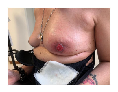
Figure 1: The mass on the left breast, which 3.0cm×3.0cm in size. The boundary is unclear, the shape is irregular, and the mass is immovable.
Keywords: Retroareolar breast tumors, Superficial infiltrating ductal carcinoma, Modified radical mastectomy, Tamoxifen.
Introduction
Retroareolar breast tumors, also known as aberrant mammary glands, represent a condition where additional mammary tissue persists instead of degenerating during embryonic development [1]. Even though this condition is quite prevalent, malignancies arising from these glands are rare [2]. A case of a superficial breast tumor in a 54-year-old female presents a unique clinical scenario, and this case report aims to add to the body of knowledge concerning the diagnosis and treatment of such cases [3].
Case Presentation
A 54-year-old female patient presented with a superficial breast mass in her left breast, which had been progressively growing for the past nine months. The mass had begun to ulcerate over the past month. Initially, the mass was approximately 1.0cm×1.5 cm. However, over time, the mass had expanded to 3.0cm×3.0 cm. The mass was hard, immovable, and had an unclear boundary with the surrounding skin. The patient reported increasing discomfort when the mass was touched. No enlargement of the ipsilateral supraclavicular lymph nodes was observed.
Diagnostic assessment Positron emission computed tomography (PET) [4] revealed a dense nodule in the left breast’s subcutaneous tissue, with the boundary unclear and some sections protruding to the skin surface. The Standardized Uptake Value max level was 9.0 [5]., suggesting a malignant tumor. No enlarged lymph nodes were observed. The histopathological examination confirmed the diagnosis of a Grade II superficial infiltrating ductal carcinoma [6]. The immunohistochemical examination revealed estrogen receptor (+++) 95%, progesterone receptor (+++) 90%, human epidermal growth factor receptor-2 (1+), ki67 (30% positive), and other markers typical for breast carcinoma [7].
Therapeutic intervention Following the confirmation of the diagnosis and after ruling out any contraindications, a decision was made to perform a modified radical mastectomy under general anesthesia [8]. This involved the resection of the tumor along with a margin of healthy tissue and dissection of the axillary lymph nodes [9]. The wound was thoroughly irrigated and dressed, and two drains were placed, one in the axilla and another near the surgical site.
Follow-up and outcomes Postoperatively, the patient was initiated on tamoxifen, an estrogen receptor modulator, as per the current guidelines [10]. The patient made an uneventful recovery and was discharged three days postoperatively. She reported minimal discomfort, and no complications such as infection or hematoma were noted. Regular follow-ups were scheduled every three to six months [11]. No signs of recurrence or metastasis were observed during the follow-up period.
Discussion
This case highlights a rare presentation of a superficial breast tumor. The differential diagnoses for such a case would include be nign breast conditions such as fibroadenomas or cysts, infections, and other malignancies [12]. Pathological and immunohistochemical examinations played a vital role in confirming the diagnosis in this case [13].
The treatment approach for such a case is primarily surgical, followed by appropriate adjuvant therapy based on the pathological and immunohistochemical findings [14]. Despite the rarity of such cases, they underscore the importance of a thorough examination and a high index of suspicion for any breast lumps, whether in the typical mammary region or in retroareolar breast tissue. Early detection is crucial in managing such cases, as timely intervention can help prevent disease progression and metastasis. As with any breast cancer, the treatment regimen typically involves surgery, chemotherapy, radiation therapy, and targeted therapy, depending on the staging and the patient’s overall health condition [15]. In our case, the patient underwent a mastectomy with axillary lymph node dissection, followed by adjuvant chemotherapy and radiation therapy. The importance of pathological examination in these cases cannot be overstated, as it not only confirms the diagnosis but also informs about the tumor’s molecular characteristics [16]. These molecular characteristics can further guide the use of targeted therapy, such as hormone therapy in hormone receptor- positive breast cancers or monoclonal antibodies in HER2-positive breast cancers. In this patient, immunohistochemical analysis revealed triple-negative breast cancer, which necessitated the use of an aggressive chemotherapy regimen due to the absence of targeted therapy options.
This case also emphasizes the importance of patient education and awareness. Women should be encouraged to perform regular self-breast examinations and seek medical advice if they notice any abnormal changes, regardless of their location.
Conclusions
This case report presents an atypical case of a superficial breast tumor in a 54-year-old woman, emphasizing the importance of maintaining a broad differential diagnosis when evaluating breast lumps [17]. Even though the superficial location of the tumor is rare, it reinforces the understanding that breast cancer can have varied presentations. The significant role of pathological and immunohistochemical examinations in confirming the diagnosis and guiding treatment decisions was highlighted in this case. Furthermore, the case underscores the importance of aggressive management in triple-negative breast cancers, given the lack of targeted therapy options [18].
Early detection remains key in managing such cases, and the necessity of public awareness and Education about regular self-breast examinations is paramount. Clinicians should maintain a high index of suspicion for breast cancer even in atypical presentations, as early diagnosis and appropriate management are crucial in improving patient outcomes. This report adds to the existing literature on the varied presentations of breast cancer and may aid in the identification and management of similar cases in the future.
Conflict Of Interest
The authors have no conflicts of interest to disclose.
References
- Macias H, Hinck L (2012) Mammary gland development. Wiley Interdiscip Rev Dev Biol 1(4): 533-557.
- Thasanabanchong P, Vongsaisuwon M (2020) Unexpected presentation of accessory breast cancer presenting as a subcutaneous mass at costal ridge: a case report. J Med Case Rep 14(1): 45.
- Sharp PC, Michielutte R, Spangler JG, Cunningham L, Freimanis R (2005) Primary care providers' concerns and recommendations regarding mammography screening for older women. J Cancer Educ 20(1): 34-38.
- Pennant M, Takwoingi Y, Pennant L, Davenport C, Fry-Smith A, et al. (2010) A systematic review of positron emission tomography (PET) and positron emission tomography/computed tomography (PET/CT) for the diagnosis of breast cancer recurrence. Health Technol Assess 14(50): 1-103.
- Lee MI, Jung YJ, Kim DI, Lee S, Jung CS, et al. (2021) Prognostic value of SUVmax in breast cancer and comparative analyses of molecular subtypes: A systematic review and meta-analysis. Medicine (Baltimore) 100(31): e26745.
- Ginter PS, Idress R, D'Alfonso TM, Fineberg S, Jaffer S, (2021) Histologic grading of breast carcinoma: a multi-institution study of interobserver variation using virtual microscopy. Mod Pathol 34(4): 701-709.
- Chand P, Garg A, Singla V, Rani N (2018) Evaluation of Immunohistochemical Profile of Breast Cancer for Prognostics and Therapeutic Use. Niger J Surg 24(2): 100-106.
- Plesca M, Bordea C, El Houcheimi B, Ichim E, Blidaru A (2016) Evolution of radical mastectomy for breast cancer. J Med Life 9(2): 183-186.
- Rao R (2015) The Evolution of Axillary Staging in Breast Cancer. Mo Med 112(5): 385-388.
- Barnadas A, Algara M, Cordoba O, Casas A, Gonzalez M, et al. (2018) Recommendations for the follow-up care of female breast cancer survivors: a guideline of the Spanish Society of Medical Oncology (SEOM), Spanish Society of General Medicine (SEMERGEN), Spanish Society for Family and Community Medicine (SEMFYC), Spanish Society for General and Family Physicians (SEMG), Spanish Society of Obstetrics and Gynecology (SEGO), Spanish Society of Radiation Oncology (SEOR), Spanish Society of Senology and Breast Pathology (SESPM), and Spanish Society of Cardiology (SEC). Clin Transl Oncol 20(6): 687-694.
- Sisler J, Chaput G, Sussman J, Ozokwelu E (2016) Follow-up after treatment for breast cancer: Practical guide to survivorship care for family physicians. Can Fam Physician 62(10): 805-811.
- Cochran JM, Leproux A, Busch DR, O'Sullivan TD, Yang W, et al. (2021) Breast cancer differential diagnosis using diffuse optical spectroscopic imaging and regression with z-score normalized data. J Biomed Opt 26(2): 026004.
- Leong AS, Zhuang Z (2011) The changing role of pathology in breast cancer diagnosis and treatment. Pathobiology 78(2): 99-114.
- Candás G, García A, Ocampo MD, Korbenfeld E, Vuoto HD, et al. (2021) Impact of immunohistochemical profile changes following neoadjuvant therapy in the treatment of breast cancer. Ecancermedicalscience 15: 1162.
- Moo TA, Sanford R, Dang C, Morrow M (2018) Overview of Breast Cancer Therapy. PET Clin 13(3): 339-354.
- Huszno J, Kolosza Z (2019) Molecular characteristics of breast cancer according to clinicopathological factors. Mol Clin Oncol 11(2): 192-200.
- Bhushan A, Gonsalves A, Menon JU (2021) Current State of Breast Cancer Diagnosis, Treatment, and Theranostics. Pharmaceutics 13(5): 723.
- Baranova A, Krasnoselskyi M, Starikov V, Kartashov S, Zhulkevych I, et al. (2022) Triple-negative breast cancer: current treatment strategies and factors of negative prognosis. J Med Life 15(2): 153-161.


 We use cookies to ensure you get the best experience on our website.