Research Article 
 Creative Commons, CC-BY
Dose Dependence of Morbidity in Persons with Thyroid Exposure Due to Radioactive Iodine Intake During Fetal Development
*Corresponding author: Alexander N Stojarov, Department of Radiation Medicine, Belarusian State Medical University, Belarus, pr. Dzerzhinskogo 83, Minsk, Belarus.
Received: July 07, 2023; Published: July 24, 2023
DOI: 10.34297/AJBSR.2023.19.002617
Abstract
The paper analyzes the morbidity of residents of Belarus, who, because of the accident at the Chernobyl nuclear power plant, received intrauterine exposure due to transplacental intake of I-131 from the mother’s body. The control cohort consisted of residents of the same regions of the country who were born a year later and, therefore, their mothers were not exposed to radioactive iodine during pregnancy. A significant difference in primary morbidity was found between the exposed and non-exposed cohorts. In the group of individuals who were irradiated in utero, maxima of pathology registration for morbidity from 1986 to 2017, are revealed 2, 10, 15, 19, and 26 years after the Chernobyl accident. At the same time, there are two peaks of incidence associated with the absorbed dose received by the thyroid gland (21 and 66 mGy). A very noticeable difference in incidence is observed between females and males. In women, the incidence is expressed after a longer period since the exposure than in men. The main peak of incidence in all cases falls on the age of individuals from 9 to 10 years. If in the initial period the dominant pathology was represented mostly by diseases of the respiratory organs and the endocrine system, then later pathology was represented by diseases of nervous, digestive, and musculoskeletal systems. The results obtained are discussed from the point of view of the sensitivity of thyroid genes expression to the effects of radioactive iodine leading to changes in the synthesis of hormones in its turn affecting the development of organs and the functioning of body systems, that is finally causing the appearance of pathology.
Keywords: Radioactive iodine, I-131, Morbidity, Incidence, Nervous system, Cardiovascular system, Genitourinary systems, Gastrointestinal tract, Thyroid, Thyroid hormones
List of Abbreviations: TG: Thyroid Gland; ICD-10: International Classification of Diseases; COD: Coefficient of Determination.
Introduction
It is well known that Thyroid Gland (TG) plays an important role in the development and functioning of many organs and systems of the body [1]. At the same time, it was found that many environmental factors, including exposure to radiation, can disrupt the function of that gland. TG is a critical organ that is sensitive to the effects of radiation, which can lead to a malignant neoplasm for mation after a certain time. That fact was well documented in the data on thyroid cancer, which began to be registered in three neighboring countries (Belarus, Ukraine, and the Russian Federation) 5-7 years after the accident at the Chernobyl nuclear power plant [2]. This dose-dependent effect was due to the action of radioactive iodine I-131 incorporated into the thyroid gland. At the same time, relatively little is known about the effect of small doses, given that thyroid genes can change their expression in the opposite way to the magnitude of radiation exposure [3]. A special case is in utero irradiation. In this case, the source of I-131 is the mother’s body, into which, during a radiation accident, it enters by inhalation and orally, overcoming the placental barrier and accumulating in the TG of the fetus. Very little is known about the consequences of such exposure [4].
We managed to form a cohort of women who received thyroid irradiation during pregnancy due to I-131. In a cohort of children born by them who were irradiated in utero, we analyzed the degree of radiation exposure due to the incorporation of radioactive iodine, as well as their incidence of various types of pathology in the long-term period after the Chernobyl accident.
Materials and Methods
As the main cohort of individuals, residents of the Stolin district of the Brest region were selected, who were born from women lived in the same area in late April-early May 1986 and were affected by a radioactive cloud that passed through this region of Belarus. The cloud contained iodine radionuclides, including I-131. The main cohort included 123 individuals, including 62 women and 61 men. Dates of birth were in the range of 06/03/1986-02/06/1987. When analyzing the effects of radiation on certain segments of the population, it is always necessary to have a cohort for comparison consisting of individuals who have not received exposure. In this study, the cohort for comparison included residents of the Stolin district of the Brest region who were born later. In other words, their mothers did not fall under the “iodine shock”. The cohort included 113 individuals from the same area, identical not only in terms of residence, but also in social status, of which 57 were males and 56 were females. Their dates of birth were in the range of 01/03/1988-12/31/1988. The comparison group was selected considering the half-life of I-131, which is about 8 days. For 10 halflives, i.e., after 80 days, only trace activities of radioactive iodine remained in the medium and, consequently, the mothers of these children did not receive thyroid exposure during pregnancy. Verified data on the state of health of exposed and non-exposed individuals were obtained from the State Register of Persons Affected by the Chernobyl Accident. Only primary morbidity was considered in the work.
Absorbed doses on the TG due to I-131 were calculated by the head of the laboratory for the reconstruction of exposure doses to the population of the State Scientific Center of the Federal Medical Biophysical Center named after A.I. Burnazyan FMBA of Russia, Doctor of Technical Sciences, Shinkarev S.M. Doses were calculated using a semi-empirical model from the year 2004 iteration. Statistical data processing was carried out using the application software Statistics 10.0 (StatSoft. Inc., USA) and SigmaPlot 12.5 (Systat Software Inc., Germany).
Results

Figure 1: Dependence of the incidence of pathology among persons irradiated in utero on the absorbed dose by the TG.

Figure 2: Dependence of the incidence of pathology among persons irradiated in utero on the absorbed doses of their mothers.
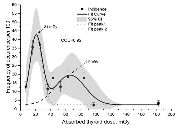
The frequency of occurrence of pathology among individuals irradiated during prenatal development is characterized by two maxima of absorbed doses by thyroid gland (Figure 1). The Figure 1 shows data for all ICD-10 classes of pathology incidence and the period from 1986 to 2017. The first, most intense, but smaller in area (43%) peak is associated with an absorbed dose of about 20mGy. The second peak, which was wider and, therefore, responsible for the appearance of a significant proportion of pathology, has a maximal absorbed dose of 66mGy. In general, both doses are relatively small. Since the developing fetus in the first trimester of pregnancy is not yet able to produce hormones (T3 and T4) itself, but uses maternal hormones, it was interesting to analyze the incidence of pathology in children with absorbed doses by the TG of their mothers [5]. Figure 2 presents these results. The dependence of morbidity among children in the post-accident period (1986-2017) on doses absorbed by their mothers is also characterized by the presence of two peaks. However, their values differ from the maximum values of doses in fetuses. Both peaks show maxima that are three times greater in magnitude than the absorbed doses of developing fetuses. This, from our point of view, should not be surprising, since, firstly, the placental barrier is not characterized by 100% permeability. Secondly, the mass of the TG of the mother is much greater than the mass of the one of fetus. In this regard, the accumulation of radioactive iodine by the gland of a mother is greater. However, the presence of two peaks in the incidence in both cases may indicate the true nature of this phenomenon (Figure 2).
The following analysis was carried out on a cohort of individuals of both sexes on whom radiation exposure was formed, as well as on a control cohort of individuals who were born later, after the decay of radioactive iodine. Figure 3 shows the incidence of pathology in the first of the cohorts. Mathematical search for peaks with a good coefficient of determination (COD=0.91) revealed five morbidity maxima among all pathology classes according to ICD-10. They were registered in 1988, 1996, 2001, 2005 and 2012, i.e., 2, 10, 15, 19 and 26 years after the Chernobyl accident. In the first two years, the main contribution to the incidence was equally made by childhood viral and infectious diseases. At the age of 10, among the victims, the most significant increase in morbidity was recorded, and it was mostly (more than 20%) due to diseases of the endocrine system (Chapter IV). Fifteen years after the Chernobyl accident, among those exposed in utero, the main contribution into the pathology was made by respiratory diseases (Chapter X). In 2005 there was an acute and small peak in incidence, and in 2012, adult victims were diagnosed with eye pathology, diseases of respiratory organs and musculoskeletal system. This picture differs sharply from the incidence of pathology in the control cohort of individuals who did not receive thyroid irradiation (Figure 3,4). According to the incidence data, two peaks are revealed among them, one of them is small and the second one is large and asymmetrical with a maximum in 1996, i.e., at 9 years of age. The prevailing types of pathology were respiratory infections (Chapter X) (30% of all cases) and viral infections. This should not be surprising, since this pathology belongs to the category of “childhood infections”. Of interest is the gender analysis of the post-accident morbidity in the exposed and control cohorts. Among the exposed females, with the help of the mathematical analysis the presence of five maxima (COD=0.88) was revealed (Figure 5). We can note the coincidence in the values of absorbed doses for most of the maxima, except for the peaks in 2001 and 2005. The third peak (2002) became more pronounced. Also, as in the previous case, diseases of the respiratory organs (Chapter X) (44%) dominated in the incidence. The peak in 2006 was due to the emergence of mental disorders (Chapter V) (43%). And at the peak of 2012, pathology of the musculoskeletal system (Chapter XIII) (50%) was registered.

Figure 5: The incidence of pathology in females who received thyroid irradiation with radioactive iodine in the post-accident period.

Figure 6: The incidence of pathology among males who received thyroid irradiation with radioactive iodine in the post-accident period.

Figure 7: Morbidity of females not irradiated because of the Chernobyl accident in the post-accident period.
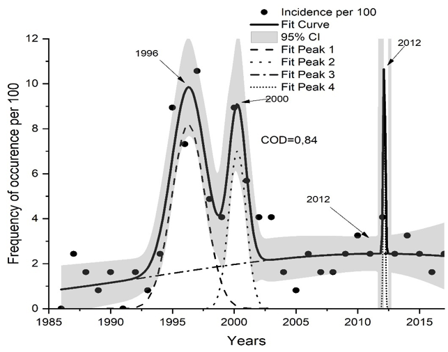
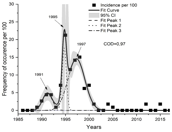
Interesting data were obtained in the analysis of the incidence of irradiated males during prenatal development (Figure 6). Among this group of victims, in fact, two peaks of morbidity stand out, which were recorded in 1996 and 2000. The second peak, apparently, is the reason for the shift in the maximum of the 3rd peak in the group of exposed females. Among the pathologies identified in 1996, diseases of the endocrine system (Chapter IV) dominated (38%). In the second maximum (2000), diseases of the respiratory organs (27%) and pathology of the gastrointestinal tract (18%) prevailed. Obviously, the age of the victims at 10 years is in both cases critical in relation to the appearance of pathology. As mentioned earlier, it is very important to compare the obtained data with the control cohort, which is identical in most parameters, but unlike the main one, did not receive radiation exposure due to the incorporation of I-131. Figure 7 shows data from the analysis of the primary incidence of the female control cohort. In contrast to the data in Figure 4, among non-irradiated women, three peaks of incidence are distinguished, which are hidden by a wide maximum of the incidence of two sexes simultaneously (from 1990 to 2004) (Figure 4).
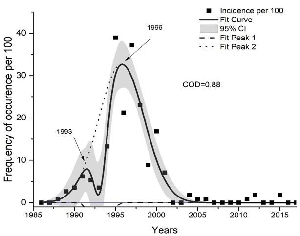
Nevertheless, the value of the coefficient of determination, equal to 0.97, gives grounds to consider the factual data on the splitting of the broad peak into several components. Consideration of the type of pathology that dominated in 1991 shows that respiratory diseases were the main type (Chapter X). Among the diseases recorded in 1995 and 1997, there were also diseases of the respiratory organs and diseases of the endocrine system (47% and 21%, respectively). Figure 8 shows the morbidity data in the post-accident period of the control cohort of males. Interestingly, in this case, their incidence coincided with that of the group of people of both sexes, characterized by one maximum in 1996. However, the dominant pathology was endocrine system diseases (Chapter IV) (27%).
Discussion
Comparison of the morbidity of persons who received thyroid irradiation during fetal development with that for a cohort of individuals identical in most respects showed quite obviously that in utero irradiation increases the morbidity of victims in subsequent years. At the same time, the registration of their pathology occurs as waves, with the detection of several morbidity maxima. Among them, a maximum of 10 years of life should be distinguished. Interestingly, the same maximum is also recorded in the group of non-irradiated individuals. The main pathology is respiratory diseases due to infectious pathology and pathology of the endocrine system. The latter is easy explanation since the Stolin district of the Brest region, whose inhabitants constituted the object of this study, belongs to the endemic region of Belarus, which is characterized by a deficiency of stable iodine. This leads to the appearance of endemic pathology [6]. Later morbidity maxima in the group of exposed victims (2000, 2001, 2002, 2006, 2012) are not detected in non-irradiated ones. And if in the early period after radiation exposure, for both irradiated and non-irradiated cohorts, diseases of the respiratory organs, mainly of infectious origin, prevail, then in the long term, diseases of other organs and systems (mental disorders, diseases of the gastrointestinal tract, musculoskeletal system) predominate. In contrast, in the group of persons who did not receive radiation during fetal development, such a pathology does not occur at all in the long-term period. There are significant gender differences in the morbidity of victims, which are characterized by a longer period of post-accident registration of morbidity in females, that was not the case in the group of non-irradiated individuals. The detection of heterogeneous, wave-like incidence cannot be explained by the thoroughness of the examination of the victims, since, according to Belarusian legislation, medical examinations of residents of the affected areas are carried out in the same volume annually.
The explanation for these phenomena may be the molecular mechanism of the genetic apparatus of thyrocytes radio damage that occurs due to the transplacental incorporation of radioactive iodine from the mother’s body. Earlier, when studying the action of I-131, more than two dozen genes in the thyroid gland were found that can reversibly regulate their activity. A few of genes (Pax8, Sic5a5, Tg, Tpo), which play an important role in the functioning of the thyroid gland, the synthesis of its hormones and the influence on the metabolism of peripheral cells, demonstrate low expression at low doses formed by I-131 and change their activity with the increase in radiation exposure. There are other examples. Thus, the mentioned gene Sic5a5 under conditions of different activity of the acting I-131 demonstrates two-phase dependence: at low and medium activities of radioiodine, it is inhibited to a greater extent than at intermediate ones, and at high activities, on the contrary, it is activated [3]. Different absorbed doses by the thyroid gland were formed by developing fetuses, which could be responsible for the change in the expression of genes. This resulted in a different intensity of the synthesis of thyroid hormones: T4, T3, rT3 and T2. And they, in turn, affect the development of the organs of the fetus and the further functioning of many body systems.
It has already been mentioned that thyroid hormones play an important role in the prenatal and postnatal development of the nervous system [7]. They are involved in several processes such as neurogenesis, glycogenesis, myelination, synaptogenesis, etc. [8]. A decrease in the level of circulating thyroxine in the mother in the first trimester of pregnancy, regardless of whether it is accompanied by an increase in the level of thyroid-stimulating hormone, can lead to irreversible mental and psychomotor disorders [9]. It is possible that this mechanism may be responsible for the increased incidence of pathology in the main group with respect to mental and behavioral disorders. In other words, the decrease in thyroxin in utero may be associated with the subsequent appearance of mental illness, which we observe. Thyroid hormones play an important role in the development of the gastrointestinal tract [10-12]. There is evidence of the effect of a deficiency or excess of thyroid hormone production on intestinal motility, as well as on the appearance of microscopic lesions of its epithelial layer [13,14]. Thyroid hormones play an important role in the functioning of the liver [15]. Changes in their production, from our point of view, can serve as the basis for the development of this pathology in the remote period. At the same time, it is known that irradiation induces instability of the cell genome [16]. Its instability is a persistent functional state leading to a violation of genetic control and being the most important factor in the development of subsequent pathology. These changes can be fixed in the child’s body, and therefore we observe these effects many years after the Chernobyl accident.
Conclusion
In the dynamics of morbidity from 1986 to 2017, in the group of individuals who were irradiated during fetal development, several maxima of pathology registration are revealed after 2, 10, 15, 19 and 26 years after the Chernobyl accident. At the same time, two peaks of incidence are revealed depending on the absorbed dose received by the TG (21 and 66mGy). The incidence in women differs from that in males. In the initial period, the dominant pathology was represented by diseases of the respiratory organs and the endocrine system, then later, most of pathology came from the side of nervous, digestive, and musculoskeletal systems.
Acknowledgements
None.
Conflict of Interest
None.
References
- Hulbert AJ (2000) Thyroid hormones and their effects: a new perspective. Biol Rev 75(4): 519-631.
- Ron E (2007) Thyroid Cancer Incidence Among People Living in Areas Contaminated by Radiation from the Chernobyl Accident. Health Phys 93(5): 502- 511.
- Rudqvist N (2015) Radiobiological effects of the thyroid gland. University of Gothenburg, Gothenburg 69.
- Buzunov VO, Kapustynska OA (2018) Epidemiological studies of cerebrovascular disease of the population evacuated from the 30/km zone of the ChNPP at the age of 18/60 years. Analysis of influence of internal ionizing radiation on the thyroid gland I-131. Probl Radiac Med Radiobiol 23: 96-106.
- Geno KA, Robert D Nerenz (2022) Evaluating thyroid function in pregnant women 59(7): 460-479.
- Gembicki M, Stozharov AN, Arinchin AN, Moschik KV, Petrenko S, et al. (1997) Iodine deficiency in Belarusian children as a possible factor stimulating the irradiation of the thyroid gland during the Chernobyl catastrophe. Environ Health Perspect 105: 1487-1490.
- Doreen Braun, Eva K Wirth, Ulrich Schweizer (2010) Thyroid hormone transporters in the brain. Rev Neurosci 21(3): 173-186.
- Сarreón Rodríguez A, Pérez Martínez L (2012) Clinical implications of thyroid hormones effects on nervous system development. Pediatr Endocrinol Rev 9(3): 644-649.
- Jean David Gothié, Barbara Demeneix, Sylvie Remaud (2017) Comparative approaches to understanding thyroid hormone regulation of neurogenesis. Mol Cell Endocrinol 25: 104-117.
- Koldovský O, Dobiásová M, Hahn P, Kolínská J, Kraml J, et al. (1995) Development of gastrointestinal functions. Physiol Res 44(6): 341-348.
- Lebenthal A, Lebenthal E (1999) The Ontogeny of the Small Intestinal Epithelium. JPEN J Parenter Enteral Nutr 23: S3-S6.
- Brown A, Simmen R, Simmen F (2013) The role of thyroid hormone signaling in the prevention of digestive system cancers. International Journal of Molecular Sciences 14(8): 16240-16257.
- Gregory CH, Gregory RF, Gregory R (1969) Effect of endocrine glands on function of the gastrointestinal tract. Am J Surg 117(6): 893-906.
- Eber EC (2010) The thyroid and the gut. J Clin Gastroenterol 44: 402-406.
- Malik R (2002) The relationship between the thyroid gland and the liver. QJM 95(9): 559-569.
- Vorobtsova IE (2006) Transgenerational transmission of radiation induced genomic instability. Rad Biology Radioecology 46(4): 441-446.


 We use cookies to ensure you get the best experience on our website.