Case Report 
 Creative Commons, CC-BY
Congenitally Corrected Transposition of the Great Arteries with Severe Tricuspid Regurgitation and Systemic Right Ventricular Dysfunction: A Case Report in Bangladesh
*Corresponding author: Saurav Das, Deputed to Chittagong Medical College, Chattogram, Bangladesh
Received: December 20, 2023; Published: December 22, 2023
DOI: 10.34297/AJBSR.2023.20.002787
Abstract
Congenitally Corrected Transposition of Great Arteries (ccTGA) is a complex congenital heart disease. It may remain asymptomatic in the first few decades of life ; therefore, diagnosis can be incidental. Symptoms arise when there is any complication or associated anomaly. We report the case of a 35-year-old man, presented with shortness of breath in Cardiology department in Chittagong Medical College Hospital. The man had progressive breathlessness for 2 months with previous episodes of similar kind of illness over the last 2.5 years. Examination revealed a pansystolic murmur (loudest at the left lower parasternal area), apex beat was diffuse, thrusting in nature, just lateral to the midclavicular line in the left fifth intercostal space. Transthoracic Echocardiography (TTE) confirmed the anatomy of ccTGA with tricuspid regurgitation.
Background
Congenitally Corrected Transposition of the Great Arteries (ccTGA) is a rare congenital heart disease; with an estimated prevalence of 1 per 33,000 live births [1,2]. The disease is characterized by atrioventricular and ventriculo-arterial discordance [1-4].
The condition can be associated with interventricular communications( 70%~80%), obstructions of the outlet from the morphologically left ventricle(25%~50%), pulmonary stenosis, AV block and anomalies of the tricuspid valve(70%) [2-9]. Generally, by the fourth decade, systemic Right Ventricle (RV) dysfunction is clinically apparent [1,3] Systemic RV dysfunction is the main cause of mortality and morbidity in adults with ccTGA [9]. We present the case of a 35-years old patient with ccTGA with severe tricuspid regurgitation, who took repeated medical consultation for his progressive breathlessness. In our centre, the medical management was given and referred for valve replacement.
Case Presentation
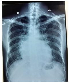
A 35-year-old male, normotensive, nondiabetic patient came with complain of progressive dyspnea on moderate to severe exertion .He had to take admission in hospital for two times in the last one month. 2.5 years back, he was diagnosed to have severe mitral valve regurgitation, due to mitral valve prolapse, and was advised for mitral valve replacement/repair. He decided to take second opinion/consultation from the specialized heart center both in Bangladesh and India. Then he was diagnosed as a case of ccTGA with moderate left atrioventricular valve regurgitation with normal biventricular function. He was advised on regular medications and routine follow-up. His last TTE was done 7 months ago. But his condition deteriorated in the last two months before the current admission. Examination revealed a 3/6 holosystolic murmur in the left lower parasternal area. Other positive findings included palpable P2, left parasternal heave, epigastric pulsation. Chest radiograph showed cardiomegaly, splaying of carina, vascular pedicle appears abnormally straight (since there is an abnormal relationship of the great arteries) (Figure 1)

Figure 1: Chest radiograph shows cardiomegaly, splaying of carina, straightening of vascular pedicle.
The ECG showed sinus rhythm, rate around 80/min, absence of Q waves in V4-6 (physiologic Qwaves in this lead ofen referred to as “Septal Q waves”), [5] S in V1+R in V6>35 mm (Figure 2).
Echocardiography showed the aorta arising from the morphological Right Ventricle (RV), which was identified by the three-leaflet tricuspid valve that inserted more apically than the mitral valve. The pulmonary trunk, identified by its bifurcation, arose from the morphological Left Ventricle (LV). The systemic RV was dilated (Figure 3).
Primary management with beta blockers and diuretics was given in the department. Then the patient was consulted accordingly and referred to a specialized centre for valve replacement.
Discussion
CCTGA comprises both atrioventricular and ventriculoarterial discordance; the right atrium enters the morphological LV, which gives rise to the pulmonary artery, whereas the left atrium enters the morphological RV, which gives rise to the aorta [7]. This defect occurs due to abnormal cardiac development during the third gestational week where Left Looping (L-loop) of the heart tube instead right looping occurs [4]. Patients without associated intracardiac lesions are usually asymptomatic early in life. Later on, patients may present with ventricular dysfunction [3,7]. 80% of patients with CCTGA have associated lesions (e.g., ventricular septal defect, pulmonary stenosis, Ebstein anomaly of tricuspid valve, and abnormalities of the conduction system) and they develop symptoms earlier [1-7]. Abnormalities of the tricuspid valve are found in up to 70% of patients with ccTGA. Ebstein-like anomaly in tricuspid valve in ccTGA represents a variable spectrum of disease [8]. Tethering and apical displacement of the leaflet hinge points, maximally at the commissure between the posterior and septal leaflets is observed [8].
The RV, normally supporting the low-pressure pulmonary circulation, when in the systemic position has to pump high-pressure systemic blood [7,9]. RV undergoes various adaptive mechanisms to enable itself to sustain the systemic load [7]. High systolic pressures inside the RV lead to eccentric hypertrophy of its free wall, dilation of the RV, increased ventricular wall stress and dysfunction of the trabecular component [9]. In complete TGA/d-loop TGA, tricuspid regurgitation is secondary to annular dilatation and RV dysfunction, and thus tricuspid valve replacement is not warranted. On the other hand, choice of intervention in CCTGA is addressing intrinsic abnormalities of tricuspid valve itself. Therefore, tricuspid replacement (repair is usually not gratifying) is ideal in ccTGA to preserve SRV function [7].
Conclusion
We presented the case of ccTGA that came in the fourth decade of life with severe tricuspid regurgitation and systemic RV dysfunction. The age of presentation and the ongoing complications are consistent with existing literatures on ccTGA. Inspite of regular medication for 2.5 years, the condition of the patient worsened. The final decision was made to refer the patient for valve replacement.
Acknowledgements
None.
Conflict of Interest
None.
References
- Auer J, Pujol C, Maurer SJ, Nagdyman N, Ewert P, Tutarel O (2021) Congenitally Corrected Transposition of the Great Arteries in Adults-A Contemporary Single Center Experience. J Cardiovasc Dev Dis 8(9): 113.
- Wallis GA, Debich-Spicer D, Anderson RH (2011) Congenitally corrected transposition. Orphanet J Rare Dis 6: 22.
- Hornung TS, Calder L (2010) Congenitally corrected transposition of the great arteries. Heart 96(14): 1154-1161.
- Connolly HM, Miranda WR, Egbe AC, Warnes CA (2019) Management of the Adult Patient With Congenitally Corrected Transposition: Challenges and Uncertainties. Semin Thorac Cardiovasc Surg Pediatr Card Surg Annu 22: 61-65.
- Baruteau AE, Abrams DJ, Ho SY, Thambo JB, McLeod CJ, et al. (2017) Cardiac Conduction System in Congenitally Corrected Transposition of the Great Arteries and Its Clinical Relevance. J Am Heart Assoc 6(12): e007759.
- Kumar TKS (2020) Congenitally corrected transposition of the great arteries. J Thorac Dis 12(3): 1213-1218.
- Brida M, Diller GP, Gatzoulis MA (2018) Systemic Right Ventricle in Adults With Congenital Heart Disease: Anatomic and Phenotypic Spectrum and Current Approach to Management. Circulation 137(5): 508-518.
- Myers PO, Bautista-Hernandez V, Baird CW, Emani SM, et al. (2014) Tricuspid regurgitation or Ebsteinoid dysplasia of the tricuspid valve in congenitally corrected transposition: is valvuloplasty necessary at anatomic repair? J Thorac Cardiovasc Surg. 147(2): 576-580.
- Filippov AA, Del Nido PJ, Vasilyev NV (2016) Management of Systemic Right Ventricular Failure in Patients With Congenitally Corrected Transposition of the Great Arteries. Circulation 134(17): 1293-1302.


 We use cookies to ensure you get the best experience on our website.