Case Report 
 Creative Commons, CC-BY
The Role of Serial Physical Examinations in Diagnosis of a Tension Pneumothorax Secondary to Alveolopleural Fistula
*Corresponding author: Mirna Kaafarani, MD candidate, Wayne State University School of Medicine.
Received: September 25, 2023; Published: October 17, 2023
DOI: 10.34297/AJBSR.2023.20.002703
Abstract
Tension Pneumothorax (TNP) is a life-threatening condition that is shown to have the best outcomes on patient mortality when it is caught early and treated immediately. The diagnosis of a TNP is usually clinical as patients will present hemodynamically unstable and delaying treatment to obtain imaging can result in death. Occasionally, some patients with TNP can present with minimal symptoms. In such cases, physical exam findings are essential in establishing a diagnosis and initiating treatment before clinical deterioration occurs. Here, we present a case of a 53year old woman with a history of COPD and >20 pack year smoking history who came to the emergency department for 2 days of cough and dyspnea. Upon presentation the patient was hemodynamically stable and a chest x-ray showed a cavitary lung lesion in the right upper lung field. On day 2 of admission the physical exam rendered harsh bronchial breath sounds and hyperresonance to percussion in the right upper lung field where the cavitary lesion was that were not present on prior examinations. The change of physical exam findings prompted the care team to obtain a chest x-ray that showed a massive right sided TNP with right lung atelectasis and mediastinal shift to the left. After the chest x-ray was obtained and TNP confirmed the patient’s oxygen saturation trended down. The patient immediately underwent chest tube placement and she remained clinically stable. In this case, the attention to changes on the physical exam led to a prompt diagnosis and the patient was able to be treated accordingly and remain stable. Typically, patients with a TNP present with signs of hemodynamic instability such as hypotension and tachycardia- but this patient had no change in her dyspnea or vitals when the physical exam was performed. As a result, the physical exam findings were the only indicator to get the repeat chest x-ray that led to the diagnosis of TNP. This case emphasises the importance of the physical exam in all patients but especially those with cavitary lung lesions as rapid clinical deterioration due to cavity rupture is possible.
Introduction
A Tension Pneumothorax (TNP) is the accumulation of air in the pleural space that leads to collapse of the lung tissue and a mediastinal shift to the ipsilateral side that can lead to decreased lung ventilation, hypoxemia, and impaired cardiopulmonary function ventilation. A good prognosis of TNP relies heavily on early diagno sis. Therefore, it is essential to recognize the signs and symptoms of TNP and to promptly initiate treatment to prevent the development of complications. Here, we present a case of a 53year old woman who had a cavitary lung lesion with an alveolopleural fistula who developed a TNP that was initially suspected based on physical exam findings.
Case Presentation
A 53year old woman with a history of alcohol use disorder, COPD, asthma and >20 pack year smoking history presented to the emergency department for two days of worsening shortness of breath and cough with rusty brown sputum and occasional streaks of blood. She denied any fever or chills but endorsed night sweats which she attributed to menopause. On initial evaluation the patient was afebrile, hemodynamically stable and saturating well on room air. Initial work up was positive only for an elevated c-reactive protein of 441. Chest x-ray showed a cavitary lesion in the right upper lobe of the lung measuring 5.7x4.3cm and bibasilar subsegmental atelectasis. Notably, the patient had a chest x-ray 6 months prior that showed no cavitary lesions. The patient was put on oxygen supplementation to assist in resolution of the atelectasis. Subsequent CT chest imaging on the same day showed a 5.1x4.3x5.7 cm thick-walled cavitating lesion in the right upper lobe with right bronchial lymphadenopathy and a small right apical pneumothorax and mild basilar effusion. The patient was put in isolation due to concerns for tuberculosis and further work up was sent including mycobacterial culture along with another infectious workup. Rheumatologic workup was also done secondary to concerns for granulamtosis with polyangitis. She was started on broad coverage with ceftriaxone, doxycycline, and metronidazole (Figures 1-3).

Figure 1: Single view chest X-ray showing thick walled cavitary leision in the right upper lobe 5.7x4.3cm; Yellow Arrows.

Figure 3: CT chest showing small pneumothorax on the anteromedial aspect of the right lung apex; Red arrow.
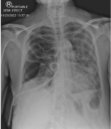
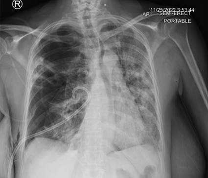
On day 2 of admission, the patient was comfortable still on supplemental oxygen to address the atelectasis and morning vital signs were all within normal limits. On routine bedside chest exam was done and yielded righ-sided, harsh, bronchial breathing and hyperresonance on percussion that were not present on admission. These findings prompted an emergent chest x-ray which showed a massive right sided pneumothorax with right lung atelectasis and mediastinal shift to the left. Ath that time, the surgery service was consulted and a pigtail chest tube was placed. Post-tube-placement chest x-ray suggested an initial slight improvement of the pneumothorax. However, after two days with her chest tube intact, serial chest x-rays failed to show any appreciable improvement of the size of the pneumothorax. To further investigate the persistence of the pneumotorax, a thorax CT was ordered and showed confluence of the lower border of the cavitary lesion with the right pleural space and tethering of tissue to the right sided chest wall pleura highly suggestive of an alveolarpleural fisutla (Figures 4-7).

Figure 4: Single view chest X-ray showing Right pneumothorax with collapsed right lung; Red arrows, and left mediastinal shift; red arrow heads. Persistent Cavitary lesion with tethering of right lung tissue to chest wall; white arrows.

Figure 5: Single view chest X-ray showing post non-surgical chest tube placement with slight improvement of right lung atelectasis.

Figure 6: Serial single view chest X-ray two days post non-surgical chest tube placement showing no significant change in the size of the pneumothorax.

Figure 7: coronal view of Chest CT showing disruption of the inferior border of the cavitary lesion and confluence with the cavitary space with the pleural space. Red arrows; Cavitary lesion, Yellow arrow; disrupted inferior border of the cavitary lesion.
With these image findings and lack of clinical improvement, she was taken to the operating room (OR) for a thoracostomy. Pleural fluid analysis yielded a lactate dehydrogenase of 5249 and protein of 4.5 indicative of an exudative effusion likely an empyema. Post-thorocostomy chest x-ray showed minimal improvement in the pneumothorax likely explained by the alveolarpleural fistula. Surgery recommended thoracotomy for further evaluation and treatment. On day seven of admission, cardiothoracic surgery took the patient for a right thoracotomy that included total decortication of the affected area, right upper lobe lobectomy, mediastinal lymph node dissection, pleural biopsy and wedge resection of the right middle lobe of the lung. Intraoperative tissue and fluid samples were obtained and stains were positive for gram positive cocci. She was subsequently started on Bactrim and Augmentin per the infectious disease service recommendations. Serial post-op chest x-rays showed. The apical chest tube was pulled on post op day 4 and the basilar chest tube pulled on post operative day 6; both were well tolerated. Repeat chest x-rays remained stable. On post op day 8 the patient was discharged with instructions to follow up at a later date.
Discussion
Literature has suggested a strong correlation between early detection of TNP with a better TNP prognosis of decreased mortality and morbidity [1]. Patients with non-traumatic spontaneous TNP typically present with complaints of severe ipsilateral pleuritic pain that in some cases radiate to the ipsilateral back or shoulder. Symptoms can also include subjective shortness of breath. Vital signs are usually a strong indicator with some patients presenting with tachycardia and increased respiratory rate. However, TNP can also present as asymptomatic as well, similar to the case we described. Our patient had no complaints of chest pain, shortness of breath, and had normal heart rate ranging from 70-80s and O2 saturation was in the high 90s on initial presentation. She was put on the supplemental oxygen the day prior to her TNP diagnosis and kept on it only as a measure to keep her alveoli patent after noting atelectasis on imaging.
It is evident from this case that conducting a daily thorough physical examination is crucial for patients with a high susceptibility to TNP. The new physical findings of harsh bronchial breathing and hyperresonance on percussion on the day following admission prompted timely imaging studies that were otherwise not clinically necessary considering the patients overall relative stability and tolerance for the infection treatment plan.
A strong history collection should also be done to determine who the high-risk patients are. Our patient had previously been diagnosed with Chronic Obstructive Pulmonary Disease (COPD), which is a significant risk factor for TNP [2]. In addition, the presence of an infective cavitary lesion in our patient keyed us into her susceptibility of developing a TNP. While the exact mechanisms behind the formation of TNP and acute pulmonary failure secondary to infection are not fully understood, it is postulated that the infective cavitary lesion weakens the lung tissue through the release of lytic enzymes increasing the chances of wall rupture [3]. Thus, it is pertinent that a high-risk assessment should be done on initial encounter to stratify the patient’s risk for TNP and serial chest exams can be used to supplement vital sign to define their clinical picture.
Acknowledgement
None.
Conflict of Interest
None.
References
- Jalota Sahota R, Sayad E (2023) Tension Pneumothorax. StatPearls. Treasure Island (FL).
- Gadkowski LB, Stout JE (2008) Cavitary pulmonary disease. Clin Microbiol Rev 21(2): 305-333.
- Briones Claudett KH, Briones Claudett MH, Posligua Moreno A, Estupiñan Vargas D, Martinez Alvarez ME, et al. (2020) Spontaneous Pneumothorax After Rupture of the Cavity as the Initial Presentation of Tuberculosis in the Emergency Department. Am J Case Rep 21: e920393.


 We use cookies to ensure you get the best experience on our website.