Case Report 
 Creative Commons, CC-BY
Unilateral Ptosis with Ipsilateral Frontalis Muscle Activation and Brow Elevation
*Corresponding author: Fabian H Rossi, Director Clinical Neurophysiology Laboratory Orlando VA Medical Center Professor Neurology UCF Medical School Orlando, USA.
Received: December 14, 2024; Published: January 03, 2024
DOI: 10.34297/AJBSR.2024.20.002790
Keywords: Ptosis, Myasthenia, Activation frontalis, Brow
Case Report
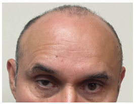
54-year-old male with several-week-history of new onset right ptosis and horizontal diplopia. Ptosis was present all the time, but worsen at the end of the day. Neurological examination revealed a right ptosis and ipsilateral wrinkling of the forehead. Pupils were reactive to light and accommodation. There was no anisocoria. Neurological examination otherwise was entirely unremarkable. Acetylcholine receptor antibodies were positive supporting the diagnosis of myasthenia gravis. Anti-musk antibodies were nega tive. Head MRI was unremarkable. Figure 1 depicts the right-sided ptosis with compensatory ipsilateral contraction of the frontalis muscle and elevation of the eyebrow arch at the highest point as an attempt to keep the eyelid open. In some patients with ptosis, frontalis muscle overactivation results in elevation of the brow and indirectivity of the eyelid to improve vision. Neurologists should be alert about this sign of frontalis muscle activation and eyebrow elevation as it might revealed a mild or unperceived ptosis [1].
Study funding
No targeted funding reported.
Disclosures
The authors report no disclosures relevant to the manuscript.
Acknowledgements
None.
Conflict of Interests
None.
References
- (2019) Lisa Moody, Tarek El Sawy, Rohit K Khoslta Ptosis Repair Global Reconstructive Surgery. Global Reconstruction Surgery. pp. 190-197.


 We use cookies to ensure you get the best experience on our website.