Review Article 
 Creative Commons, CC-BY
Neurobiomics the New Paradigm of Neuronal Conception
*Corresponding author:Bruno Riccardi, Biologist freelancer, 56022 Castelfranco di Sotto (Pisa), Via dei lazzeri, 33, Italy.
Received: September 15, 2023; Published: February 02, 2024
DOI: 10.34297/AJBSR.2024.21.002844
Abstract
The extraordinary complexity of the brain has stimulated the rise of numerous conceptions that compete for the primacy for the solution of its mysterious functioning. With the aim of simplifying and solving the complex matter, the researchers have divided and identified in different structural and functional entities those responsible for brain activity. The result produced by the antithetical and contradictory proposals has fed confusion and uncertainties instead of providing shared solutions. The extravagant theories that contend for the solution of the neurological enigma have proposed the most daring hypotheses, among which, the Microbiomics, the Nutrigenomics, the genetic and environmental factors, the quantum mechanics, and so on. The latter is the most accredited hypothesis and supported by physical laws and rigorous mathematical formulas. This article is going to present a provocative critical review of the literature of all these theories. The critical examination follows the proposal of a holistic unitary conception of neuronal functioning that I enclosed in the neologism NEUROBIOMA. This proposal was inspired by the authoritative European organization Human Brain Project that aims to recompose in a single overall view all the matter.
Keywords: Neurobiom, Nutrigenomics, Microbiomics, Quantum mechanics, Nerve and synaptic conduction
Introduction
Neurobiology is the science that more than any other has collected countless multidisciplinary theories and research that impact the functioning of our brain. And it is understandable that so many disciplines have ventured into the description of brain activity because it is this cognitive capacity that depends on all knowledge and the possibility of knowing ourselves and the universe around us. The incredible number of disciplines that have addressed the complex subject of the brain include Biology, Psychology, Ethology, Artificial Intelligence, Quantum Mechanics, and so on. The functioning of neuronal networks, from the monosynaptic circuit to the complex brain structure, presents an extraordinary intrinsic difficulty for its understanding, for this reason so many theories propose a different interpretation to solve this mysterious phenomenon. The difficulty of interpreting neurological mechanisms has stimulated sectoral research approaches with the idea that dividing the study of phenomena into elementary processes can simplify research. This process has produced contradictory results. What changes in the different investigations are the interpretative keys adopted by the individual researchers, which as mentioned are very discordant for the thesis supported by each and although apparently plausible, they often present a logical inconsistency on critical examination. The courageous attempt to collect the complex neurobiological mechanisms under the aegis of a single unifying law has so far produced so far unsuccessful and disconcerting results.
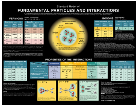
The discipline that more than any other has imposed itself with a unifying theory for the understanding of fundamental biological mechanisms, and in particular neurological ones, has been quantum mechanics, followed by all other sciences that directly or indirectly deal with the topic. Quantum mechanics has acquired scientific dominance over the interpretation of universal matter, based on its theories and the standard model of elementary particles and the forces that unite them. [1-3], and later it also imposed itself in the interpretation of neurological mechanisms. Thanks to the scientific hegemony acquired on the laws that regulate matter, quantum mechanics has extended it to living matter. He used the Aristotelian logic of syllogism, starting from the fundamental premise that his laws and theories control the atoms from which universal matter is constituted and being living matter formed by the same atoms, it follows that those laws also control living matter (Figure 1).
This scientific supremacy obtained the consent of all other sciences and was generally adopted. It has been adopted by Biology which has since become Quantum [4-9]. Today there is no discipline that does not follow a quantum approach. All cultural life is permeated by the epistemic omnipresence of quantum probability, which has become the viaticum that gives authority to universal knowledge. I remember quietly that the task of scientific research is to investigate natural phenomena and interpret them with unitary logical theories and confirm them with experimental evidence, in the absence of these requirements the theories must be rejected. In this work I am going to examine the theories in the literature that contend for different interpretations on the structure and functioning of the neuronal apparatus and the Central Nervous System, CNS. I am going to summarize in Table 1 the various concepts which I am going to examine later individually.
Already from the brief comparison of the theories set out in the table emerges the inconsistency and contradiction of the individual proposals (Table 1) [10-46].
The detailed examination of the individual theories and the description of the most salient contradictions are described in the text.
List of mechanisms held responsible for the operation of the SNC:
Genetic and environmental factors
Nutrigenomics Single Nucleotide Polymorphysms snps
Dysfunction of the Microbiome
Quantum Mechanics effect on microtubules
Electromagnetic effect on DNA
Morphic resonance
Genetic and Environmental Factors
An examination of the copious literature on the subject puts first, among the morphogenetic and pathogenetic causes that control the brain mechanisms, genetic and environmental factors. A first list of papers on the web on these topics includes 59 articles, and it is so vast that it is impossible to consider them all. These factors are all clearly plausible, among them genetic determinants, and in particular epigenetic ones, play an important role in conditioning and directing the formation and development of the brain, and it is equally clear that environmental and pathological factors are equally important. The problem of interpretation arises when the most unlikely and disparate causes are identified between genetic, morphogenetic and environmental factors, or when a single factor is held solely responsible for the morphogenesis and pathogenesis of different diseases [10-15]. Interpretative uncertainty and contradictions arise when in the morphogenetic or pathogenetic determinism of a neurological disease the factors deemed responsible are different and antithetical.
Nutrigenomics Diet and Brain Activity
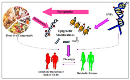
Nutrigenomics and brain activity: Nutrigenomics and Nutrigenetics provide tools to study the interaction of the diet with genes and their products, to alter the phenotype and, conversely, how genes and their products use nutrients and bioactive compounds to produce a certain phenotype. So according to these disciplines, through the study of genetic polymorphism and gene mapping it is possible to identify foods that can have positive or negative effects on our health, helping us to protect the body.
Single Nucleotide Polymorphysms (SNPs): Single Nucleotide Polymorphisms (SNPs) are the result of mutations in nucleotide sequences and are used to evaluate the effects produced at the level of gene expression, as the various types of mutation can have very different effects. Predicting the effects of a mutation at the level of phenotypic expression is a complex process, in which all the factors involved in the process and their reciprocal interactions must be considered. The following Figure 2 shows the correlations between different environmental factors and the consequences at the neuronal level (Figure 2).
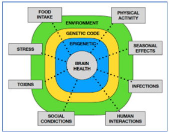
Figure 2: Correlations between different environmental factors and the consequences at the neuronal level.
We know that gene expression is the process by which the instructions in DNA are converted into a functional product, which will help determine the specific characteristics of the phenotype. However, there is no direct relationship genes - functional product as the "central dogma of Biology" states. We know that in the long journey from the gene to its phenotypic effect there are numerous intermediate stages that can profoundly change the final result. This path is the subject of study of Epigenetics.
Epigenetics: studies what and how many factors are involved in the ultimate formation of the functional product. These factors are the following Figure 3.
DNA Methylation: DNA methylation works by adding a chemical group to DNA. Typically, this group is added to specific places on the DNA, where it blocks the proteins that attach to DNA to “read” the gene.
Histone modification: DNA wraps around proteins called histones. DNA wrapped tightly around histones cannot be accessed by proteins that “read” the gene.
Non-coding RNA: Your DNA is used as instructions for making coding and non-coding RNA (Figure 3).
Nutrition and Neuronal Plasticity
This investigation procedure is also used to study the effects of food on neuronal expression.
The results obtained with this method are disconcerting, the perplexities derive from the numerous and contradictory food factors that are considered responsible for the neuronal development and the same pathologies, with a singular coincidence of factors contributing to the ontogeny and pathogenesis of common diseases [16-21].
Microbiota and Brain activity
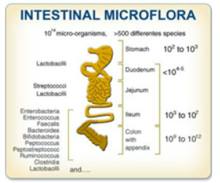
Microbiomics has a mainly bacterio-centric research method, which focuses on the study of the bacterial flora present in the intestine, known as the Microbiota as a whole, and its effects on health or disease conditions [22-26]. The Microbiota contained in the intestine consists of several microorganisms, bacteria, viruses, fungi, parasites, etc. In particular, the intestine has about 3000 prokaryotic bacterial species, composed mainly of Proteobacteria, Firmicutes, Actinobacterias and Bacteroidetes, which are the specific object of study of Microbiomica. The composition of the intestinal flora contains the bacterial species indicated before that are normally present in quantity and quality in balance with each other, and is characteristic of each person. When this balance is altered it is called "Intestinal Dysbiosis". Microbiomics attributes to specific "dysbiosis" of the intestinal flora the cause of numerous metabolic and chronic-degenerative diseases, the list of which is constantly growing. The study of the microbiota within the intestine called Metagenomica, makes use of advanced technologies for the specific species determination of bacterial strains present, the study allows:
The detection of a genetic set of bacterial population presents in the intestine, and in case of dysbiosis,
The restoration of homeostatic balance, with the implantation of specific bacterial strains, called probiotics
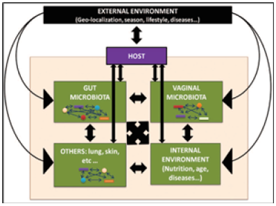
The Metagenomic Genetic Analysis consists in the sequencing of the DNA and in the study of the ribosomal Operon, of the gene coding the rRNA 16S present in the Microbiome. That genetic mutations are responsible for many diseases is safe, as is certain that some pathogenic bacteria cause gastroenteric infections. But by what inexplicable coincidence can altered polymorphisms or intestinal dysbiosis cause the same identical pathologies? While acknowledging the importance and reciprocal influence exerted between environment, diet and microbiota as a whole, Figure 4, however the various researchers in this discipline, attribute to the alteration of the gut microbiome a primary role on the health or disease status of patients [26,27] (Figure 4).
The most basic living species, such as bacteria, fungi and viruses, reproduce faster and adapt quickly to their environment, with the known mechanism of mutation and selection. Therefore, it is quite natural to find different microbial species (bacteria, fungi, viruses, etc.) that we have defined as a microbiota complex, in the intestine and in other apparatuses of different people [28] (Figure 5).
We also know that the microbiota changes in relation to changing eating habits and over the life of the individual. Attributing to a certain, recognizable and measurable cause the origin of diseases, such as the presence of a particular set of bacteria, is a loophole that satisfies our scientific ego, even if the causal link is certainly not provable. The only causal relationship demonstrated by traditional microbiology is between pathogenic bacteria and the infectious diseases they produce. Below is a synopsis of the criticisms levelled at the causal role of the microbiome for many pathologies: What Are Current Limitations to Establishing a Healthy Gut Microbiome-Host Relationship [29]?
Causality has not been established between changes in gut microbiome structure and function and markers of human health.
It is not established if dysbiosis is a cause, consequence, or both of changes in human gut epithelial function and disease.
Microbiome communities are highly individualized, show a high degree of interindividual variation to perturbation, and tend to be stable over years.
The complexity of microbiome-host interactions requires a comprehensive, multidisciplinary research agenda to elucidate relationships between gut microbiome and host health.
Biomarkers and/or surrogate indicators of host function and pathogenic processes based on the microbiome need to be determined, along with normal ranges, and validated.
Future studies measuring responses to an exposure or intervention need to combine validated microbiome-related biomarkers and surrogate indicators with multiomics characterization of the microbiome.
Because of human gut microbiome dynamics, static genetic sampling misses important short- and long-term microbiome-related changes to host health, so future studies should be powered to account for inter- and intraindividual variation and should use repeated measures within individuals.
Quantum Conception of Brain Activity
We have already mentioned the cultural dominance of quantum mechanics over the other sciences, and how it has profoundly modified concepts historically considered consolidated. It revolutionized classical physics, Biology that became Quantum, Astronomy, and so on. Here we are going to examine the transformation of neurobiology from classical to quantum. According to the new conception all neurobiological phenomena would be under the influence of electromagnetic waves, which with the various expressions of resonance, entanglement, and collapse of wave function, etc., control and regulate their operation. This theoretical setting reaches its peak in the interpretation of phenomena when it’s described neuronal activity and consciousness. In the description of these mysterious functions, the quantum-biologists fall into an embarrassing logical conflict between the conflicting conceptions of the individual authors, which attribute to different cellular structures, from the cytoskeleton to DNA, the origin of the same cognitive activities. Let us summarize the terms of the question by examining the various hypotheses proposed for example on consciousness, each of which is based on consolidated quantum bases:
Francis Crick formulated the idea of consciousness as an effect of tuning to the same frequency as neurons that constitute specific anatomical-functional areas of the human brain: the Neural Correlates of Consciousness (NCC) [33].
Penrose, Hameroff, Higgins, Lambert, Al-Kahalili, and others attribute brain functioning, and functions related to neurotubule activity [30-39,43].
Several other authors propose alternatively that the guiding force of all cellular activities, consciousness and cognitive functions included, originates in DNA, conceived not only as a coordinating center of genetic replication and protein synthesis, but also as a centre for the transmission of electromagnetic frequencies capable of directing and coordinating all cellular activities [44-46].
I have closed the quotations extracted from the "garden of wonders" of scientific production on the subject, citing the incredible hypothesis of Rafi Letzter reproduced in the article: “An Ancient Virus May Be Responsible for Human Consciousness, Live science February 02, 2018”. The author argues in this article, that consciousness would originate from an ancestral virus. It obviously ignores the elementary fact that viruses, as well as first-year biology and medicine students, know, mutate and evolve continuously and with remarkable frequency, how it is possible to identify in such a wide continuum the original virus is a mystery.
The Morphic Resonance
A scientific corollary has flanked the complex theoretical building of quantum mechanics, and it is the conception of formative causality and of the "morphogenetic field", which is the place where the consciences and thoughts of all humanity are gathered. It is a sort of icloud or otherwise defined "Field of Shared Consciousness", the place of the gnoseological events that inspire the activity of the brain when it collapses in the neuronal structures of individual thinkers (Figure 6).
In this imaginary Hyperuranium of Platonic memory, reside all the thoughts that inform the gnoseological activities of all present and future humanity. This is the extravagant hypothesis supported by the biologist Rupert Sheldrake and others [47-50], according to which in the field of "morphic resonance" resides the "formative causality" of biological evolution, which has shaped all living forms today and will give life to future ones, since embryonic development, and is postulated as the primary cause of the thoughts, behaviors and consciousness of each of us.
There is even a mathematical model of the morphogenetic fields that defines the targets through which the systems defined as attractors by René Thom (1975,1983) develop. These fields would derive from the strength of habits through the repetition of stereotypical behaviors. Thus, an immense reservoir collects and preserves the morphogenesis of biological evolution and informs the ontogenesis of each of the current eight billion people who populate the earth, all their thoughts and behaviors, not to mention animals, because they too have a share of sensitivity and ability to interact with the environment. Some people find the ability of this theory to explain the functioning of thoughts illuminating. I personally wonder how it is possible that in this chaotic "Field of Shared Consciousness" of cosmic dimensions that welcomes all thoughts, consciences and universal behaviors, then they can find the right path to reach the brain of every single living being. Moreover, according to this logic, thoughts, consciousness and behaviors do not belong to individual thinking beings, they are not the result of the laborious research and studies of every scientist, but are transmitted to them passively.
According to this original conception in the morphic fields are deposited all the information that will shape the body of every living being and form all its knowledge, it will then be a matter of identifying the body and brain of individual owners to be able to settle there. The brain is only the passive executor of the extended mind. It basically gives us all the license of perfect idiots. This also implies that all the knowledge of any individual present in the morphic fields must then be expressed in his brain, in the absence of which all will be lost.
Critical Examination of Quantum Conceptions
The quantum biology exam presented above presents many contradictions. Among these, the supposed regulatory activity of the cytoskeleton and neuro tubules, considered the coordinating and motor center of all nerve activity, takes on particular importance. Thanks to this new conceptual tool it is possible to explain the function of our brain, and even the origin of thought and consciousness that is all enclosed in some organelles that make up the cytoskeleton. Let’s go explain it.
The cytoskeleton is the texture that forms the network present in all eukaryotic cells (they are those with nucleus) and is formed by a complex scaffold of three different types of fibrous proteins: which are microtubules, microfilaments and intermediate filaments. This scaffold has a double structural and functional activity because it forms the substrate that maintains and modifies the shape of the cells and is responsible for some fundamental cellular activities. These include the movement of intracellular organelles, cellular movements, and the control of all phases leading to cell division. Microtubules undoubtedly contribute to the development of axonal and dendritic processes of neurons and are the basis of support for the movement of many organelles, synaptic vesicles, receptors of neurotransmitters, signal molecules, etc. However, they require the binding of supporting proteins, Kinesin and Dinein for their activity. In addition, for their synthesis require the association with accessory proteins (bridge proteins), essential for the assembly of cytoskeletal structures and for their operation, (Table 2).
None of the three classes of proteins that form the cytoskeleton have a stable structure, but are in continuous dynamic change in order to carry out the specific activities necessary in the life cycle of the cells.
The description of the properties of individual proteins requires a specific and complex treatment that goes beyond the objectives of this paper. Instead, we will pay particular attention to microtubules, which have been the focus of research in recent years. The reason for this interest lies in the fact that many scientists, in particular quantum physicists have identified the structures of the cytoskeleton as the main responsible for nerve activities. The morphological transformations, the transmission of the nervous impulse, the movement of the vesicles of the neurotransmitters, the intellectual function and even the consciousness, are all due to the presence of the neurotubules that direct the main brain functions.
According to this conception, if our brain works, we owe it to the neurotubules. And how does this miracle happen? Thanks to the electromagnetic waves that direct the whole device. The new paradigm of the neurobiological conception has been elaborated by some eminent scholars, in particular by quantum physicists, among whom stand out the names of the already mentioned Higgins, Penrose, Lambert, Al-Kahalili, etc. mentioned above. According to the explanations of this discipline, neurotubules are solely responsible for all neuronal activity and act under the synchronized direction of electromagnetic waves. [35-37] (Figure 7).
Actually, the neurotubules are not the factotum of the whole neuronal physiology, since their assembly is the epigenetic result of numerous stages of protein synthesis of the constituent monomeric proteins, tubulins, and proteins that accompany them, and other fundamental metabolic processes. Moreover, they act in close association with the other components of the cytoskeleton, the actins of the intermediate filaments, Table 2.
This theoretical approach overlooks the fact that upstream of the functioning of nerve cells there are two fundamental events extremely laborious and complex: The transmission neuro synaptic and protein synthesis of neuro tubules and neurofilaments of actin. But the question that arises is: even assuming that the trigger stimulus originates from the electromagnetic frequencies emitted on the microtubules, and from these transmitted to the entire brain, from which source do the electromagnetic stimuli that activate the cytoskeleton and to which other structures are transmitted and decoded, and how do they interact with the integrated complex and interconnected brain system? And in particular, how does the conduction of stimuli and synaptic transmission propagate? We know that there are networks and nervous systems that express a different activity in specific brain areas, some are excitatory, some are motor, others are modulatory, but all depend on the fundamental function of synapses. So that many pathologies depend on genetic defects in the synthesis of ion channels and ion exchange pumps present at the synaptic level, while they do not compromise the synthesis of neuro tubules. In contrast to the theories proposed earlier, I recall the concepts of classical neurobiology that in my opinion retain all their value.
Neuronal and Synaptic Transmission
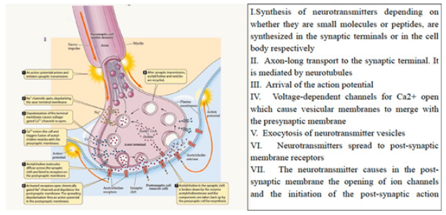
Nerve and Synaptic Transmission can be Synthetically Described in the Following Steps (Figure 8)
Synapses are the fundamental structures where nerve activity is processed. They are distinguished into electrical synapses and chemical synapses. With the type of neurotransmitters that carry in the post-synaptic membrane determine the neurological effect, excitation, inhibition, modulation. They therefore represent the true effectors of neuronal functioning.
Protein Synthesis of the Neuro Tubules and Actin Filaments Occurs according to the Following Steps (Figure 9)
The protein synthesis of neurofilaments and neuro tubules follows the classic rules typical of all protein synthesis, which lead to the formation of monomers (tubulin) and the control of their assembly. So that it must be considered only a fundamental stage of neuronal metabolism, and not the responsible for its functioning. Starting from this observation, we recall that as proteins microtubules and microfilaments are subject to protein synthesis, which provides for the known sequences according to the central dogma of biology: DNA- RNA-Protein synthesis.
Moreover, it is also essential to remember the role of the cell membrane, which ensures the exchange of substances with the outside, maintains the energetic homeostasis and the electrolytic and osmotic composition of cells, using transport structures present in the cell membrane, in the form of protein channels and receptors.
Finally, to communicate with each other neurons produce simple and complex molecules with high informational content, represented by neurotransmitters and hormones (Figure 10).
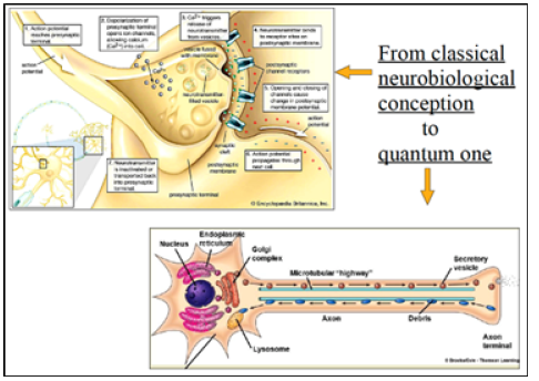
From Classical Neurobiology to Quantum Neurobiology
The concept of quantum neurobiology described above reduces all neurological functions to the activity of the protein structures of the cytoskeleton or other cellular components. The logical short-circuit thus produced greatly facilitates the description of the extraordinary complexity of neuronal networks, leading it back to elementary structures and a circumscribed space-time phenomenon. Many biologists converted to the new quantum belief, have denied the classic conceptions of neurobiology and shelved the theories that we thought consolidated not only on neurobiology but on all cell biology. So, all the cellular structures described by molecular biology have lost their functions, and we have to think about how to relocate them into new quantum systems. The cell membrane has lost its function due to electrical activity and nerve transmission and the fundamental exchange of neurotransmitters at synaptic levels. The mitochondria and the Golgi apparatus have given way and have been supplanted by the cytoskeleton main creator of nerve activity. What will be the fate of receptors, complex transport systems and ion channels present on the cell membrane, specialized in function and arrangement?
The Central Role of Synapses
In any case, if we wanted to prioritize individual structures, it is undeniable that the synapses play the role of the most important end effectors and the neuronal functioning Figure 3. They are the regulatory center where the synthesis of all signals that are carriers of information between cells takes place and where the adaptive response to environmental stimuli is processed. At the level of synapses occurs the expression of various systems: cholinergic, adrenergic, gabaergic, serotoninergic, and so on, which preside over the most important nerve functions. They also represent the most important target of the pharmacological activity of therapeutic and toxicological substances. A similar function is performed by the neuronal membrane that has the dual function of protecting the transmitting complex, forming the contact between adjacent neurons and generating the nerve conduction that frees the chemical messages, the neurotransmitters, at the synaptic level. In summary, the neuronal membrane with its branchings creates contacts between contiguous neurons, generates the action potential that releases neurotransmitters in synapses, protects the thin texture of the cytoskeleton that transports and directs the traffic of synaptic vesicles (Figure 11).
In all this complex activity the cytoskeleton has the ancillary function of guiding the branches of the cell membrane, transporting the neurotransmitters and cellular organelles, which synergistically maintain the nerve function, finally preside over cell division which in neurons is almost totally absent. In this complex organization chart in constant dynamic equilibrium each component is essential for the functioning of the whole system and there are no parts that play an indispensable primary role. Disruption at any point of the metabolic and functional process compromises and alters the vital activity of the whole apparatus. From the advent of the new quantum conceptions, all the neurobiology until now considered valid and essential in the study of the brain has been overcome and replaced by the neurotubules that, according to the new conceptions, have assumed the role of protagonists for neuronal functions. The fathers of neurology Charles Sherrington, Camillo Golgi, Ramon y Cajal’s, etc. were buried with a tombstone!! But the new description presents numerous conceptual flaws, which we try to highlight. The reductive conception that compresses the complex nerve activity to a single aspect is a mental deformation, which leads us to identify the complex natural phenomena, as a sequence of instantaneous manifestations, each of which assumes the primary role and is considered the creator of the totality of the phenomena themselves.
Whereas in reality every single manifestation is the effect of other concatenated causes and events that have had an important previous role in the determinism of the observed phenomena. Only a short-sighted and reductionist theoretical conception can force the extraordinary complexity of the functioning of neuronal synapses, still not completely understood, to an elaborate molecular quantum mechanism, with mathematical formulas by suffrage. The neurotubules thus have to shoulder the heavy responsibility of making the whole brain work. Focusing attention on these complex proteins, and on their Orch-OR (Orchestrated Objective reduction) according to Penrose’s already mentioned theory [31] is anti-scientific, because it does not take into account the previous causes that produced them and the resulting epigenetic effects. So that an infinitesimal event in the myriad of other activities taking place in a cell becomes solely responsible for the essence of life. Then of all an infinitely complex process and in continuous transformation and evolution, of which it is difficult to determine the boundaries of space-time, it is examined and describes only a single instant, a frame, and then extrapolate the obtained data to the whole system in its entirety.
It’s the same logic adopted by quantum mechanics according to which from the instantaneous Big Bang and singularity, it has derived all the matter of the universe. For any cell to function and live it is necessary that all the structures described, plus others not mentioned by synthesis, act synergistically and synchronically in a coordinated way. It is sufficient that any of the above processes does not work to compromise cell life and function.
The Fundamental Role of Mitochondria and Cerebral Oxygenation
In all the conceptions examined, the absence of any reference to the essential role of circulation and oxygenation for brain functioning stands out, as if it were a secondary phenomenon. Oxygenation has a key role in the functioning of nerve cells, all other mechanisms invoked as responsible for neuronal activity actually have an accessory role. The CNS is the organ that absorbs the greatest amount of energy and oxygen compared to all other organs of our body. And this is understandable in consideration of the indescribable amounts of interactions that occur at every moment at the level of synapses, for the maintenance of which it is necessary to provide energy.
The Mitochondria
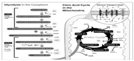
In all eukaryotic organisms, the cellular organelles that provide energy are mitochondria (Figure 12).
The mitochondria perform multiple fundamental roles in all cells:
They derive energy from food in the form of ATP.
Allow the use of oxygen for cellular respiration.
Regulate their duplication according to cellular activity.
Promote neuronal activity by electron transport.
Promote neuroprotection.
So that the malfunction of the mitochondria is held responsible for some nerve pathologies.
This explains the presence of their high number in neurons and in all cells of the brain, and in particular their concentration in the areas of greatest activity. The primary function of mitochondria is to generate energy in the form of ATP: a role by which these organelles are considered the "power plant" of the cell. The brain is an organ with very high metabolic activity, and the neurons of the CNS have an intense energy demand that requires a perfect mitochondrial function. Mitochondrial dysfunction involves neurodegeneration, because a defective mitochondrial functioning causes a deficit in cellular respiration and energy production, with obvious deleterious consequences on neuronal vitality. The mitochondria are constantly moving in the cell, they merge and divide, fragment and separate, they exist both in clusters and as individual entities. These processes are called Mitochondriogenesis. In the neurons the mitochondria are concentrated in the presynaptic terminations, at the extremity of the axon, and in the postsynaptic portions, at the extremity of the dendrites, where the energetic demand is particularly high. They are the site of aerobic oxidation of metabolism products. In the mitochondria, in fact, occurs the oxidation of pyruvate and fatty acids, nitrogen metabolism and heme biosynthesis. Respiration is the primary activity that keeps all cells alive, especially brain cells. As is known, the absence of oxygen of a few minutes is sufficient to determine death.
The Respiratory Chain and the Flow of Electrons
Of extreme importance is the function of the respiratory chain which is the place where the electron transport takes place and the oxidative phosphorylation system that provides the energy, in the form of ATP, necessary for cellular functions (Figure 13).
What animates, activates and keeps the nerve cells and all other cells alive is the flow of ions that transmit the nerve impulse through the neuronal membrane and neuritic fibers. If this flow ceases, the neuron dies.
All nerve activity, and more generally cellular activity, is moved and driven by an exchange of ions between the inside and outside of the cell membrane, the passage of ions generates a difference in transmembrane concentration, the membrane potential, which represents the vital energy of the cell. And it is by virtue of this that neuron, and more generally all cells, extract energy for all their activities through the respiratory chain. All this is possible because there is that incessant flow of transmembrane ions with the exchange of electrons within the mitochondria associated with it. But what keeps this flow of electrons active? The flow of electrons exists and is fed into the mitochondria by a final acceptor that collects them and disperses them into the environment in the form of water. And it is precisely this final acceptor that subtracts electrons and requires their continuous supply, to determine the flow of electrons, is a thermodynamically favored process with negative free energy (G<0). In aerobic organisms the final acceptor is oxygen, in anaerobic organisms the final acceptor is a different molecule that has a high affinity for electrons (Figure 14).
Ultimately all the mechanism that maintains the electronic vital flow is actually an electron-greedy molecule, which captures them at the end of the metabolic process and disperses them into the environment in the form of water.
Nerve Conduction
A mechanism similar to that described for oxidative phosphorylation is used to produce the action potential and conduction along nerve fibers. The ion flow through the cell membrane follows the gradient of electrochemical concentration between the two sides of the membrane. The passage of transmembrane ions modifies its potential to produce the action potential that is responsible for nerve transmission. This depends on the flow produced by the movement of ions. In fact, ions move along their concentration gradient across the membrane and along nerve fibers (Figure 15).
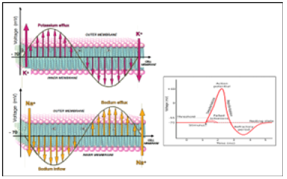
Figure 15: The action potential begins with the influx of sodium followed by the discharge of potassium according to their electrochemical gradients. The flows of the two ions are reciprocal, so that the increase of the one corresponds the decrease of the other they seem to follow a sinusoidal wave course.
In practice, the flow of ions seems to follow a complementary wave pattern between a few ions, as one grows the other is reduced. Could the flow of electrically charged ions generate an electromagnetic field?
But who feeds the flow of ions along the nerve fibers? There must be a motor that attracts the flow. In mitochondria this is formed by oxygen, the final acceptor of electrons, which continuously subtracts them. In neurons the continuous flow of action potentials is thermodynamically favored by the work they produce on target organs, muscles, glands, sense organs, neuronal circuits, and so on, which keep the process active.
Even in the rest conditions the whole system is constantly active to maintain the overall functional homeostasis. Neurons autonomously discharge action potentials like Pacemakers even if they are not stimulated.
All this extraordinary and complex mechanism serves the neuronal circuits to communicate with each other. How is information transferred and translated between neurons? The fundamental mechanism is represented by the action potentials or spikes, in short, the information between neurons takes place through frequency codes of the action potentials. And everything we’ve described about neuronal mechanisms, ultimately comes from the orchestrated exchange of ions that happens across membranes in the different ion channels that they contain.
New Perspectives in the Study of Neuronal Circuits
New technological approaches are opening up interesting developments in the study of neuronal networks.
Human Brain Project, which is a European organization that embraces many nations and researchers in different branches that directly or indirectly study the brain, has opened new perspectives that seem to be very promising for the future. The Human Brain Project (HBP) ran from 2013 to 2023 as one of the European Future and Emerging Technologies (FET) Flagship projects. https://www.humanbrainproject.eu/en/. The results obtained have brought so far numerous acquisitions that have improved the understanding of the brain.
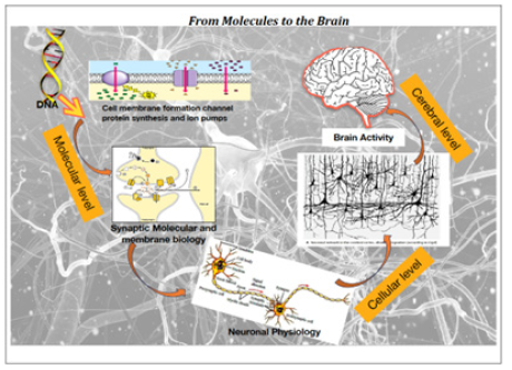
Although the approach followed is of the Top-Down type, and divides the investigation from the molecular level, and from the small neuronal networks to reach the most complex structures, the fundamental aspect that emerges is the irreducible functional unit of the brain. The innovative aspect that characterizes this investigation procedure is not to favor a single mechanism or structure and make it unique cause of the functioning of the brain, but to recognize the intrinsic indivisible functional unit (Figure 16).
Defining
NEUROBIOMICS is the science that studies NEUROBIOM which is the set of properties that possess the excitable substrate of living systems and neuronal networks in particular, the only one capable of collecting and recognizing external and internal environmental stimuli and of developing an effective adaptive response. The neologism proposes in a unitary key the theories of classical neurobiology. This property belongs to all living systems from prokaryotes to complex eukaryotic organisms and is capable of communicating with the environment and manifesting its existence in life. A system is only alive if it is able to interact and communicate with others and the environment.
Conclusion
Starting from the final considerations and totally sharing the goal pursued by the Human Brain Project to achieve a unifying understanding of the brain, I hope that the piecemeal method of investigation, which produces only logical conflicts and inconclusive diatribes on what is the most authoritative and rewarding conception for the scientific ego of individual researchers, will be overcome. I conclude these notes by proposing a neologism that best expresses the substantial unity of the nervous system and its ability to communicate.
Acknowledgement
None.
Disclosure
The Authors declare that they have no conflicts of interest in the present work.
References
- GBD2015 LRI Collaborators (2017) Estimates of the global, regional, and national morbidity, mortality, and a etiologies of lower respiratory tract infections in 195 countries: a systematic analysis for the Global Burden of Disease Study 2015. Lancet Infect Dis 17(11): 1133-1161.
- Walker CLF, Rudan I, Liu L, Harish Nair, Evropi Theodoratou, et al. (2013) Global burden of childhood pneumonia and diarrhea. Lancet 381(9875): 1405-1416.
- (2007) Pneumococcal conjugate vaccine for childhood immunization. WHO positions paper. wkly Epidemiol Rec 82(12): 93-104.
- Igor Rudan, Katherine LO Brien, Harish Nair, Li Liu, Evropi Theodoratou, et al. (2013) Epidemiology and etiology of childhood pneumonia in 2010: estimates of incidence, severe morbidity, mortality, underlying risk factors and causative pathogens for 192 countries. J Glob Health 3(1): 010401.
- Bartlett JG (2011) Diagnostic tests for agents of community-acquired pneumonia. Clin Infect Dis 52(Suppl 4): 296-304.
- UNICEF (2006) Pneumonia the forgotten killer of the children pp. 1-42.
- Cohen N Ouldali, E Varon, C Levy (2019) A germ and its prevention. Pneumococcus, Pediatric realities: 233.
- Bogaert D (2011) Variability and diversity of nasopharyngeal microbiota in children: a metagenomic analysis. 6(2): e17035.
- T Goulenok (2014) Pneumococcal vaccination in adults: how to improve vaccination coverage. Journal of Anti-infectives 89: 10.
- PO Lang (2012) Anti-pneumococcal vaccination: current situation and perspectives, NPG Neurology - Psychiatry - Geriatrics 12: 111-120.
- Deirdre A Collins, Anke Hoskins, Jacinta Bowman, Jade Jones, Natalie A Stemberger, et al. (2013) High Nasopharyngeal Carriage of Non-Vaccine Serotypes in Western Australian Aboriginal People Following 10 Years of Pneumococcal Conjugate Vaccination 8(12): e82280.
- S Oukid, H Tali Maamar, L Laliam, A Belboul, B Boutareg, et al. (2015) Streptococcus pneumoniae carriage and frequency of serotype: in children less than 25 months: a preliminary study in Algeria, 33nd Annual Meeting of the European Society for Peadiatric Infectious Diseases Leipzig.
- H Ziane (2015) Microbiology department chu Mustapha Algiers, thesis for obtaining the degree of doctor in medical sciences. Supported in 2015: Streptococcus Pneumoniae: Antibiotic resistance, frequency of circulating serotypes and main genotypes involved in invasive infections and nasopharyngeal carriage.
- Ahmed A El Nawawy, Soad F Hafez, Marwa A Meheissen, Nehal M Shahtout, Essam E Mohammed, et al. (2015) Nasopharyngeal Carriage, Capsular and Molecular Serotyping and Antimicrobial Susceptibility of Streptococcus pneumoniae among Asymptomatic Healthy Children in Egypt. Journal of Tropical Pediatrics 61(6): 455-463.
- KL Ravi Kumar, Vandana Ashok, Feroze Ganaie, AC Ramesh (2014) Nasopharyngeal Carriage, antibiogram & serotype distribution of Streptococcus pneumoniae among healthy under five children. Indian journal of medical research 140(2): 216-220.
- M Bouskraoui Soraa, K Zahlane, L Arsalane, C Doit, P Mariani, et al. (2011) Study of nasopharyngeal carriage of Streptococcus pneumoniae and its sensitivity to antibiotics in healthy children aged less than 2 years in the Marrakech region (Morocco). Archives de Pédiatrie 18: 1265-1270.
- MA Charvériat, M Chomarat, M Watson, B Garin (2005) Study of nasopharyngeal carriage of Streptococcus pneumoniae in healthy children aged 2 to 24 months in New Caledonia. Med Mal Infect 35(10): 500-506.
- Miwako Kobayashi, Laura M Conklin, Godfrey Bigogo, Geofrey Jagero, Lee Hampton, et al. (2017) Pneumococcal carriage and antibiotic susceptibility patterns from two cross-sectional colonization surveys among children aged <5 years prior to the introduction of 10-valent pneumococcal conjugate vaccine-Kenya, 2009–2010. BMC Infect Dis 17(1): 25.
- Richard A Adegbola, Rodrigo DeAntonio, Philip C Hill, Anna Roca, Effua Usuf, et al. (2014) Greenwood. Carriage of Streptococcus pneumoniae and Other Respiratory Bacterial Pathogens in Low and Lower-Middle Income Countries: A Systematic Review and Meta-Analysis. PLOS ONE 9(8).
- Murad Chrysanti, Eileen M Dunne, Sunaryati Sudigdoadi, Eddy Fadlyana, Rodman Tarigan, et al. (2019) Pneumococcal carriage, density, and co-colonization dynamics: A longitudinal study in Indonesian infants. Int J Infect Dis 86: 73-81.
- N Ramdani Bouguessa (2001) Streptococcus pneumoniae: resistance to penicillin and serotypes in children. Doctor of Medical Sciences thesis 2001. Unidversity of Medicine of Algiers, Algiers, Algeria.
- H Tali Maamar, R Laliam, C Bentchouala, D Touati, K Sababou, et al. (2012) Serotyping and antibiotic susceptibility of Streptococcus pneumoniae strains isolated in Algeria from 2001 to 2010. Med Mal Infect 42(2): 59-65.
- Abla Hecini Hannachi (2014) Thesis With a view to obtaining a DOCTORATE IN SCIENCE degree in Applied Microbiology, Streptococcus pneumoniae in invasive infections: identification, antibiotic resistance and serotyping.
- N Ramdani Bouguessa, H Ziane, S Bekhoucha, Z Guechi, A Azzam, et al. (2015) Evolution of antimicrobial resistance and serotype distribution of Streptococcus pneumoniae isolated from children with invasive and noninvasive pneumococcal diseases in Algeria from 2005 to 2012. New Microbes and New Infections 6: 42-48.
- Melody Kasher, Hector Roizin, Adi Cohen, Hanaa Jaber, Sharon Mikhailov, et al. (2020) The impact of PCV7/13 on the distribution of carried pneumococcal serotypes and on pilus prevalence; 14 years of repeated cross-sectional surveillance. Vaccine 38(19): 3591-3599.
- C Wintenberger (2010) Pneumococcus in 2010: from genomics to the clinic Proceedings of Congress. Medicine and infectious diseases 40: 605-609.
- Marcus HY Leung a, Ndekya M Oriyo, Stephen H Gillespie, Bambos M Charalambous (2011) The adaptive potential during nasopharyngeal colonization of Streptococcus Pneumoniae. Infect Genet Evol 11(8): 1989-1995.
- H Tali Maamar Laliam, C Bentchouala, D Touati, K Sababou, S Azrou, et al. (2012) Serotyping and antibiotic susceptibility of Streptococcus pneumonia strains isolated in Algeria from 2001 to 2010. Med Mal Infect 42(2): 59-65.
- Benouda, S Ben Redjeb, A Hammami, S Sibille, M Tazir, (2009) Antimicrobial resistance of respiratory pathogens in North African countries. J Chemother 21(6): 627-632.
- N Elmdaghri, M Benbachir J Najib, H Belabbes (2009) Invasive pneumococcal infections in children in Morocco: antibiotic resistance and fluctuation of responsible serotypes before introduction of conjugated vaccines. French-speaking laboratory review - supplement to n°
- P Chavanet, A Atale, S Mahy, C Neuwirth, E Varon, et al. (2011) Nasopharyngeal carriage, sensitivities and serotypes of Streptococcus Pneumoniae and Haemophilus influenzae in nursery children Medicine and infectious diseases 41: 307-317.
- J Raymond, R Cohen, F Moulin, D Gendrel P, P Berche, et al. (2002) Factors influencing the carriage of Streptococcus pneumoniae, Med Ma and Infect 32(1): 13-200.








 We use cookies to ensure you get the best experience on our website.