Research Article 
 Creative Commons, CC-BY
Comparative Analysis of Malondialdehyde Levels and Ferric Reducing Ability of Plasma in Pregnant and Non-Pregnant Women
*Corresponding author: Manisha Shukla, Department of Medical Biochemistry, Integral Institute of Medical Sciences & Research, Lucknow, India.
Received: May 29, 2024; Published: June 03, 2024
DOI: 10.34297/AJBSR.2024.22.003002
Abstract
During pregnancy, the increased oxygen demand by tissues can lead to oxidative stress, which is an imbalance between the production of reactive oxygen species (free radicals) and the body's detoxification mechanisms. Insufficient maternal antioxidant status to counter the elevated free radicals can result in complications such as infertility, miscarriage, preeclampsia, fetal growth restriction, and preterm labor. Free radical scavenging and chain-breaking antioxidants are the two main mechanisms the cell employs to counteract reactive oxygen species. Enzymatic antioxidants like dismutase, catalase, and peroxidases act as free radical scavengers, while exogenous antioxidants like Vitamin C and alpha-tocopherol function as chain-breaking antioxidants. Adequate antioxidant support during pregnancy is crucial to reduce oxidative stress and promote a healthy pregnancy and fetal development.
Method: The study comprises of a total of 60 subjects (30 cases and controls) and was selected based on inclusion and exclusion criteria. The estimation of MDA and FRAP was done by using spectrophotometer.
Results: Study shows rise in MDA level highest in pregnant (3.39±68) women that showing significant increase in levels of MDA (p < 0.001). Whereas normal level of MDA was seen in non-pregnant women. TAC measured by FRAP significantly decreased in pregnant women (1074.33±53.46) as compared to non-pregnant women that was statistically significantly (p < 0.001).
Conclusion: Oxidative stress is a well-documented occurrence during pregnancy and can lead to serious complications like pre-eclampsia or miscarriage if left unaddressed. MDA is a preferred biomarker for measuring oxidative stress, and studies have shown that oxidative stress increases with advancing stages of pregnancy. However, relying solely on FRAP for diagnosing total antioxidant capacity is not sufficient; it should be complemented by other established protocols such as SOD activity and glutathione reduction. Further research is required to enhance the clinical usefulness of these parameters in diagnosis.
Keywords: Pregnancy, Oxidative stress, Total Antioxidant Capacity, Reactive Oxygen Species, Superoxide, Dismutase
Introduction
Pregnancy is a normal physiological phenomenon accompanied by dynamic changes in women's bodies, making them susceptible to oxidative stress. Oxidative stress results from an imbalance between prooxidant and antioxidant defense systems [1].
The energy and oxygen demands during pregnancy lead to increased oxidative stress, affecting both the mother and fetus. Deficiencies in antioxidant activities related to micronutrients like selenium, copper, zinc, and manganese can lead to poor pregnancy outcomes [2]. Epidemiologically, live birth rates have declined in India, with some states reporting lower rates. Abortion rates among adolescent women in Telangana have increased during specific periods. [3]. Pregnancy is divided into three trimesters:
1) First trimester: Marked by increased minute breathing and pregnancy indicators such as nausea and breast development.
2) Second trimester: Women experience more energy, and the uterus grows significantly, leading to a noticeable baby bump.
3) Third trimester: The largest weight gain occurs, and the fetus's growth can be disruptive. Oxygenation for the fetus improves when the mother lies in the lateral position. [4].
Oxidative stress during pregnancy can lead to various complications, such as pre-eclampsia, gestational diabetes, and fetal hypoxia [5]. Excessive free radical production causes lipid peroxidation, leading to tissue damage. Antioxidant substances in the body help scavenge free radicals and reduce oxidative damage [6].
ROS (Reactive Oxygen Species) are essential for cellular function but can lead to pathological disorders when present in excess. Various factors contribute to excessive ROS generation, including UV radiation, alcohol, ischemia-reperfusion injury, infections, and inflammatory disorders [7]. Maintaining a balance between ROS and antioxidants is crucial for a healthy pregnancy. ROS plays a significant role in ovarian cell activity, and their concentration is indicative of ovulation [8]. Antioxidants are vital in protecting the body from oxidative damage caused by free radicals [9]. The body employs several strategies to counter such damage, including prevention, repair, and physical defense [10]. During pregnancy, free radicals can lead to oxidative stress, making pregnant women more susceptible to infections and health issues [11]. Low antioxidant levels can result in adverse pregnancy outcomes and hinder fetal and childhood development. To address this, prenatal care should focus on reducing free radical generation and increasing antioxidant levels in pregnant women [12]. Recent research has focused on studying oxidative stress and antioxidants in pregnant women to better understand their roles during pregnancy.
Materials and Methods
This study was carried out in the department of Biochemistry in Integral University, Lucknow. 30 pregnant women and 30 non- pregnant women (Controls) were taken for the study.
Inclusion Criteria
Healthy non-pregnant women and pregnant women with age group between 18-40 years.
Exclusion Criteria
Pregnant women with smoking and alcoholic history, diabetes, hypertension, infections, hepatic disorders, endocrine disorders, and any other causes of anemia such as thalassemia, haemolytic disease etc. were excluded from the study.
Collection of Samples
Under aseptic conditions, 4ml venous blood was obtained 2ml in PLAIN and 2ml in EDTA vials for the determination of malondialdehyde and FRAP assay respectively. EDTA was used as an anticoagulant to help preserve our sample for FRAP assay.
Sample Storage
The sample was refrigerated at -20°C at the central clinical laboratory for preservation.
Investigation
1) Malondialdehyde (MDA) was estimated by Satoh k method. Deproteinized serum is treated with TBA at about 90℃ for about 1 hour. The pink color formed gives the measure of TBARS which was read at 530nm using a spectrophotometer.
2) Antioxidant status by frap assay Benzie, I. F., &Strain, J. J. In FRAP assay antioxidants are used as reductants using a colorimetric method where ferric tripyridyltriazine to ferrous tripyridyltriazine i.e. colorless to blue color observed 593 nm.
Statistical Analysis
Statistical analysis was performed using IBM-SPSS software (version 16), Graph Pad (Prism 6.0) and Microsoft-Excel (version 2013). All the data were expressed as mean ± standard deviation. An unpaired t-test was performed to compare the study parameters between cases and controls. Karl Pearson’s correlation analysis was employed to determine the relationship between variables. p-value<0.05 was considered statistically significant.
Result
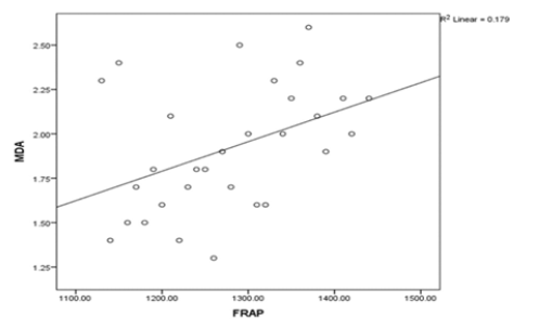
A total of 30 subjects were enrolled in this case control study. The results of statistical analysis have been summarized in Table 1 shows the comparison of clinical parameters between the study groups. According to statistical analysis there was no significant difference between cases and controls with regards to MDA and FRAP. However, it was found that levels of MDA and FRAP were raised significantly in cases as compared to controls (p=0.0001 and p= 0.0001 respectively). Table 2 shows Pearson correlation between variables in subjects with pregnant women. (Table 1, Table2, Figure1).
Discussion
Due to the elevated metabolic load and increased tissue oxygen requirements, oxidative stress increases throughout a typical pregnancy. MDA is a suitable marker for the evaluation of free radical-induced harm to tissues since it is a stable by-product of free radicals created by lipid peroxidation in the body [13].
In the investigation, pregnant women and non-pregnant women had their marker values compared to MDA and FRAP. According to this study, the average plasma level of MDA in pregnant women is 3.39±0.68 (µmol/L), while in non-pregnant women, the value is 1.92±0.35 (µmol/L), value and FRAP 1074.3±53.46 (µmol/L) in 1276.3±88.7 (µmol/L), in pregnant women and non-pregnant women respectively.
Because they are fragile and fleeting, reactive oxygen species can be hard to directly quantify. It has been utilized for indirect measurement of their ability to trigger lipid peroxidation. The development of a typical pregnancy has been accompanied by a rise in lipid peroxidation markers (MDA) [14].
Chamy, et al., 2006 [15] deduced that healthy pregnant women have greater lipid peroxidation levels than normal pregnant women. In response, the body tips antioxidant defense system to restore hemostatic balance. Consequently, oxidative equilibrium might endure the entire pregnancy.
According to [16] the maternal antioxidant system regulates placental lipid formation during a healthy pregnancy. ROS serves as signal transducers in physiology, but their overproduction can lead to a variety of health issues in people. While the body's own mechanisms play a critical part in regulating the amounts of these free radicals, the antioxidant levels that serve as a counterweight to these oxidative radicals themselves deteriorate. The aim of the study was to investigate the difference in levels of MDA within pregnant women compared to non-pregnant women.’
Reduced AOA is a sign of an issue with the antioxidant system and may be caused by fewer individual antioxidants. In a normal pregnancy, we observe a drop in each person's antioxidant status. According to this hypothesis, the lower AOA found in our research is due to a fall in the number of specific antioxidants in pregnancy (Bainbridge, et al., 2005).
The dynamic equilibrium between different antioxidants is what it is. Therefore, even though total antioxidant capacity might decrease while individual antioxidant levels increase during pregnancy [17].
In our investigation, there was a statistically significant decrease in FRAP and a spike in malondialdehyde.
Conclusion
In conclusion, it is a proven fact that oxidative stress occurs in pregnancy, the results of which can lead to complications such as pre-eclampsia or worse miscarriage if left unchecked. MDA is a preferred biomarker for oxidative stress. According to SB Patil, et al., (2007) with increasing stages of pregnancy, there is increased oxidative stress. Though increase OS triggers a reduced antioxidant response, FRAP cannot be sufficiently used to diagnose total antioxidant capacity it should be accompanied by other well-established protocols such as SOD activity and glutathione reduction. More research needs to be conducted along these parameters for them to be more useful clinically in the diagnosis.
Ethical Approval
Permission from the Institutional Ethics Committee was taken (IEC/IIMS&R/2023/64), Integral Institute of Medical Sciences and research, Lucknow, U.P.
Conflict of Interest
None.
Source of Funding
None.
Acknowledgement
We Thank the Department of Medical Biochemistry for providing all the support and facilities in this work.
References
- Shah AM, Channon KM (2004) Free radicals and redox signalling in cardiovascular disease. Heart 90(5): 486-487.
- Giles GI, Jacob C (2002) Reactive sulfur species: an emerging concept in oxidative stress 383(3-4): 375-88.
- Kuppusamy P, Prusty RK, Chaaithanya IK, Gajbhiye RK, Sachdeva G (2023) Pregnancy outcomes among Indian women: increased prevalence of miscarriage and stillbirth during 2015-2021. BMC Pregnancy and Childbirth 23(1): 150.
- Stacey T, Thompson JM, Mitchell EA, Ekeroma AJ, Zuccollo JM, et al. (2011) Association between maternal sleep practices and risk of late stillbirth: a case-control study. Bmj, 342.
- Torres Cuevas I, Parra Llorca A, Sánchez Illana A, Nuñez Ramiro A, Kuligowski J, et al. (2017) Oxygen and oxidative stress in the perinatal period. Redox biology 12: 674-681.
- Tiwari AKM, Mahdi AA, Zahra F, Chandyan S, Srivastava VK, et al. (2010) Evaluation of oxidative stress and antioxidant status in pregnant anemic women. Indian Journal of Clinical Biochemistry 25(4) 411-418.
- Bhattacharyya A, Chattopadhyay R, Mitra S, Crowe SE (2014) Oxidative stress: an essential factor in the pathogenesis of gastrointestinal mucosal diseases. Physiological reviews 94(2): 329-354.
- Shkolnik K, Tadmor A, Ben Dor S, Nevo N, Galiani D, et al. (2011) Reactive oxygen species are indispensable in ovulation. Proceedings of the National Academy of Sciences 108(4): 1462-1467.
- Pereira RD, De Long NE, Wang RC, Yazdi FT, Holloway AC, et al. (2015) Angiogenesis in the placenta: the role of reactive oxygen species signaling. BioMed research international 2015: 814543.
- Hussain T, Murtaza G, Metwally E, Kalhoro DH, Kalhoro MS, et al. (2021) The role of oxidative stress and antioxidant balance in pregnancy. Mediators of Inflammation 2021: 9962860.
- Wang, L, O Kane AM, Zhang Y, Ren J (2023) Maternal obesity and offspring health: Adapting metabolic changes through autophagy and mitophagy. Obesity Reviews 24(7): e13567.
- Evans P, Halliwell B (2001) Micronutrients: oxidant/antioxidant status. Brit J Nutr 85(2): 567574.
- Victora CG, Adair L, Fall C, Hallal PC, Martorell R, et al. (2008) Maternal and child undernutrition: consequences for adult health and human capital. The lancet 371(9609): 340-357.
- Wickens D (1981) Oxidation (peroxidation) products in plasma in normal and abnormal pregnancy. Ann ClinBiochem 18: 158-162.
- Chamy VM, Lepe J, Catalán Á, Retamal D, Escobar JA (2006) Oxidative stress is closely related to clinical severity of pre-eclampsia. Biological research 39(2): 229-236.
- Walsh SW (1994) Lipid peroxidation in pregnancy. Hypertension in pregnancy 13(1).
- Adiga U, Adiga MNS (2009) Total antioxidant activity in normal pregnancy. Online J Health Allied Scs 8(2): 8.


 We use cookies to ensure you get the best experience on our website.