Research Article 
 Creative Commons, CC-BY
Correlations Between Selected Pelvic Traits in Habitual Posture Among Female Individuals of Urban and Rural Environments from 3 to 20 Years Old
*Corresponding author: Mirosław Mrozkowiak, Physiotherapy Practice Akton, Poland.
Received: April 19, 2024; Published: April 29, 2024
DOI: 10.34297/AJBSR.2024.22.002944
Summary
Introduction: The pelvis is a part of the skeletal system. It consists of the pelvic girdle formed by the sacrum, the coccygeal bone and the two pelvic bones, right and left. During walking, it undergoes fluctuations of angular displacement in the frontal, sagittal and transversal planes.
Material and Method: The study was conducted in randomly selected kindergartens and schools, urban and rural environments of Warmian-Masurian and Pomeranian Region among 1,974 female individuals aged 3 to 20 years.
Tools and Subject of the Study: A photogrammetric method was used to diagnose the left and right pelvic torsion angle in the transversal plane and the left and right tilt in the frontal plane.
Results Obtained: The overall analysis including positive and negative relationships shows that KNM and KNM- ranges from -0.37 to -0.45, KNM- and KSM- from 0.17 to -0.11, and KSM- and KSM- from 0.11 to -0.75. All analyzed results were highly significant.
Conclusion
a) The relationships between the angles of pelvic torsion in the transversal plane and tilt in the frontal plane are highly significant.
b) The interrelationships of torsion and pelvic tilt angles should be considered in postural error routines at the age from 3 to 20 years.
Keywords: Angle of Pelvic Torsion and Tilts, Projection Moiré
Introduction
The pelvis is a part of the skeletal system. It consists of the pelvic girdle formed by the sacrum, the coccygeal bone and the two pelvic bones, right and left. It shows major gender differences. The female pelvis is low, wide, and spacious, while the male pelvis is high, narrow, and tight. In both sexes, the smaller pelvis is shaped like a section of a cone: in women it is cut off closer to the base, in men closer to the apex [1].
In the skeletal system, two supporting "beams" of the lower limbs are symmetrical components, on which the horizontal "beam" of the pelvis is propped up and the "axial" spine anchored in it through the sacrum. It is the upright spine that deviates from the vertical only in the sagittal plane. Any asymmetrical positionings of the pelvis lead to changes in the spine orientation in space, and these permanent changes result in pathology. Torsion of the pelvis in the transversal plane due to, for example, asymmetric tensions of the muscles rotating the pelvis regarding the thighs, causes the sacrum and the higher located vertebrae to rotate in this plane. Such a rotation may cause the vertebrae to tilt off the side in the frontal plane and lead to scoliosis. In turn, lowering one of the pelvic ala of ilium in the frontal plane with asymmetric tension of the adductor muscles or shortening of one of the lower limbs leads directly to the deviation of the vertebrae from the vertical in this plane, to functional scoliosis [2]. However, the orientation of the pelvis in the sagittal plane determines the compensatory angular values of the physiological curvatures lying above. Studies carried out using Posturometer S in a group of 2,413 girls and 2,446 boys aged 6-16 years showed that males were more likely to have one of the hip bones lower than females. There is a tendency to increase the frequency of the above-mentioned postural error with age. It can be said that lateral curvatures are more common in women, and pelvic rotation and lowering are more common in boys [3]. In turn, the symmetry of the pelvic position improves slightly in older age groups. The obtained results confirm the results of investigations by other authors [4].
Mrozkowiak's research [5] showed that the angle of the pelvis tilt to the left has a positive and significant effect on the total length of the spine, its percentage (DCK%, the length of thoracic kyphosis and its percentage (DKP%), the height of thoracic kyphosis and its percentage (RKP%)), the length and angle of lumbar lordosis, deviation of the vertebra along the line of the spinous processes of the spine to the left, and the location of the atlas vertebra deviation. It influences negatively and significantly the angle of inclination of the lumbar-sacral section, the sum of angles, the height of lumbar lordosis and its DCK percentage (RLL%), and deviation of the atlas vertebra to the right along the line of the spinous processes. The angle of the pelvis tilt to the right (KNM-: 24) influences positively and significantly the total length of the spine and its percentage (DCK%), the depth of lumbar lordosis, as well as its length, height, and percentage (RLL%), and deviation of the atlas vertebra to the right along the line of spinous processes. It has a negative and significant impact on the percentage, and height of thoracic kyphosis (RKP%), and deviation of the atlas vertebra to the left along the line of spinous processes.
The aim of the research is to determine the mutual dependence of the angles of pelvic rotation to the left and right in the transversal plane and the angles of pelvis tilt to the left and right in the frontal plane among female individuals aged 3 to 20 years.
Material and Research Method
The research was carried out in randomly selected kindergartens and schools in urban and rural environments of the Warmian-Masurian and Pomeranian Region like 10 kindergartens, 20 primary schools, 6 junior high schools, 1 upper-secondary school after obtaining the consent of the Local Education Authority in Olsztyn, the school or kindergarten principal, and the teacher in charge of the given branch, parent and child and students of the Kazimierz Wielki University in Bydgoszcz. The general criteria for qualifying for the study were based on the selection of a sufficiently large number of similar healthy body postures during the study. The research involved 21,356 female individuals from both environments, aged 3 to 20 years, Table 1.
Note*: Source: Mrozkowiak M. [5].
Based on interviews with parents and school medical charts, all students with documented structural abnormalities in the musculoskeletal system were excluded. On the first day of the study, in the light of the medical diagnosis, the subjects were generally healthy, therefore it was assumed that the results and any postural errors found later would be within the limits of physiological deviations appropriate for the represented population and age range. This concerns small functional deviations of the vertebral process lines from the anatomical axis of the spine and the angle of trunk flexion in the frontal plane, the angle of flexion and extension in the sagittal plane, and the longitudinal arch of the foot (Table 1).
Tools and Subject of Research
The measurement site consisted of a computer and a card, a programme, a monitor and a printer, a projection and reception device with a camera for measuring selected parameters of the pelvis-spine complex. Obtaining a spatial image is possible by displaying lines with precisely defined parameters on the child's back and feet. The lines falling on the skin are distorted depending on the surface configuration. Thanks to the use of a lens, the subject's image can be received by a special optical system with a camera and then transmitted to a computer monitor. The distortions of the line image recorded in the computer memory are converted by a numerical algorithm into a contour map of the examined surface [6]. The obtained image of the surface of the trunk and pelvis allows for a multi-aspect interpretation of body posture. The accuracy of measurement and analysis of recorded spatial parameters means that the conclusions drawn may differ from those previously published.
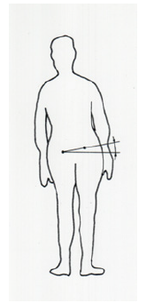
The short duration of recording the subject's figure allows to avoid postural muscle fatigue that occurs during examinations by somatoscopy. The most important in this method is the simultaneous measurement of all actual values of the spatial location of individual body sections. During the research, generally accepted principles and procedures were followed [7]. The angle of pelvic rotation to the left (KSM-) and to the right (KSM), Figure 1, was measured in the transversal plane, together with pelvic tilt angle to the right (KNM), and to the left (KNM-), Figure 2. Unfortunately, the method used does not measure the pelvic rotation angle in the sagittal plane, in the axis of the hip joints (Figures 1,2).
Statistical Methods Used
Four features describing the pelvis were analyzed: KNM angle of pelvic tilt to the left in the frontal plane, KNM- angle of pelvis tilt to the right in the frontal plane, KSM angle of pelvic rotation to the right in the transversal plane, and KSM- angle of pelvic rotation to the left in the transversal plane. Results of the statistical analysis, Spearman's coefficient, classified here as a measure of the strength of monotonicity between two variables, i.e., an increase in the size of one variable usually corresponds to an increase in the other. The coefficient ranges from -1 (this corresponds to the strongest negative relationship between the variables) to +1 (this corresponds to the strongest positive relationship), (Table 2).
Note*: Source: Mrozkowiak M [5].
Note*: Source: Mrozkowiak M., [5].
Note*: Source: own research.
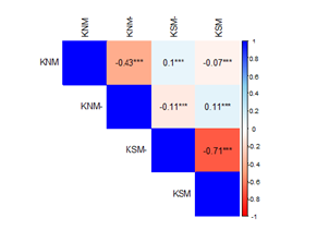
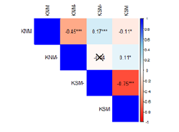
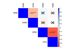
The values of Spearman's coefficients for the tested pairs are presented in the form of matrix colours, according to the scale on the right side of the figure. A totally red color corresponds to a coefficient value of -1, and a totally blue color corresponds to a coefficient value of +1. Additionally, the numerical values of the coefficient are entered in the corresponding field with the appropriate number of "stars" indicating the significance of the relationships.
Results Obtained
Among the analyzed characteristics of subjects aged 3 to 6 years, all analyzed results turned out to be highly significant. However, they differ in terms of the Spearman’s coefficient. Thus, the correlation coefficient between the features of KNM and KNM- is -0.45, between KNM and KSM- is 0.17, between KNM and KSM is -0.11, between KNM- and KSM- does not occur, between KNM- and KSM is 0.11, and between KSM- and KSM is -0.75, (Figure 3).
Among the analyzed characteristics of respondents aged 7 to 13, all analyzed results turned out to be highly significant. However, they differ in terms of the Spearman’s coefficient. Thus, the correlation coefficient between the features of KNM and KNM- is -0.43, between KNM and KSM- is 0.01, and between KNM and KSM is -0, (Figure 4).

Figure 4: Relationships between pelvic characteristics in female subjects aged from 7 to 13(n) 18675.
Among the analyzed characteristics of respondents aged 14 to 20, all analyzed results turned out to be highly significant. However, they differ in terms of the Spearman’s coefficient. Thus, the correlation coefficient between the features of KNM and KNM- is -0.37, between KNM and KSM- does not occur, between KNM and KSM does not occur, between KNM- and KSM- does not occur, KNM- and KSM does not occur, and between KSM- and KSM is -0.72, (Figure 5).
Comparison to Other Authors
The available literature on the subject does not contain any studies describing the interdependence of pelvic features. The measurement results of other researchers very often concern body posture in a general sense and do not refer to individual features describing it. Analysis of research results by Burdukiewicz, et al., Bibrowicz, et al., Cieśla, et al., Kabsch, et al., Lewit, et al., Saulicz, et al., Tylman, et al., Walker, et al. and Wielkie, et al. [2,3,8-14] on the influence of the angle of inclination and rotation of the pelvis on the characteristics of the spine showed that this influence is definitely negative and multidirectional.
Conclusion
a) Correlations between angles of pelvic rotation in the transversal plane and pelvic tilt in the frontal plane are highly important.
b) Postural error routines should include mutual dependencies of the angles of pelvis rotation and tilt at the age from 3 to 20.
Acknowledgement
None.
Conflict of Interest
None.
References
- Marecki B (1991) Functional Anatomy. 1: 81-82.
- Kabsch A (1999) Biomechanical and Biocybernetic Foundations of Axial Exercise symmetrical according to Hoppe. Provincial Methodological Centre N Sącz: 11-18.
- Burdukiewicz A (1995) Posture variability of Wrocław children from 7 to 15 years of age in longitudinal research. Studies and Monographs: 3-4.
- Górniak K (1999) The incidence of postural defects in rural and rural children starting primary education. [In:] Zagórski J. et al. [ed.], Determinants of the physical development of rural children and youth. Institute of Physical Education and Sport Biała Podlaska 1: 245-250.
- Mrozkowiak M (2007) Determinants of selected parameters of children's body posture and youth and their variability in the light of projection moiré. Gorzów Wlkp: 227-288.
- Świerc A (2006) Computer diagnostics of body posture-user manual, Czernica Wrocł
- Mrozkowiak M (2015) Modulation, influence and relationships of selected parameters of body posture children and adolescents aged 4 to 18 years in the light of projection moiré. Bydgoszcz I, II.
- Bibrowicz K, Skolimowski T Occurrence of body posture symmetry disorders in frontal plane in children from 6 to 9 years of age. Physiotherapy 3(2): 26-29.
- Carpenter T Some compensation asplets in lateral curvatures of the spine. Methodological and Scientific Journal (3): 29-38.
- Lewit K (1984) Manual treatment of musculoskeletal disorders. PZWL Warsaw: 56-57.
- Saulicz E (2003) Pelvic spatial alignment disorders in low-grade scoliosis and the possibility of their correction. AWF Katowice: 4-6.
- Tylman D (1974) Compensatory pelvic lesions in lateral curvatures spine, [In:] Trześniowski T., Maszczak T. [eds.], Correction and compensation in development of school youth, SiT, Warsaw: 107-111.
- Walker AP, Dickson RA (1984) School screening and pelvic tilt scoliosis. Lancet 2(8395): 152-153.
- Wielki Cz (1987) Shoulder and hip asymmetry in school-age children (6 - 14years), In: Wyżnikiewicz Knopp [ed.], Physical education and sport of children in University of Szczecin: 203-213.


 We use cookies to ensure you get the best experience on our website.