Case Report 
 Creative Commons, CC-BY
Incarceration of the Donor Ureter into the Inguinal Canal Following Kidney Transplantation: A Case Report
*Corresponding author: Atta Nawabi MD, MBA, FACS, FICS, Associate Professor of surgery division of HPB, and transplant, Department of surgery, 3901 Rainbow Boulevard Kansas City, KS 66160, USA.
Received: March 21, 2024; Published: May 24, 2024
DOI: 10.34297/AJBSR.2024.22.002995
Abstract
Introduction: Inguinal hernias present as a common surgical problem in the general population. These anatomic defects are especially common among men with the most feared complications including strangulation and incarceration of bowel. Transplant patients have additional risk factors contributing to hernia formation with regards to the surgery itself as well as the immunosuppression involved. Obstruction of the ureter secondary to inguinal herniation is a rare complication of renal transplantation that often leads to graft loss. Our case study demonstrates how early diagnosis and treatment of obstructive uropathies in transplanted patients improve outcomes of the allograft kidney transplant.
Presentation of Case: Here we report the case of a 62-year-old male with development of a ureteral obstruction, hydronephrosis, and urosepsis secondary to an inguinal hernia following a renal transplantation. The patient required PCNU for decompression and broad-spectrum antibiotic therapy followed by inguinal hernia repair.
Discussion: Abdominal hernias develop when intraabdominal contents protrude through a fascial defect due to increase intraabdominal pressure and wall weakness. Several types of abdominal wall hernias have been named including inguinal, femoral, umbilical, incisional, Spigelian, and parastomal. Amongst inguinal hernias, indirect hernias account for most cases thought to arise from a patent processes vaginalis. Direct inguinal hernias account for roughly 30% of inguinal hernias and are typically caused by loss of abdominal wall strength. While many inguinal hernias are asymptomatic here, we present a unique case of a patient who developed an obstructive uropathy as their inciting symptom. Transplanted ureters have a rare, but known risk of obstruction, but this was a rare case of ureteral obstruction secondary to incarceration within an inguinal hernia of a patient with a previous renal allotransplantation.
Conclusion: Early detection of ureter obstruction secondary to inguinal herniation is key to ensuring continued renal allograft function. Consistent follow-up is essential for the timely diagnosis and treatment of patients following renal allotransplantation to assess for threats of renal graft function, both common and rare.
Keywords: Kidney transplant, Inguinal hernia, Ureteral obstruction
Introduction
As the treatment of choice for end-stage renal disease, renal allotransplantation is a well-studied procedure with known late-presenting complications of ureteric leak and ureteral obstruction that can lead to graft failure [1-3]. Late ureteral obstruction following renal transplantation is most often caused by stricture at the ureterovesical junction or distal ureter. [2,3]. In this report, we discuss a rare cause of ureteral obstruction in a post-renal transplant patient of ureteral obstruction secondary to inguinal herniation. To our knowledge, less than 30 reported cases of transplant ureteral obstruction due to inguinal hernia have been identified.
Presentation of Case
A 62-year-old male presented to the Emergency Department with the chief complaint of urinary retention. He reported that he had been having pain with urination for multiple days with associated muscle aches and lower abdominal pain. His past medical history was significant for End Stage Renal Disease (ESRD) presumed to be secondary to hypertension status post deceased donor renal transplant in 2018. The patient reports having been adherent to his immunosuppression regimen until 5-6 days prior when he began having difficulty obtaining his medication from his pharmacy; however, per records, he had missed multiple appointments with transplant nephrology in the two years leading up to this encounter. He endorsed urinary retention and when he was able to urinate, it was foul-smelling and contained frank blood. On exam, he was ill-appearing and had a catheter in place from Emergency Department staff filled with bright red urine. He had significant lower abdominal and right inguinal tenderness. However, no inguinal hernia was appreciated on exam.
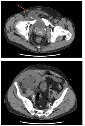
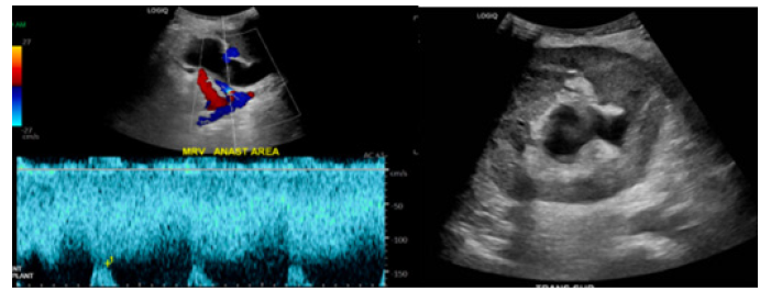
Computed Tomography (CT) upon admission demonstrated the transplanted ureter coursing into the right inguinal hernia resulting in obstruction with marked transplant hydroureteronephrosis (Figure 1). Both urine and blood cultures were found to be positive for Proteus mirabilis for which he was subsequently treated. Ultrasound of the transplanted kidney the following day continued to demonstrate hydroureteronephrosis with no change from prior CT. Fortunately, transplant vasculature was found to be patent with only mild tapered narrowing at the donor renal vein anastomosis (Figure 2). Interventional radiology was consulted for the placement of a percutaneous nephroureteral catheter that was successfully placed on hospital day 1.

Figure 1: CT imaging demonstrating obstruction of the ureter secondary to incarcerated right inguinal hernia.
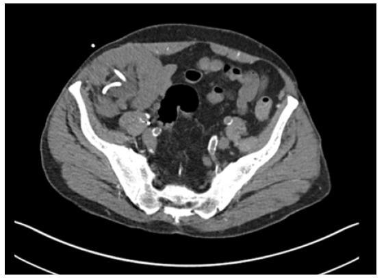
CT one week later demonstrated significant improvement in the hydroureteronephrosis of the transplanted kidney (Figure 3). He was continued on antibiotic therapy and scheduled for follow up for the repair of his inguinal hernia upon resolution of his urosepsis. His PCNU catheter was to remain in place until surgical repair of the hernia.
Discussion
Transplanted ureters have a known risk of obstruction, but this was a rare case of ureteral incarceration within an inguinal hernia of a patient with a previous renal allotransplantation. Most often, ureteral obstruction in this patient population comes secondary to stricture at the site of the ureterovesical anastomosis or the distal ureter [1-3] However, this patient’s ureteral obstruction, and resulting hydronephrosis and urosepsis, were secondary to incarceration within an inguinal hernia.
Our patient was five years from surgery with a history of poor follow-up. On admission, his immunosuppression was below therapeutic levels, and he had a history of missed appointments. It is unclear how long this hernia had been present, although the patient’s report indicated this had been bothering him for some time.
Conclusion
Obstruction of the ureter secondary to incarcerated inguinal herniation is a rare cause of obstructive uropathy. Inguinal herniation must be considered in post-renal transplant patients who present with hydronephrosis and poor graft function. While percutaneous nephrostomy tube insertion and proper antibiotic treatment are critical to the stabilization of a patient with resulting hydronephrosis and urosepsis, herniorrhaphy is the only definitive treatment for the obstructed ureter. Consistent follow-up is essential for the timely diagnosis and treatment of patients following renal allotransplantation to assess for threats of renal graft function, both common and rare.
Acknowledgement
None.
Conflict of Interest
None.
References
- Furtado CD, Sirlin C, Precht A, G Casola (2006) Unusual cause of ureteral obstruction in transplant kidney. Abdom Imaging 31(3): 379-382.
- Ingber MS, Girdler BJ, Moy JF, Frikker MJ, Hollander JB (2007) Inguinal herniation of a transplant ureter: rare cause of obstructive uropathy. Urology 70(6): e1-3.
- Shoskers DA, Hanbury D, Cranston D, Morris PJ (1995) Urological Complications in 1,000 Consecutive Renal Transplant Recipients. J Uro 153(1): 18-21.


 We use cookies to ensure you get the best experience on our website.