Review Article 
 Creative Commons, CC-BY
Review on Biomedical Applications of Textile Surface Forms of PES Polymers
*Corresponding author: Ömer Fırat Turşucular, R&D Chief, Hatin Textile, and Hatin Tex Weaving Companies, DOSAB, Bursa Province, Turkey.
Received: May 06, 2024; Published: May 15, 2024
DOI: 10.34297/AJBSR.2024.22.002980
Abstract
In this the review study consists of 3 subheadings. The definition of bio composite materials, design criteria, raw materials used, application areas, biomedical expectations in the human body, the properties of chitin and chitosan, and their applicability to textile surfaces are mentioned in the first subheading. The chemical structures, production methods, derivatives, mechanical, chemical, thermal, and biological properties of types of PES polymer are mentioned in the second subheading. The weaving, braiding, and knitting technologies, design criteria, and biomedical applications of various textile structures from the PES and its derivative polymers, and the other polymers are mentioned in the last subheading. In conclusion, the types of PES polymer such as PET, PCL, PLL, PLA, PGA, and PLGA, polymeric materials such as PA, PP, and PTFE, and shape memory metallic materials such as Ni are widely used in biomedical applications. Textile surfaces should be selected according to the specific needs of the biomedical application area. Fiber cross-section profile, yarn type, yarn structure, yarn biological, chemical, thermal, and mechanical properties, yarn count, filament number, density values, and construction should be determined. It provides high biological, chemical, thermal, and mechanical properties of the yarn (the ideal type is PET), fine yarn count (range 88 dtex to 220 dtex), number of filaments (range 108 to 272), high pore number, and low pore size in biomedical applications. In addition, the diameter over 6 mm, 3-D braiding textile surface, 1x1 diamond braiding construction, 30° braid angle, minimum 1 (center)+24 (braid)=25 total yarns, PET yarn type, triangular geometry fiber cross-section profile is used for its dimensional stability in artificial vascular vessel applications. After, it should be produced by fixing at 150℃ and for 1 minute, too. Moreover, it can also be coated with chitin, chitosan, PU, or PC polymers by dip coating to improve biological properties.
Keywords: Bio composite materials, Biomedical, PES polymers, Textile structures, Biomedical applications
Introduction
Biocomposite Materials and Biomedical
Biocomposite materials are materials produced by the compatibility of biological and mechanical properties of at least 2 materials with a heterogeneous structure [1]. Biocomposite materials provide a support mechanism that helps the growth of immature cell groups and enables the integration of surrounding tissues by turning into complex tissue networks [2,3]. Moreover, FDA approval is required for the reconstruction of biocomposite materials into the human body [2]. They have become widespread since the 1960s [4]. The design criteria of biocomposite materials are pH, biocompatibility, biodegradability, antimicrobial, antifungal, non-toxic, non-allergic and non-carcinogenic, high dimensional stability, high corrosion resistance, high fatigue resistance, high impact strength, low weight/strength ratio, high fiber-to-volume ratio, high modulus of elasticity, high bending stiffness, low coefficient of thermal expansion, low thermal and electrical conductivity, high fiber orientation, high crystallinity, viscoelastic behavior, homogeneous fiber distribution, anisotropic structure, fiber and matrix interfacial strength, high tensile strength, high compressive strength, high modulus of elasticity, high wear resistance, crease recovery behavior, and creep resistance [1-3,5-20]. The production methods of biocomposite materials are compression molding, injection molding, resin transfer molding, sheet molding, hand lay-up, filament winding, extrusion, pultrusion, electrospinning, impregnation, coating, CVD, non-woven surface, and textile-based such as weaving, braiding, and knitting methods [3,5-9,11-24]. The raw materials used in biocomposite materials are shape memory polymer, polymeric, metallic, ceramic, and biocomposite materials [1-30]. Natural fibers such as cotton, linen, hemp, jute, ramie, sisal, enset, bamboo, silk, and wool are used in biomedical applications thanks to their biological, health, ecological, and low-cost properties. They should have pre-treatment processes to react -OH, amino acid groups, and Van der Waals as their chemical bonds. They can be used in biomaterial applications by activating the bonds with various chemicals [7,9, 21,22].
Ni, PC, PU, PE, PP, PS, PEG, PEA, PEO, PVA, PGS, PVP, PEEK, PTFE, PDMS, PMMA, PEGDM, PEDOT, and Polyester (PET) and its derivatives such as PDO, PVL, PHA, PHB, PHBV, PBAT, PMLA, PPC, PPF, PTT, PBT, PBS, PMD, PLA, PLL, PCL, PGA, PLLA, PLGA, PLAGA, PDLLA, PEGMA and PHEMA are widely used thanks to their high biological and mechanical properties [2,3,5-20,22-30]. Usage areas of biocomposite materials are cardiovascular graft, heart valve, dental, cancer treatment, drug release, tissue engineering, tissue scaffold, wound dressing, hernia treatment, artificial liver, artificial kidney, artificial lens, artificial heart, artificial nerve, artificial skin, artificial cartilage, artificial ligament, artificial tendon, artificial surgical sutures, catheter, artificial joints, and artificial bone [1-3,5-20,24-29]. While the pH of healthy human tissue is 7.4, the pH is around 6.5, especially in cancerous and infected tissues. Moreover, the cell divisions occur less between pH 5 and pH 6 in cells such as lysosomes, and endosomes [28,29]. The biocomposite materials are generally expected to support the surrounding tissue within 6 to 8 weeks thanks to their biological, chemical, and mechanical properties and to exhibit their biodegradable properties in biomedical applications [2]. They cover the period from 8 weeks to 20 weeks in the in-vivo environment for heart valve application. Moreover, the complete integration of collagen tissues consisting of myofibroblast, and endothelial cells is usually completed by the 17th week in another source [11]. Keeping the diameter constant on all textile-based surfaces produced cannot provide the required increasing and decreasing diameter values in systole and diastole behaviors in artificial vascular applications. For this purpose, they are recommended to produce products from shape-memory polymeric materials. Moreover, they must be also produced with a diameter over 6mm, too [20]. Chitin and chitosan are widely used in biomedical applications by applying them to textile surfaces with a finishing process (usually by coating them (in biofilm form)) thanks to their biodegradability, biocompatibility, mucoadhesion, hemostatic, analgesic, adsorption enhancing, antimicrobial, antioxidant, anticholesterolemic and cell adhesion enhancing properties [5,7,13,22,24].
Polyester (PES)
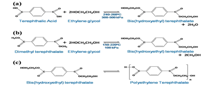
The most commonly used type of Polyester (PES) is PET. PET can be produced with TPA (esterification), or DMT (trans-esterification), and EG chemicals. It is a synthetic based product. It is a thermoplastic, and semi-aromatic chemical structure that can be produced as a result of a two-stage step polycondensation reaction with comonomer, metal catalyst, hardener, and stabilizer chemicals. Its production process parameters such as pH, temperature, time, pressure, concentration, relative viscosity, and molecular weight are important for its various properties. Moreover, bis(hydroxyethyl)terephthalate (catalyzed polycondensation) is a thermoplastic polymer that can be produced because of a single stage polycondensation reaction [24,28,30]. The chemical structure of PET has 1 piece benzene ring, 2 piece -C=O, 1 -OH, and 1-piece OCH2CH2OH chemical bonds [11,24]. The PET polymer syntheses are presented in Figure 1 [24].
It is a synthetic-based thermoplastic polymer that has been used in many areas, especially the textile industry, since the 1950s. The properties of PES fibers are excellent tensile and impact strength, high acid resistance, clarity, processability, chemical resistance, and thermal stability [3,11,12-14,24]. In 1977, a mechanical and chemical-based recyclable PES polymer method was developed because it is the most widely used polymer type in the world thanks to its qualities such as lightness, cheapness, easy availability, and low energy consumption [24]. The reasons for using PES in biomedical applications are its bioinertness, its ability to provide drug release, and its suitability for more advanced medical and regenerative applications [11,13,14,20,24-30]. It has also low cell adhesion, hydrophobicity and a high risk of infection in biomedical applications, too. Effective production process parameters in the production of PES derivatives are chemical structure (especially comonomer type), orientation, crystallinity, porosity, temperature, time, pressure, relative viscosity, and molecular weight differences. These differences distinguish the biological, chemical, thermal and mechanical properties of PES derivatives [11,24-30]. Especially in the types of PES fibers with aliphatic (linear structure, that is, straight chemical chain) chemical structure such as PLA, PGA, PLLA, PGLA, PBS, PHA, PHB, PHBV, PMLA, PEGMA, and PHEMA have lower mechanical, chemical and chemical properties compared to PET. While they have lower thermal properties but they have higher biological properties compared to PET [11,25-29].
PES polymers are polymers that can be easily hydrolyzed by the presence of esterase in the body, thanks to the ester groups that are characteristic groups in their chemical structures. Thus, they provide the transfer of tension and functionality to newly cultured tissues in the in-vivo environment, depending on the biodegradability behavior and time. While this occurs, mass loss, and dimensional reduction occur [19]. Thermal and biodegradability behaviors of some of the most commonly used types of PES polymer in biomedical applications are presented in Table 1 [29]. Some of the most commonly used types and chemical structures of PES polymer in biomedical applications are presented in Figure 2 [29].

Table 1: Thermal and biodegradability behaviors of some of the most commonly used types of PES polymer in biomedical applications [29].
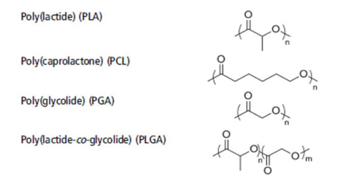
Figure 2: Some of the most commonly used types and chemical structures of PES polymer in biomedical applications [29].
Various textile structures and biomedical applications from PES, its derivatives, and other polymers
The textile-based surfaces such as 2D or 3D structured braiding, woven, knitted, and non-woven surfaces are widely used for biomedical applications of PES and its derivative polymers by developing textile surfaces from yarn forms and allowing their combined use [3,12-20]. PET is the most commonly used type of PES in biomedical applications. Especially in PET polymer as its yarn form, it is used by coating it with metal yarns in artificial vascular graft biomedical applications [14,17,19,20]. Important design criteria in biomedical applications are polymer type, fiber diameter, fiber distribution, fiber spacing, cross-sectional shape of the fiber, yarn count, yarn structure, biological, mechanical, thermal, and mechanical properties of the yarn, textile surface construction and density values, pore size and number of pores [3,12-20]. The textile surfaces used in biomedical applications and some important design criteria are presented in Figure 3 [3].
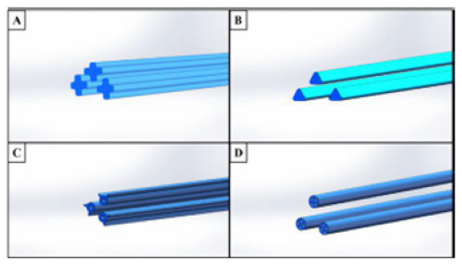
As the fiber diameter increases and the surface area of the textile surface construction due to yarn-yarn friction increases, cell adhesion, and proliferation increase. The fiber gap is necessary for cell adhesion and proliferation, but it should not be too wide to prevent this situation. Pore size does not affect cell adhesion and proliferation. Cell adhesion and proliferation increase when the cross-section of the fiber has non-round profiles such as triangles, hollow, and sea islands [3]. It is known that monofilament yarns have higher elasticity mossdulus, tensile, compressive, and bending strength values compared to multifilament yarns [17,18]. Examples of cross-sectional profiles of fibers are presented in Figure 4 [3].
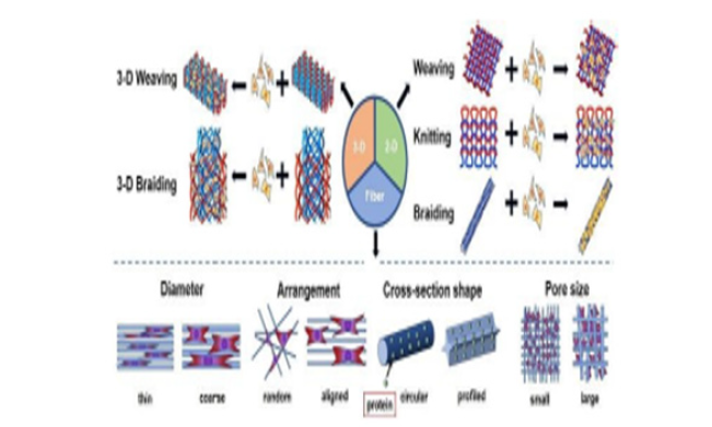
Figure 3: The textile surfaces used in biomedical applications and some important design criteria [3].
The yarn type, yarn structure, yarn count, number of yarn, and fabric layers, density values of warp, and weft yarns, woven fabric construction, and number of woven fabric layers are the most important design criteria on woven fabric surfaces [3,12-14,20]. Woven fabric surfaces produced with the yarn count as thick as possible, multifilament structure, and maximum warp, and weft density values on woven fabric surfaces offer the ideal solution for biomedical applications [3,12-14,20]. 1x1 plain, 2x1 twill, 2x2 twill, 1x3 twill, 3x1 twill, 3x3 twill, 5-warp satin, and honeycomb woven fabric constructions are used. As a result, that woven fabric production parameters such as yarn type, yarn structure, yarn count, yarn mechanical properties, woven fabric construction, and woven fabric density values are effective factors on porosity, and woven fabric mechanical properties in biomedical applications. [3,12-14,20]. The effectiveness of woven fabric constructions for increasing cell adhesion and proliferation is as follows 1x1 plain > (2x1, 3x1, or 3x3) twill > (5 warp, or weft). The reasons for this situation are yarn floating behavior and the number of yarn intersection points. In conclusion that when 1x1 plain woven fabric construction is compared to other woven fabric constructions thanks to the decrease in yarn floating behavior and the increase in the number of yarn intersection points, the pore size decreases, the number of pores increases, and cell adhesion, and proliferation increase. In addition, tensile strength increases, and shear strength decreases [3,12-14,20]. As warp and weft density values increase, cell adhesion, and proliferation increase on woven fabric surfaces [3,13,20]. While the recommended warp density value is 55, the weft density value is 100. Honeycomb woven fabric construction provides the optimum solution for myocardial treatment thanks to its high mechanical properties and dimensional stability [3].
It is recommended to use woven fabric in PET yarn type, 88 dtex yarn count, 272 filaments, 34 warp/cm as warp density, and 32 weft/cm as weft density in 1x1 plain woven fabric construction so it supports cell adhesion and proliferation of woven fabric textile surfaces for heart valve biomedical application [13]. PES derivative yarn types such as PLA, PLL, and PCL can also be used in heart valve biomedical applications when their yarn counts are between 110 and 220 dtex [13]. PET yarn type, the yarn count ranges from 100 denier with 36 filament counts to 179 deniers with 108 filament counts can be used for biomedical application of artificial vascular graft. Moreover, it is recommended to use woven fabric textile surfaces in 1x1 plain, 2x2 twill and 1x3 twill woven fabric constructions at a warp densities value of 50 warp/cm, and a weft densities range of 38 weft/cm to 40 weft/cm so they support cell adhesion and proliferation [14]. Moreover, 3-D structures have higher mechanical properties and dimensional stability compared to 2-D structures [12,13]. Artificial vascular graft in 3-D woven fabric structure is presented in Figure 5 [14].
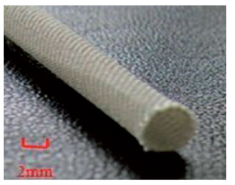
The yarn type, yarn structure, yarn count, number of yarn and braiding structure layers, braid angle (°), density values of core and braid yarns, braiding structure construction, and number of braiding structure layers are the most important design criteria on braiding building surfaces [3,15,20]. It is used in biomedical applications because its biaxial (fiber axis and directions perpendicular to the fiber axis) mechanical property supports cell adhesion and proliferation by providing the appropriate pore size and number. 1x1 diamond, 2x2 double-layer, and 3x3 hercules constructions are used in braiding structural surfaces [3,20]. A 30° braid angle is recommended [3,15,20]. Because 30° braid angle provides maximum axial tensile strength, minimum radial tensile strength, maximum axial facial elongation at break, minimum radial facial elongation at break, maximum axial kink strength, and minimum radial kink strength [15]. Moreover, 3-D structures have higher mechanical properties and dimensional stability compared to 2-D structures [3]. Braiding structures were produced in nitinol yarn type, 5 mm braiding diameter, 2-D structure, 30°, 45°, and 60° braid angles, 24 braid yarn count, 1x1 diamond braiding structure constructions for biomedical application of artificial vascular graft. Afterward, as a post-process, textile surfaces with a 2-D braiding structure, which were first fixed at 500 °C for 5 minutes and then coated with PC-based silicone elastomer by the bottom coating method, were recommended to be used as they support cell adhesion and proliferation [15]. Examples of artificial vascular grafts in 2-D braiding structures with variable braid angle (°) are presented in Figure 6 [15].

Figure 6: Examples of artificial vascular grafts in 2-D braiding structures with variable braid angle (°) [15].
The yarn type, yarn structure, yarn mechanical properties, yarn structure, yarn count, number of yarn, and fabric layers, loop yarn length, needle height, density values of wale, and course yarns, knitted fabric construction, and number of knitted fabric layers are the most important design criteria on knitted fabric surfaces [3,16-20]. Warp, weft, and spacer knitted fabric structures are available. Monofilament, and multifilament yarn structures are used [3,16-20]. Monofilament PP yarn with a yarn count of 244 denier with 4 different knitted fabric constructions with a density of 12 inches can be produced by fixing it at 150 °C for 1 minute for the biomedical application of hernia treatment [18].
Monofilament, or multifilament structured PU, PA, PET, PAN, and PTFE yarns with a diameter range of 6 mm to 20 mm can be produced with different knitted fabric constructions, usually with a thickness of 12 inches for heart valve biomedical application [18-20]. Plain, rib, purl, interlock, tricot, pin-hole-net, quasi-sandfly, sandfly, and quasi-marquisette knitted fabric constructions are used in biomedical applications. As a result, that the knitted fabric production parameters such as yarn type, yarn structure, yarn count, yarn mechanical properties, knitted fabric construction, knitted fabric density values are effective factors on their porosity, and their mechanical properties in their biomedical applications [16-20]. The mechanical properties of the knitted fabrics are higher in constructions such as interlock produced in double-cylinder knitted fabric constructions. It is tight and rigid. It also has a high porosity number, and small pore size, too [29]. Knitted fabric surfaces provide higher cell adhesion and proliferation since it has higher elasticity, handle, bursting strength, and porosity compared to woven and braiding textile surfaces [3,16,17,19,20]. An example of an artificial vascular graft in a warp-knitted fabric structure (left) and an example of an artificial ligament in a circular-knitted fabric structure (right) is presented in Figure 7 [16].
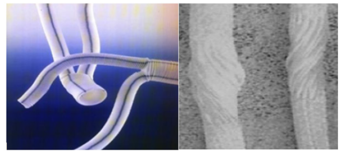
Figure 7: An example of an artificial vascular graft in a warp-knitted fabric structure (left) and an example of an artificial ligament in a circularknitted fabric structure (right) [16].
The 3-D braiding construction of textile surfaces is recommended to be used in biomedical applications thanks to supports cell adhesion and proliferation due to its advantages such as high mechanical properties in various axes, dimensional stability, pore size, and number for biomedical applications of textile surfaces [3,13,15].
Concluding Remarks and Future Research
Biocomposite materials, which began to become widespread in the 1960s, provide a support mechanism that helps the growth of immature cell groups and also enables the integration of surrounding tissues by turning into complex tissue networks. Moreover, FDA approval is required. The design criteria of biocomposite materials must provide pH, biological, chemical, thermal, and mechanical properties according to specifically for biomedical applications. The raw materials used in biocomposite materials are shape memory polymer, polymeric, metallic, ceramic, and biocomposite materials. PES derivatives such as PET, PLA, PLL, PGA, and PLLA are especially widely used. Moreover, polymeric materials such as PA, PP, PTFE, and shape memory metallic materials such as Ni are also used. The production methods of biocomposite materials are molding methods, hand lay-up, filament winding, extrusion, pultrusion, CVD, non-woven surface, textile-based weaving, braiding, and knitted textile surfaces are used. Braiding textile structures is the most ideal method.
In conclusion, the types of PES polymer such as PET, PCL, PLL, PLA, PGA, and PLGA, polymeric materials such as PA, PP, and PTFE, and shape memory metallic materials such as Ni are widely used in biomedical applications. Textile surfaces should be selected according to the specific needs of the biomedical application area. Fiber cross-section profile, yarn type, yarn structure, yarn biological, chemical, thermal, and mechanical properties, yarn count, filament number, density values, and construction should be determined. It provides high biological, chemical, thermal, and mechanical properties of the yarn (the ideal type is PET), fine yarn count (range 88 dtex to 220 dtex), number of filaments (range 108 to 272), high pore number and low pore size in biomedical applications. In addition, the diameter over 6 mm, 3-D braiding textile surface, 1x1 diamond braiding construction, 30° braid angle, minimum 1 (center) + 24 (braid) = 25 total yarns, PET yarn type, triangular geometry fiber cross-section profile is used for its dimensional stability in artificial vascular vessel applications. After, it should be produced by fixing at 150 °C and for 1 minute, too. Moreover, it can also be coated with chitin, chitosan, PU, or PC polymers by dip coating to improve biological properties.
Acknowledgement
None.
Funding
None.
Conflicts of Interest
The author declares that there is no conflict of interest.
References
- Egbo MK (2021) A fundamental review on composite materials and some of their applications in biomedical engineering. Journal of King Saud University-Engineering Sciences 33(8): 557-568.
- Huyer LD, Zhang B, Korolj A, Montgomery M, Drecun S, et al. (2016) Highly elastic and moldable polyester biomaterial for cardiac tissue engineering applications. ACS biomater Sci Eng 2(5): 780-788.
- Jiao Y, Li C, Liu L, Wang F, Liu X, et al. (2020) Construction and application of textile-based tissue engineering scaffolds: a review. Biomaterials Science 8(13): 3574-3600.
- Linsenmeier RA, Saterbak A (2020) Fifty years of biomedical engineering undergraduate education. Ann biomed Eng 48(6): 1590-1615.
- Islam S, Bhuiyan MAR, Islam MN (2017) Chitin and chitosan: structure, properties and applications in biomedical engineering. Journal of Polymers and the Environment 25(1): 854-866.
- Ghalia MA, Dahman Y (2017) Biodegradable poly (lactic acid)-based scaffolds: synthesis and biomedical applications. J Poly Res 24(1): 1-22.
- Makvandi P, Wang CY, Zare EN, Borzacchiello A, Niu LN, et al. (2020) Metal‐based nanomaterials in biomedical applications: antimicrobial activity and cytotoxicity aspects. Adv Fun Mater 30(22): 1910021.
- Falde EJ, Yohe ST, Colson YL, Grinstaff MW (2016) Superhydrophobic materials for biomedical applications. Biomaterials 104(1): 87-103.
- Elahi F, Lu W, Guoping G, Khan F (2013) Core-shell fibers for biomedical applications-a review. J Bioeng Biomed Sci 3(1): 1-14.
- Da Silva AC, De Torresi SIC (2019) Advances in conducting, biodegradable and biocompatible copolymers for biomedical applications. Frontiers in Materials 6(1): 98.
- Ye H, Zhang K, Kai D, Li Z, Loh XJ, et al. (2018) Polyester elastomers for soft tissue engineering. Chemical Society Reviews 47(12): 4545-4580.
- Giordano M, Schmid S, Arjmandi M, Ramezani M (2017) Wear evaluation of three-dimensionally woven materials for use in a novel cartilage replacement. Wear 386-387(1): 179-187.
- Liberski A, Ayad N, Wojciechowska D, Kot R, Vo DMP, et al. (2017) Weaving for heart valve tissue engineering. Biotechnol Adv 35(6): 633-656.
- Yang X, Wang L, Guan G, King MW, Li Y, et al. (2014) Preparation and evaluation of bicomponent and homogeneous polyester silk small diameter arterial prostheses. J Biomater Appl 28(5): 676-687.
- McKenna CG, and Vaughan TJ (2020) An experimental evaluation of the mechanics of bare and polymer-covered self-expanding wire braided stents. J Mech Behav Biomed Mater 103(1): 103549.
- Zhang X, Ma P (2018) Application of knitting structure textiles in medical areas. Autex Res J 18(2): 181-191.
- Gokarneshan N, Dhatchayani (2017) Mini Review: Advances in Medical Knits. J Textile Eng Fashion Technol 3(2): 621-625.
- Mirjavan M, Asayesh A, Asghar A, Jeddi A (2017) The effect of fabric structure on the mechanical properties of warp knitted surgical mesh for hernia repair. J Mech Behav Biomed Mater 66(1): 77-86.
- Liberski A, Ayad N, Wojciechowska D, Zieliska D, Struszczyk MH, et al. (2016) Knitting for heart valve tissue engineering. Glob Cardiol Sci Pract 2016(3): e201631.
- Singh C, Wong CS, Wang X (2015) Medical textiles as vascular implants and their success to mimic natural arteries. J Funct Biomater 6(3): 500-525.
- Tavares TD, Antunes JC, Ferreira F, Felgueiras HP (2020) Biofunctionalization of natural fiber-reinforced biocomposites for biomedical applications. Biomolecules 10(1): 148.
- Zille A, Almeida L, Amorim T, Carneiro N, Esteves MF, et al. (2014) Application of nanotechnology in antimicrobial finishing of biomedical textiles. Materials Research Express 1(3): 032003.
- Chen HM, Wang L, Zhou SB (2018) Recent progress in shape memory polymers for biomedical applications. Chinese J Polymer Sci 36(1): 905-917.
- Park SH, Kim SH (2014) Poly (ethylene terephthalate) recycling for high value-added textiles. Fashion and Textiles 1(1): 1-17.
- Manavitehrani I, Fathi A, Badr H, Daly S, Shirazi AN, et al. (2016) Biomedical applications of biodegradable polyesters. Polymers 20-8(1): 1-32.
- Dirauf M, Muljajew I, Weber C, Schubert US (2022) Recent advances in degradable synthetic polymers for biomedical applications‐Beyond polyesters. Progress in Polymer Science 129(1): 101547.
- Gigli M, Fabbri M, Lotti N, Gamberini R, Rimini B, et al. (2016) Poly (butylene succinate)-based polyesters for biomedical applications: A review. Eur Poly J 75(1): 431-460.
- Urbánek T, Jäger E, Jäger A, Hrubý M (2019) Selectively biodegradable polyesters: Nature-inspired construction materials for future biomedical applications. Polymers 11(6): 1061.
- Washington KE, Kularatne RN, Karmegam V, Biewer MC, Stefan MC, et al. (2017) Recent advances in aliphatic polyesters for drug delivery applications. Wiley Interdiscip Rev Nanomed Nanobiotechnol 9(4).
- Gonçalves FAMM, Fonseca AC, Domingos M, Gloria A, Serra AC, et al. (2017) The potential of unsaturated polyesters in biomedicine and tissue engineering: Synthesis, structure-properties relationships and additive manufacturing. Progress in Polymer Science 68(1): 1-34.



 We use cookies to ensure you get the best experience on our website.