Research Article 
 Creative Commons, CC-BY
The Right Coronary Artery Agenesis Case Report, Identified by Computed Tomography Coronary
*Corresponding author: Sami Smerat, Istshari Arab Hospital, Radiologic Technologist, Ramallah, Palestine.
Received: August 16, 2024; Published: August 23, 2024
DOI: 10.34297/AJBSR.2024.23.003130
Abstract
The Right Coronary Artery (RCA) is a vital blood vessel that serves the right side of the heart and the inferior portion of the left ventricle. Even while some may not manifest any symptoms, irregularities in their genesis or evolution might have a significant impact on cardiovascular health. Our case study involves a 42-year-old female patient who did not exhibit any symptoms. However, an activity test indicated ST segment depression, which could potentially be a sign of myocardial ischemia. The initial echocardiography and biochemical evaluations yielded normal results. To investigate this further, a Computed Tomography Coronary Angiography (CTCA) was conducted. By using CT scanning, an unusual anatomical discovery was made: there was no artery originating from the right sinus of Valsalva. The beginnings and paths of the Despite this curvature, the Left Anterior Descending artery (LAD) and Left Circumflex Artery (LCX) were normal, and a calcium score of 0 revealed the lack of significant coronary artery calcification. The patient was treated conservatively with anti-platelet medication. Recommendations for preventing coronary atherosclerosis included leading a healthy lifestyle and refraining from physically demanding activities. The imaging data available at the time and the clinical status did not support the need for surgery. The importance of accurate imaging in the detection of cardiac anomalies and its implications for patient management are highlighted by this example. The patient’s condition had nothing to do with coronary artery disease, even though it was unusual for a right coronary artery to be absent from the Valsalva sinus. Optimizing results requires thorough assessment and customized management.
Keywords: Computed tomography coronary angiography (CTCA), Left circumflex artery (LCX), Left anterior descending artery (LAD), Right coronary artery (RCA)
Introduction
Congenital absence of the Right Coronary Artery (RCA) is a rare congenital malformation of the cardiovascular system with risk that may lead to death [1]. Congenital absence of (RCA) estimated to be approximately 0.014% to 0.066% [2]. Congenital absence of the Right Coronary Artery (RCA) is a distinctive case of abnormal coronary anatomy [1]. RCA supplies the right myocardium with blood from the circumflex branch (LCX). These patients are usually found by coronary angiography (CAG) or Computed Tomography Coronary Angiography (CTCA) [3,1] Congenital absence of the right coronary artery may result from an absence of the right coronary artery or its congenital obstruction during fetal development [3,1]. It has also been proven to be associated with congenital heart disease. In most cases, congenital absence of the right coronary artery is asymptomatic [4,5]. One of the most serious malformations is the Right Coronary Artery (RCA) which arises from the left main coronary artery (LM) and runs between the aorta and the pulmonary artery, which can lead to severe myocardial ischemia due to compression of the artery by the aorta and pulmonary artery [6,1]. Congenital coronary anomalies are the second most common cause of sudden death in young athletes after hypertrophic cardiomyopathy [1]. We present the case of a patient with a history of chest pain in which a congenital absence of RCA with CTO was confirmed by selective Coronary Angiography (CAG) [7].
Case Presentation
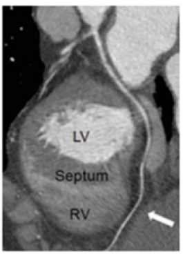
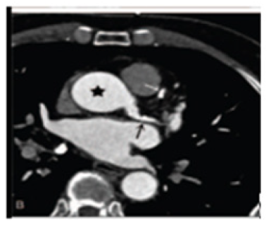
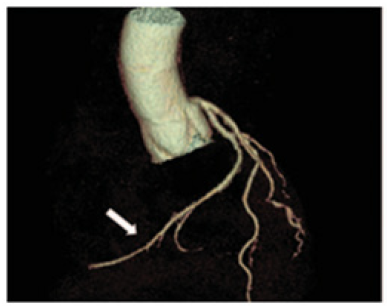
In accordance with AHA guidelines, a 42-year-old lady who was asymptomatic was brought to our institution with an exercise test suggestive of myocardial ischemia (ST segment depression) (Buja, 2005; Fletcher et al., 2013). She wasn't receiving any medication. Biochemical values and echocardiography results were within normal ranges. A CTCA was then scheduled to rule out CAD. The examination revealed that there isn't an artery that emerges from Valsalva's right sinus. There was no indication of CAD in the Left Anterior Descending artery (LAD) or Left Circumflex artery (LCX), which both showed regular origin and course (Calcium score=0). As a result, the patient was observed over time without undergoing surgery while being kept under pharmacological control with anti-platelets. In addition, she was counseled to keep away from vigorous physical activity and to maintain a healthy diet and way of life in order to prevent the onset of coronary atherosclerosis. A second assessment was conducted using the phase with the least amount of residual motion. Multiplanar Reformations (MPR) and Maximum Intensity Projection (MIP) are shown in (Figures 1-3).

Figure 1: Angiographic MPR replicating the LCX course: It passes through the crux cordis and reaches the acute edge of the heart (white arrow) after running in the left atrioventricular groove and supplying the cardiac inferior wall.
Discussion
The characteristic of coronary artery disease (CAD), one of the world's leading causes of morbidity and death, is the formation of atherosclerotic plaques in the coronary arteries, which causes decreased blood flow and ischemia. The Right Coronary Artery (RCA) supplies blood to the right atrium, right ventricle, and inferior portion of the left ventricle. Anomalies in the RCA's origin or course, such as its disappearance or strange branching patterns, might have major clinical consequences and even induce ischemia events, even in the absence of traditional CAD risk factors [10,11].
In our situation, the 42-year-old female patient did not have an artery originating from the right sinus of Valsalva, and there were no indications of a significant ailment on the LCX or LAD. This discovery is significant because it deviates from the way RCA anomalies are usually described in the literature, which usually lists symptoms or effects associated with the anomalies. For instance, 2002 research by [12] describes several coronary anomalies, including those in which the RCA originates incorrectly and frequently presents as symptoms due to reduced blood flow, such as angina or myocardial infarction [12]. However, our patient showed no symptoms even after the anatomical abnormalities, indicating that if CAD is absent, this specific aberration could not be dangerous.
Many coronary anomalies are inadvertently discovered during imaging for unrelated purposes and often have no link with coronary artery disease, according to research by [5]. This is consistent with our case, in which anatomic variation was detected by CTCA in the absence of a notable history of coronary artery disease. This suggests that certain abnormalities, such the absence of the RCA from the right sinus of Valsalva, would not always have a negative impact on clinical outcomes, particularly if the other coronary veins are normal and there isn't any serious atherosclerosis.
With the recent advancements in technology, multidetector computed tomography has become increasingly important in the detection of cardiovascular disease and may now be used to collect more patients with lower ambient radiation doses [13]. According to the 2016 NICE guideline update, CTCA should always be the initial course of treatment for any patient exhibiting typical or atypical angina symptoms, as well as for those who are asymptomatic but have recommended ECG abnormalities for ischemia [14]. Congenital heart disease is being diagnosed and monitored more often with CTCA, which offers superior visibility of anomalies, extra-cardiac blood vessels, systemic veins, and coronary arteries [15]. Even though invasive coronary angiography only displays the arterial lumen and does not reveal information about the vessel wall or surrounding tissues, it is still regarded as the gold standard for the diagnosis and treatment of coronary disorders [16]. Additionally, attempting to catheterize a non-present coronary ostium becomes more difficult for the operator when a coronary absence comes place [17].
Regarding patient management, there isn't a set protocol or evidence-based medical guideline in place at the moment for treating congenital absence of the RCA. Treatment options range from conservative measures like anti-platelets, lipid-lowering, anti-hypertensive medicine, etc. to interventional measures like pacemaker implantation, coronary artery revascularization, and other heart surgery procedures [17]. Additionally, current studies on the care of patients with coronary anomalies indicate conservative treatment for asymptomatic individuals with normal coronary arteries, including our patient [18]. This therapy method includes lifestyle modifications and regular monitoring; in our case, these steps were also recommended to prevent future atherosclerotic changes.
Acknowledgements
None.
Conflict of Interest
None.
References
- Smerat S, Dwaik M (2023) Review of clinical presentations, pathogenesis, and treatment for myocarditis associated with Covid-19. J Clin Med Img Case Rep 3(6): 1602.
- Lipton MJ, Barry WH, Obrez I, Silverman JF, Wexler L (1979) Isolated single coronary artery: diagnosis, angiographic classification, and clinical significance. Radiology 130(1): 39-47.
- Alanís Naranjo JM, Granados Casas ÓM (2023) Congenital absence of the right coronary artery with chronic total occlusion of the left coronary artery: a rare clinical situation. Cardiovascular and metabolic science 34(1): 16-20.
- Angelini P (2007) Coronary artery anomalies: an entity in search of an identity. Circulation 115(10): 1296-1305.
- Yamanaka O, Hobbs RE (1990) Coronary artery anomalies in 126,595 patients undergoing coronary arteriography. Cathet Cardiovasc Diagn 21(1): 28-40.
- Del Torto A, Baggiano A, Guglielmo M, Muscogiuri G, Pontone G (2020) Anomalous origin of the left circumflex artery from the right coronary sinus with retro-aortic course: A potential malign variant. J Cardiovasc Comput Tomogr 14(5): e54-e55.
- Harky A, Noshirwani A, Karadakhy O, Ang J (2019) Comprehensive literature review of anomalies of the coronary arteries. J Card Surg 34(11): 1328-1343.
- Buja LM (2005) Myocardial ischemia and reperfusion injury. Cardiovasc pathol 14(4): 170-175.
- Fletcher GF, Ades PA, Kligfield P, Arena R, Balady GJ, et al. (2013) Exercise standards for testing and training: a scientific statement from the American Heart Association. Circulation 128(8): 873-934.
- Gentile F, Castiglione V, De Caterina R (2021) Coronary artery anomalies. Circulation 144(12): 983-996.
- Nedkoff L, Briffa T, Zemedikun D, Herrington S, Wright FL (2023) Global trends in atherosclerotic cardiovascular disease. Clin Ther 45(11): 1087-1091.
- Angelini P (2002) Clinical articles: coronary artery anomalies-current clinical issues: definitions, classification, incidence, clinical relevance, and treatment guidelines. Texas Heart Institute Journal 29(4): 271-278.
- Forte E, Monti S, Parente CA, Beyer L, De Rosa R, et al. (2018) Image quality and dose reduction by dual source computed tomography coronary angiography: protocol comparison. Dose Response 16(4): 1559325818805838.
- Moss AJ, Williams MC, Newby DE, Nicol ED (2017) The updated NICE guidelines: cardiac CT as the first-line test for coronary artery disease. Current Cardiovasc Imaging Rep 10: 1-7.
- Kulkarni A, Hsu HH, Ou P, Kutty S (2016) Computed tomography in congenital heart disease: clinical applications and technical considerations. Echocardiography 33(4): 629-640.
- Desmet W, Vanhaecke J, Vrolix M, Van de Werf F, Piessens J, Willems J, De Geest H (1992) Isolated single coronary artery: a review of 50 000 consecutive coronary angiographies. Eur Heart J 13(12): 1637-1640.
- Yan GW, Bhetuwal A, Yang GQ, Fu QS, Hu N, et al. (2018) Congenital absence of the right coronary artery: A case report and literature review. Medicine 97(12): e0187.
- Alam MM, Tasha T, Ghosh AS, Nasrin F (2023) Coronary Artery Anomalies: A Short Case Series and Current Review. Cureus 15(5): e38732.


 We use cookies to ensure you get the best experience on our website.