Review Article 
 Creative Commons, CC-BY
Ecomorphological Analysis of The Trochoid Joint and Its Evolutionary Significance
*Corresponding author: Norihisa Inuzuka, Palaeo-Vertebrate Laboratory, Saiwai-cho 45-25-303, Itabashi-ku, Tokyo 174-0034, Japan.
Received: September 30, 2024; Published: October 04, 2024
DOI: 10.34297/AJBSR.2024.24.003181
Abstract
Observation and interpretation of the shape of joints is important for restoring the posture and movement of extinct animals. In human anatomy, the atlantoaxial joint and the superior/inferior radioulnar joints are given as examples of trocoid joints, but this does not necessarily match the definition. In fact, when bones are articulated in animals, it has been found that there are two different types of trocoid joints. Therefore, we decided to classify trocoid joints with one set of head and fossa and two sets of head and fossa into type I and type II, respectively. Type I trocoid joints include the atlantoaxial joint of most mammals and the midcarpal joint of elephants, while type II includes the proximal/distal radioulnar joints of primates and carnivores, the antebrachiocarpal joint of large ungulates, and the talocalcaneal joint of primates. The correlation between habitat, mode of locomotion, feeding habits, and wrist and ankle shape and locomotion in primates, carnivores, and ungulates can be said to be an example of the law of correlated evolution, and serves as the basis for skeletal and ecological restoration.
Definition and Problems of the Trocoid Joint
Observation of joint shape and functional interpretation are important for restoring the posture and locomotion of extinct animals. Here, we compare the form and function of the trocoid joint, which has not been studied much so far, between different lineages and consider its evolutionary significance. The morphology of the joint is described in detail osteology and syndesmology in anatomy as follows:
In human anatomy, the trocoid joint is defined as having a cylindrical or disc-shaped head that coincides with the long axis of the bone, and the fossa is a curved notch according to its side, and examples include the atlantoaxial joint and the proximal/distal radioulnar joint [1].
“Trochoid joint; trocoid joint (articulatio trochoidea; rotary joint) This joint, which is limited to rotational movement, consists of an axial process that rotates within a ring, or a ring of bones and ligaments around the axial bone. In the proximal radioulnar joint, the ring is formed by the radial notch of the ulna and the radial annular ligament, and the head of radius rotates within the ring. In the articulation between the atlas and the odontoid process (median atlantoaxial joint), the ring is formed in front by the anterior arch of the atlas and in back by the transverse atlantoaxial ligament, and it rotates around the odontoid process [2].”
“In the proximal radioulnar joint, for example, the wheel-shaped head of radius rotates about an axis of symmetry within an annulus formed by the radial annular ligament and a small glenoid fossa on the ulna. In the atlantoaxial joint, however, the bony annulus rotates about a pin, the odontoid axis. This type of joint is also called a trocoid joint [3].” In veterinary anatomy, “Trochoid, or pivot joint. In these the movement is limited to rotation of one segment around longitudinal the axis of the other. Example: atlantoaxial joint [4].”
“The trocoid joint (articulatio trochoidea) is a joint that has its primary movement around the long axis of the bones that compose it. The median atlantoaxial joint and the proximal radioulnar joint are examples of trocoid joints. The atlantoaxial joint (articulatio atlantoaxialis) is a trocoid joint that allows the head and atlas to rotate about their longitudinal axis. The distal radioulnaris joint (articulatio radioulnaris distalis) is a distal extension of the radius and ulna. It is a distal trocoid joint that allows rotational movement between the bones of the forelimb [5].”
The part of the atlantoaxial joint that articulates with the odontoid process of the axis is the median atlantoaxial joint. On the other hand, in another forearm, in the anatomical correct position, the radius and ulna run parallel to each other (Figure 1). This position is called the supination position. In this position, the palm faces upwards. The action of rotating the palm 180° from there so that it faces down is called pronation, and this position is called the pronation position. In the pronation position, the radius crosses over the ulna. The radius and ulna articulate at two points, proximally and distally. The proximal one is called the proximal radioulnar joint, and the distal one is called the distal radioulnar joint. Although they have different names, neither joint moves independently and shares a single axis of rotation, so the radioulnar joint can in fact be considered a single trocoid joint.
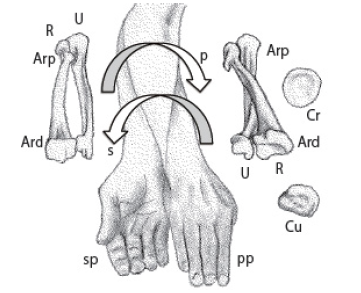
Figure 1: Pronation (p) and supination (s) of human antebrachial skeleton. Ari: Articulatio radioulnaris superior, Ars: Artictulatio radioulnaris inferior, Cr: head of radius, Cu: head of ulna, pp: pronated position, R: radius, sp: supinated position, U: ulna.
The proximal end of the radius is called the head of radius. It is a low cylinder that articulates with the radial fossa of the ulna on the side. The distal end of the ulna is called the head of ulna. It is a thin cylinder compared to the head of radius, and articulates with the ulnar fossa of the radius on the side. The line connecting the centers of the cylinders of the head of radius and head of ulna is the axis of rotation of this trocoid joint. Therefore, it intersects obliquely with the long axes of both the radius and ulna, and at the same time moves around each other, which does not match the previous definition. For this type of trocoid joint, the important thing is not the direction of the axis of rotation, but the movement of the two convex and concave articular surfaces sliding around each other’s articular heads. Even in textbooks on veterinary anatomy and comparative anatomy, which are basically the same as human anatomy, this problem remains unresolved.
Redefinition and Examples of Trocoid Joints
Therefore, we decided to classify trocoid joints with one set of head and fossa and those with two sets of head and fossa as type I and type II, respectively.
Type I trocoid joints: the atlantoaxial joint of most mammals, and the midcarpal joint of elephants.
Type II trocoid joints: the radioulnar joint of primates and carnivores, the antebrachiocarpal joint of rhinoceros and desmostylians, and the talocalcaneal joint of primates.
The focus here is on bones of large mammals, particularly ungulates, carnivores, and primates, whose articular surface shapes can be easily observed with the naked eye. Specifically, we look at Asiatic elephants, hippopotamuses, and sea lions from the Ashoro Museum of Paleontology, and white rhinoceros, brown bear, lion, human, and Japanese macaque from the Palaeo-Vertebrate Laboratory. The bones include the atlas, axis, radius, ulna, proximal carpal bones (scaphoid, lunate, and cuneiform), distal carpal bones (trapezoid, capitate, hamate), talus, and calcaneus, which all have trocoid joints. We also look at fossils of the desmostylian Paleoparadoxia and Desmostylus, and the proboscidean Palaeoloxodon.
The method involves observing the articular surfaces of the bones and then actually articulating them and moving them relative to one another without dislocating them. By comparing the joints of the same part with those of other taxa, it is possible to consider the evolutionary background, such as differences in the lifestyles of each taxa.
Atlantoaxial Joint
In most mammals, the atlas and axis are separate, and the atlantoaxial joint is formed between the odontoid process of the axis and the atlas. In humans, the spine is upright, so the head rests on the cervical vertebrae, and rotation around the odontoid process of the axis results in left or right rotation of the face. In ungulates, the median atlantoaxial joint and the lateral atlantoaxial joint are connected. In graviportal ungulates, the vertebral bodies are short and the odontoid process is thick. In cursorial ungulates, the vertebral bodies are long from front to back, the diameter of the vertebral foramen is relatively large, and the vertebral foramen is indented on the dorsal side of the odontoid process. The axis of rotation passes through the center of the long axis of the odontoid process. For this reason, in most mammals, the head can be swung around the body axis. For example, the movement of a dog when shaking off water from its body.
Midcarpal Joint in Elephants
Owen (1866) [6] wrote about the carpal bones of proboscideans: “In the Scaphoid, the small surface for the radius is remote from that which joins the trapezium, and trapezoids. The single surface of the pisiforme has two facets, the smaller of which joins the ulna. The trapezium extends along half metacarpal of the index.”
Flowers (1885) [7] states only that “In the Elephant the manus is short and broad, the carpal bones are massive and square, and articulate by very flat surfaces ; they consist of scaphoid, lunar and cuneiform, a pisiform and the usual four bones of the distal row, all distinct, without the centrale.” However, he does not mention the midcarpal joint between the proximal and distal rows.
Gregory (1910) [8] included Proboscidea among “the principal orders in which the lunar-unciform contact is greatly reduced or absent”. He also described the carpal bones from the perspective of the evolution of the hand, saying, “In connection with the growth of the ulna and the reduction of the radius, the cuneiform and especially the lunar, become very broad, the lunar overspreading the trapezoid and causing the reduction of the scaphoid.” without any regard for the shape of the articular surfaces. Reynolds (1913) [9] very roughly states “In the Proboscidea the manus is very short and broad, with large somewhat cubital carpals which articulate by very flat surfaces and do not interlock at all.”
Kingsley (1925) [10] divided the structure of the carpal bones in ungulates into two types: taxeopodous and diplarthrous. Regarding proboscideans, he wrote, “The carpus is completely taxeopod; the centrale fuses with the radiale in the young, but the other carpal bones remain separate.” Max Weber (1928) [11] also described the arrangement of the carpal bones as “partially in series; that this is a primary taxeopode, and not a secondarily acquired one, as has been claimed, is evident from the general structure, and especially from the appearance of the centrale, which is fused only with the scaphoid in young animals.”
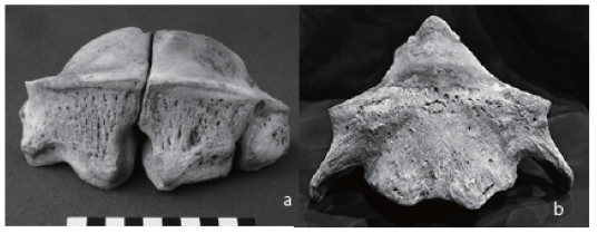
In the fossil record, Cooper (1928) [12] reported on Elephas antiquus from Upnor, in which he described, measured, and drew the six carpal bones, excluding the cuneiform and the unciform, and compared them with the E. africanus, E. maximus, and E. primigenius. Cornwall (1974) [13] provides the most detailed description of the proboscidean hand: “The manus is extremely short and stout, with 8 massive carpals, including a pisiform. The centrale is absent. The trapezium projects distally beyond the rest of the carpus as a whole. The 5 digits, being symmetrically developed about III, the longest and strongest, the axis of the manus runs from the radius, through lunar and magnum to m/c III. Metacarpal IV is the next in order of development and its axis runs from the ulna through cuneiform and uncinate.” However, there is no mention of the shape of the articular surfaces or movement. Among the major comparative anatomy books, the only one that writes about the carpal joint is Bolk, et al. (1938) [14], but it also does not describe the intercarpal joint in proboscideans. As we have seen, the midcarpal joint is unique, although it is not mentioned in the descriptions of the carpal bones of proboscideans. The proximal surface of the distal carpal bone has a high hill-like shape at the center, and overall resembles the odontoid articular surface of the axis (Figure 2).
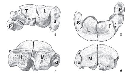
In addition to palmar flexion as in normal ungulates, this joint allows the proximal carpal bones to rotate above the distal carpal bones. While the trocoid joint is uniaxial, the midcarpal joint of the elephant is a biaxial joint capable of palmar flexion and rotation (Figure 3,4).
The trigger for this discovery was the excavation of Naumann’s elephant at Lake Nojiri. During the fifth excavation at Lake Nojiri, four carpal bones articulating with each other of the elephant, namely the trapezoid, capitate, hamate, and cuneiform, were discovered. As this was the first time such bones had been found, they were identified and compared with bones of modern Asiatic and African elephants (Figure 5).
The wrists of most ungulates are capable of palmar flexion only, and are uniaxial hinge joints with a dorsiflexion stopper. Why do only proboscideans rotate at the midcarpal joint? This question was answered by fossil footprints of sauropod dinosaurs. An off-tracking has been observed in sauropod tracks [15]. An off-tracking is like the internal off-tracking of a car, and when a car turns, the rear wheels pass on the inside of the track of the front wheels. In dinosaur footprints, the tracks of the hind limbs are also on the inside compared to the tracks of the forelimbs. This is the opposite for elephants, so it can be determined which leg is used when turning. While sauropods turn with their forelimbs, elephants turn with their hind limbs like a forklift. This explained why the glenoid fossa of the scapula of ungulates is circular, while only elephants have a long truck-like shape fore-and-aft [16]. While the shoulder joint is generally a ball-andsocket joint, an elephant’s forelimbs can only flex and extend parasagittally, and cannot abduct. For example, when an elephant bends to the right, after the right forelimb touches the ground, the left hind limb touches down more laterally than the forelimb. At this time, the right forelimb twists clockwise along with the body, but because neither the shoulder joint nor the elbow joint can rotate, it is thought that rotation is only carried out by the midcarpal joint.
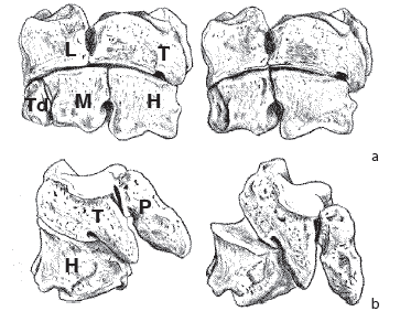
Figure 3: Elephant. left carpal bones. Inuzuka (2017) [22] a: anterior view showing rotation, b: lateral view showing palmiflexion. H: hamatum, L: lunar, M: magnum, P: pisiform, T: triquetral, Td: trapezoideum.
Radioulnar Joint
In fact, many of the limb bone joints in mammals are involved in the change in posture from the lateral type of reptiles to the inferior type. In inferior type mammals, the elbow rotates from the lateral to the caudal, so the manus pronates 180° with the forearm. This rotational movement is preserved in primates and carnivores, which also use their forelimbs for manipulation. On the other hand, in mammalian hind limbs, the knee rotates cranially and the pes faces forward, so the rotation function of the crus is lost, and the fibula degenerates and comes out of the knee joint. In human anatomy, it is common to be able to turn the palm of the hand, so the pronation and supination action of the radioulnar joint of the antebrachial skeleton is taken for granted. However, in many large mammals, this action seems to be limited to species that use the forelimbs as hands, and in ungulates the two bones of the forearm tend to be immobile and fused together. In primates, the radius and ulna are the same thickness. Primates are arboreal by nature, and their hands and feet are opposable. When brachiating, they hang by one hand and, staying flexed elbow, they can freely change the direction of movement by pronating and supinating their forearm.
They are omnivorous, but because they keep their spine upright when sitting, they use their hands to eat. As a result, they are able to move their forelimbs with the greatest freedom of movement. At the radioulnar joint, the outline of the head of radius in carnivores is oval, whereas in primates it is nearly circular, indicating a larger angle of rotation of the wrist. On the other hand, the head of ulna is flat, and is separated from the triangular bone by an articular disc. This allows the wrist to bend towards the little finger (ulnar flexion). Even in carnivores, the radius and ulna remain the same thickness and can rotate. For running purposes, it would be more efficient to fix the bone in a pronated position like in ungulates, but in order to capture prey with the palm, it is necessary to supinate the bone. This allows the claws to dig into the prey at the optimal angle depending on its size. The outline of the head of radius is not a perfect circle due to the low degree of rotation, and the head of ulna is conical because it is the distal axis of rotation. Pinnipeds among carnivores, have limbs that form flippers for swimming, and have a wide and flat radius (Figure 6). The head of the radius is closer to a perfect circle than that of lions and bears, suggesting that the fins have a wider range of rotation. The forearm of pinnipeds is wider for its length, so the force twisting the fin at the radioulnar joint would be greater than that of terrestrial species. The tip of the head of ulna is sharply conical, and fits snugly into the bearings of the cuneiform and pisiform bones.
Antebrachiocarpal Joint
The human wrist joint is a biaxial elliptical joint called the radiocarpal joint. In the anatomical position, the carpal bones extend in extension to the radius, so bending to either side results in flexion, which is distinguished between palmar flexion and dorsiflexion, and radial flexion (radial deviation) and ulnar flexion (ulnar deviation). Monkeys are similar to humans in that the head of ulna, where the articular disc is attached, is flat. The free movement of the human hand can be said to be a legacy inherited from arboreal monkeys. In carnivores, the head of the ulna is cone-shaped and pointed, and articulates with the cuneiform and pisiform bone. As a result, ulnar flexion is not possible, and the radiocarpal joint is a uniaxial hinge joint. The proximal carpal bones, the scaphoid and lunar, are fused to form the scapholunar [7]. The scapholunar articulates with the distal carpal bones, including the unciform, and transmits forces to all five fingers via the distal metacarpals.
On the other hand, ungulates are herbivorous and are basically unguligrade adapted to running. The metacarpals grow long, and in artiodactyls, as the number of digit decreases, the third and fourth metacarpals eventually fuse to form the cannon bone. In horses of perissodactyls, the carpal bone that bears the weight expands only with the third metacarpal bone, while bilateral bones shrink. The forearm carpal joint is a hinge joint, with the latter half curved and only capable of palmar flexion, while the area near the front edge is flat and acts as a dorsiflexion stopper.
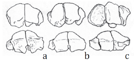
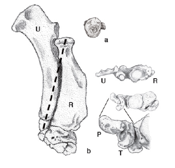
Of the antebrachiocarpal joints, only the graviportal perissodactyl, the rhinoceros, has a type II trocoid joint. The distal surface of the radius in the rhinoceros is concave, and the corresponding proximal surfaces of the scaphoid and lunate are convex (Figure 7). In contrast, the distal surface of the ulna is convex, and the cuneiform is concave. If the bones are slid against each other without dislocating any of them, the only movement possible is around the axis of rotation connecting the center of curvature of the articular surface of the scaphoid and the center of curvature of the distal surface of the ulna. In graviportal ungulates such as rhinoceroses, the shoulders are broad, the legs are not as long as in cursorial ungulates, and the point of contact of the foot with the ground is located closer to the median line, so the zygopodium is in an adducted position and a carrying angle is formed at the wrist (Figure 8).
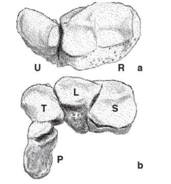
Figure 7: Rhinoceros. a: antebrachial skeleton in distal view, b: proximal carpal bones in proximal view, L: lunar, P: pisiform, R: radius. S: scaphoid, T: triquetral, U: ulna.
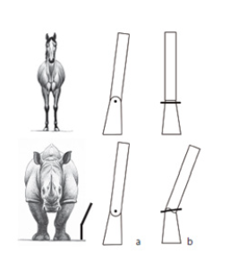
Figure 8: Comparison of antebrachiocarpal joint. a: lateral view, b: cranial view. Inuzuka and Hirono, (2023) [24] revised.
The reason why only the antebrachiocarpal joint in rhinoceroses becomes a type II trocoid joint is that as rhinoceroses grow larger and the width of the shoulder joint increases, the point where the limb touches the ground remains near the median line, causing the forearm to tilt into an adducted position. In hippos, the tilt is weaker, making it a spiral joint, while in elephants, which have long legs, it remains a hinge joint. In fossils, the distal surface of the radius of Paleoparadoxia is concave and the ulna is convex [17]. The corresponding proximal surfaces of the carpal bones, the scaphoid and lunate, are convex, and the cuneiform is concave. When these are articulated and moved without dislocating each other, it can be seen that it is a trocoid joint with the axis of rotation being the center of curvature of the convex surface, i.e., the line connecting the scaphoid and the head of ulna. Although the two joints are closer together than in the radioulnar joint of the human forearm, the basic structure is a type II trocoid joint, which is the same in both the Keton specimen [18] and the Utanobori specimen [19] of Desmostylus. The essence of a type II trocoid joint is that it has two articular surfaces that are inversely convex and concave, rather than in the direction of the axis of rotation.
The human knee joint is not a strict hinge joint like the elbow joint of the upper limbs. The range of motion cannot be determined by simply placing the femur and tibia together. The contour of the femoral condyle is not a simple arc, but also includes sliding movement. The human knee has a carrying angle like a rhinoceros’ wrist. This is because humans, who walk on two legs, must align the midline and center of gravity with each step that involves contact with the ground. During the stance phase, the knee is locked to support the body weight. When the knee is flexed, it is unlocked to rotate the crus. At this time, the center of the medial tibial condyle becomes the axis of rotation and the lateral part twists, so the concave curvature of the medial condyle is stronger and the articular surface of the lateral condyle is flatter. Although to a lesser extent, it is possible to expect similar movement to certain types of type II trocoid joints.
Primate Talocalcaneal Joint
According to Kapandji (1977) [20], “The human subtalar joint as a whole should be considered as a planar joint, because it is geometrically impossible for a combination of spherical and cylindrical surfaces to exist in a single mechanical joint and for these surfaces to move simultaneously while always remaining in contact with each other.” This is not correct. Generally, when bones are in contact with each other, they move together to create a joint surface; if there was no movement at all, the bones should be fused. On the other hand, Aiello and Dean (1990) [21] write: “The subtalar axis is the axis of the subtalar joint, or the joint between the talus and the calcaneus. This joint has reciprocal concave and convex articular surfaces at either end of the talus. The convex head of the talus articulates with the concave anterior (and medial) facets on the calcaneus. At the same time the concave posterior facet on the talus articulates with the convex posterior facet on the calcaneus. The axis of rotation of the joint is the line passing through the centres of the circles of which the joints are arcs. It is this axis around which the foot inverts and everts.” However, they do not say that this is a trocoid joint. In anatomical terms, the human talocalcanal joint is divided into three, but actually it is divided into two surfaces. On the superior surface of the calcaneus, the anterior and middle articular surfaces are concave, while the posterior articular surface is convex. In contrast, on the inferior surface of the talus, the anterior and middle articular surfaces are convex, while the posterior articular surface is concave. As shown in Figure 9, the anterior and posterior articular surfaces are in contact with each other, so in order to move without dislocating each other, some rotation is possible around an axis of rotation that runs from the taler head on the anterior and dorsal side to the calcaneal eminence on the posterior and plantar side. The bones of the human foot can be divided into medial and lateral groups. The first to third metatarsals are articulated to the head of the talus via the navicular and the three cuneiformi, and the fourth and fifth metatarsals are articulated to the calcaneus via the cuboid, so when viewed from the front of the foot, the movement between the talus and calcaneus at the ankle is such that the side of the first or fifth toe is higher than the opposite side. In other words, this is inversion and eversion (Figure 9).
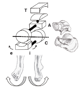
Figure 9: Human. rotation of ankle joint. Inuzuka (2010) [25] A: astragalus, C: calcaneus, T. tibia, e: eversion, i: inversion.
In humans, the upper and lower ends of the crus are limited to flexion and extension at the hinge joints at the knee and ankle, and so left and right balance is achieved through movement at the talocalcaneal joint. The talocalcanal joint is a type II trocoid joint. The peroneal muscles acts to evert. This movement is much greater in arboreal monkeys. When monkeys climb tree trunks, they press the soles of their feet against the tree, then straighten their knees and evert their feet. On the other hand, humans who walk on the ground do not need to evert their feet when walking on flat ground. Flexion and extension of the talocrural joint is sufficient for normal walking. The human talocalcaneal joint can be said to be a degenerate organ.
Evolutionary Significance of the Trocoid Joint
The three types of type II trocoid joints defined by the author in this paper are the radioulnar joint in primates and carnivores, the antebrachiocarpal joint in large ungulates, and the talocalcaneal joint in primates. A correlation is observed between the morphology of the limb bones and the ecology of large mammals such as primates, carnivores, and artiodactyls (Table 1). Why are type II trocoid joints so commonly observed in the wrists and ankles? In fossil amphibians, the carpus and tarsus are usually unossified. At the developmental stage of the hand of a two-year-old human child, most of the carpal bones are cartilage. The reason trocoid joints are concentrated in the wrists and ankles is thought to be due to delayed ossification of the carpal and tarsal bones, which connect long bones of the stylopodium and zygopodium among the three limb segments, and the autopodium that contacts the substrate. Ossification of the carpal bones, which do not present in amphibians when they first come on land, are delayed also in human development. This can be seen as a recapitulation phenomenon.
Generally, the strength of bones is proportional to their cross-sectional area. Body weight is the cube of the length, so the larger an animal is, the greater the proportion of bones in relation to leg thickness. Joints connect bones and form the skeleton. The shape of the joint surfaces reflects the movement between bones. For this reason, it is possible to reconstruct an animal’s movement from the shape of bones preserved in fossils. If correlations are found between habitat, mode of locomotion, diet, and the shape and movement of limb joints, this can be used as an example of the law of correlated evolution, and will surely be useful in reconstructing ecology even when only part of a fossil is discovered.
Conflict of Interest
None.
Acknowledgements
I have examined the specimens held at the Ashoro Museum of Paleontology. I was given the opportunity to present this paper at the Fossil Research Society of Japan and the Palaeontological Society of Japan.
References
- Mori, O Mori, T (1969) Anatomy I Kanehara,
- Goss CM (1973) Gray’s Anatomy of the human body. Lea & Febiger, Philadelphia.
- Ferner H, Staubesand J (1980) Benninghoff/Goerttler Lehrbuch der Anatomie des Menschen. Bd.1. (Urban& Schwarzenberg, Mü
- Sisson S, Grossman JD (1953) The anatomy of the domestic animals. 4th
- Miller ME, Christensen GC, Evans HE (1964) Anatomy of the dog.
- Owen R (1866) On the anatomy of Vertebrates. Vol. 2 Birds and mammals.
- Flower WH (1885) An introduction to the osteology of the Mammalia.
- Gregory WK (1910) The Orders of mammals. Bull. Amer. Mus. Nat. Hist 27: 1-524.
- Reynolds SH (1913) The vertebrate skeleton.
- Kingsley JS (1925) The Vertebrate skeleton from the developmental standpoint.
- Max Weber (1928) Die Säugetiere Einführung in die Anatomie und Systematik der recenten und fossilen Mammalia.
- Cooper CF (1928) On a specimen of Elephas antiquus from Upnor.
- Cornwall IW (1974) Bones for the archaeologist.
- Bolk L, Göppert E, Kallius E, Lubosch W (1938) Handbuch der vergleichenden Anatomie der Wirbeltiere. (Urban & Schwarzenberg, Berlin und Wien).
- Ishigaki S (2011) Restoring Living Organisms from Dinosaur Footprints. In Fossil Research Group (ed) Solving the Mysteries of Life from Fossils: From Dinosaurs to Molecules, Asahi Shimbun Publications, Tokyo 161-175.
- Inuzuka N, Hanzawa S (2013) Morphology and Functional Significance of Mammalian Carpal Bones. Abstracts of the 2013 Meeting of the Japanese Society of Paleontology Yokohama.
- Inuzuka N (2005) The Stanford skeleton of Paleoparadoxia (Mammalia: Desmostylia). Bull Ashoro Mus Paleont 3: 3-110.
- Inuzuka N (1982) The skeleton of Desmostylus mirabilis from Sakhalin. V. Limb bones. Chikyu Kagaku 36: 117-127.
- Inuzuka N (2009) The skeleton of Desmostylus from Utanobori, Hokkaido. II. Body bones. Geological Survey Research Report 60 [5/6]: 257-379.
- Kapandji IA (1977) Physiologie articulaire. Schémas commentés de mécanique humaine. Fascicule II Membre infeerieur. (Librairie maloine S. A, Paris.
- Aiello L, Dean C (1990) An introduction to human evolutionary anatomy.
- Inuzuka N (2017) Carpal bones. In the Society of Biomechanisms (eds) Encyclopedia of the Hand.
- Fossil Mammal Research Group for Nojiri-ko Excavation (1980) Fossil Mammal Research Group for Nojiri-ko Excavation Fossil remains of the Naumann Elephant from the Nojiri-ko Formation. The Memoirs of the Geological Society of Japan 19: 167-192.
- Inuzuka N, Hirono K (2023) Restoring Dinosaurs. Many Mysteries, 465, Fukuinkan.
- Inuzuka N (2010) The structure and function of the foot. In Takao M (ed) Pictorial essential guide to the latest foot care. All Japan Hospital Publishing, Tokyo, 1-11.



 We use cookies to ensure you get the best experience on our website.