Research Article 
 Creative Commons, CC-BY
Histomorphometric and Biochemical Effect of The Liver of Animal Models Treated with Aqueous Extract of Musa Paradisiaca
*Corresponding author: Kebe E Obeten, Department of Anatomy, Faculty of Medicine, Lusaka Apex Medical University, Zambia and Department of Anatomy and Forensic Anthropology, University of Cross River State, Okuku, Nigeria.
Received: October 16, 2024; Published: November 13, 2024
DOI: 10.34297/AJBSR.2024.24.003243
Abstract
This research was conducted to assess the histomorphometric and biochemical effect of the liver of wistar rats treated with aqueous extract of Musa paradisiaca. Thirty (30) Wistar rats were randomly redistributed into three (3) groups of control (animals received water and food only), low dose (animals received water, food and aqueous extract of musa paradisiaca at a low dose of 1.0mg/kgBw) and high dose (animals received food, water and aqueous extract of musa paradisiaca at a dose of 3.0mg/kgBw) respectively. At the end of the three (3) weeks experimental period, animals were sacrificed and the liver harvested and washed with 10% formalin and processed for histological studies. Our results shows morphological observable significant (P<0.05) increase in the final mean body weight when compared with the initial body weight and the control and also showed significant biochemical and histological deteriorations in the liver of experimental animals, and treatment with musa paradisiacal at high dose, reduced the elevated levels of the serum liver enzymes [Aminotransferase, Alanine Transaminase, and Alkaline Phosphatase] evidenced the hepatoprotective activity of stem of Musa paradisiaca. The present study has been able to show that at high dose, the extract of Musa paradisiaca have significant effect on the liver histomorphology and biochemical profile of the treated animal models.
Keywords: Musa paradisiaca, Histomorphometric, Liver enzymes
Introduction
A significant portion of our natural resources are medicinal plants. They are useful as a therapeutic agent and as raw materials for the production of several conventional and contemporary medicines (Motaleb, et al., 2018). Since prehistoric times, people have found plants and used them in traditional medical practices. Traditional medicine continues to be the backbone of healthcare in many underdeveloped nations, with the majority of medications and treatments derived from plants and other natural resources. The perennial herbaceous plant Musa paradisiacal (Plantain) is a member of the genus Musa and family Musaceae. It is a perennial that resembles a tree and has an underground rhizome. The fruit is also very nutrient-dense, with high concentrations of vitamins A and C, minerals including phosphorus, calcium, and potassium, and carbs. Plantains and bananas are sometimes referred to together by the word "banana" (Gawel, 2015).
Musa paradisiacal is the current scientific name that is recognized for all of these crosses. Plantain crops are grown around the world, not just in Nigeria (Gawel, 2015). Because they are available year-round due to their perennial nature, plantains have come to be recognized as a frequent and dependable all-season staple meal in the locations where they are located. In Africa, the appeal of plantain farming to the populace stems partly from its role in promoting food security, creating jobs, varying sources of income in both rural and urban regions, and requiring less labor in production when compared to the contributions of cassava, maize, rice, and yam to the continent's gross national product (GNP) [15] (Nkendah, 2020; Marriott and Lancaster, 2019).
The liver, which is the second largest internal organ, is situated above the stomach, right kidney, and intestine in the upper right section of the abdominal cavity, under the diaphragm. The liver receives its blood supply from two different sources. The hepatic artery brings in oxygenated blood, and the portal vein brings in blood that is rich in nutrients. There are two primary lobules in the liver. The liver's functions are well-known and include producing bile, some blood plasmas, storing excess glucose as glycogen, controlling blood levels of amino acids, which are the building blocks of protein, and more.
Materials & Method
Sample Collection
Fresh Musa paradisiaca bunch (plantain) were gotten from local market of Okuku in, Yala local government area of Cross River State of Nigeria. The unripe plantain was peeled, slide and properly washed to clean debris, sliced and dried in an air–dried room temperature of about 270C for two weeks. They were blended and kept in an air-tight container from where they were used for extraction.
Extract Preparation
The plantain powder was dispensed in a 9,000 mls of distilled water in a plastic container. The mixture was vigorously stirred intermittently with a stick and allowed to stand for 24hours before it was filtered with a cloth-sieve. The filtrate was evaporated at 45ºC with water both to obtain the crude solid extract for one month and the extract obtained was stored in a refrigerator until the commencement of the administration.
Experimental Design
Thirty (30) animals for group A were redistributed into three (3) groups of control, low dose and high dose according to their weight respectively.
Group A (Control) animals received water and food only.
Group B (Low Dose) animals received water, food and aqueous extract of musa paradisiaca at a low dose of 1.0mg/kgBw.
Group C (High Dose) animals received food, water and aqueous extract of musa paradisiaca at a dose of 3.0mg/kgBw.
Histological Studies
The Liver were removed and preserved in labeled bottles containing 10% buffered formalin. These were allowed to stand for 72 hours to achieve good tissue penetration and effective fixation. After these, they were placed in ascending grades of ethanol for dehydration. First, they were treated with two changes of 70% ethanol each lasting for one (1) hour followed by 95% ethanol and then absolute alcohol for the same duration. Following dehydration, the tissues were cleared in three changes of xylene each lasting for thirty (30) minutes. Impregnation in molten paraffin wax at 580C was carried out overnight and the following morning, the tissues were embedded in wax to form blocks. This tissue blocks were trimmed and sectioned using a rotary microtome. The sectioning was floated in warm water (280C) and taken up on albuminised glass slides. They were air-dried and stain using the Haematoxylin and Eosin (Harris, 1900).
Tissue Processing
Tissue blocks were sectioned at 5µ-6µ with a rotary microtome then was dewaxed in xylene for two (2) minutes per two changes. Xylene was cleared in 95% alcohol for one (1) minute per two (2) changes and 70% alcohol for another minute. The sections were then hydrated in running tap water until sections turned blue. They were thereafter counterstained with 1% alcohol eosin for one (1) minute, followed by a rapid dehydration through ascending grades of alcohol, cleared in xylene and mounted with DPX mutant. Stained sections were viewed under a light microscope and photomicrograph.
Statisitical Analysis
Statistical analysis was done using Statistical Package for Social Sciences (SPSS) Version 16 Chicago Inc., one-way ANOVA, followed by Bonferroni’s Multiple Data Comparison. Test was used to perform the analysis. Result of descriptive statistics of the experimental data was presented as Mean standard error of the mean (Mean±SEM). Paired sample T-test were considered statistically significant at P<0.05.
Results and Analysis
Effect of Aqueous Extract of Musa Paradisiaca on the Body Weight
Morphological observation from the study shows an observable significant (P<0.05) increase in the final mean body weight when compared with the initial body weight observable in control vs low dose, control vs high dose and low dose vs control but not observable in low dose vs high dose and high dose vs control dose. The final body weight of the control animals (115.0±3.310) was significantly (P<0.05) higher than its initial body weight (91.40±5.467). however, the mean final body weight of the low dose group (152.4±6.723) and high dose group (160.2±6.018) were significantly (P<0.05) higher than their initial body weights (133.8±3.094) and (135.1±6.125) respectively (Table 1).
Histological Examination
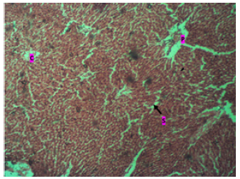
The histological result of the study in control animals, shows a portal triad (P) and central vein (C) with many surrounding hepatocytes with sinusoids. And the tissue appears normal as seen in (Plate 1).
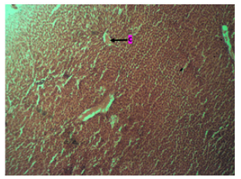
The administration of Musa Paradiasical extract at Low dose shows central vein (C). revealing many surrounding hepatocytes with sinusoids, with the parenchyma showing pseudorosettes tissue. (Plate 2).
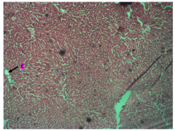
The result from the high dose of Musa Paradiasical extract shows central vein (C) with many surrounding hepatocytes with sinusoids. And the tissue appears normal. (Plate 3).

Plate 1: Photomicrograph of the liver in the control group showing a portal triad (P) and central vein (C). There are many surrounding hepatocytes with sinusoids. Tissue appears normal. H & E. X200.

Plate 2: Photomicrograph of the Liver in a Low Dose group with many surrounding Hepatocytes with sinusoids. Parenchyma shows Pseudorosettes tissue. Stain with H & E. X200.

Plate 3: Photomicrograph of the liver in high dosage administration of Musa Parasidiaca with the Tissue appears normal. H & E. X200HD.
Effect of Aqueous Extract of Musa Paradisiaca on the Serum Liver enzymes
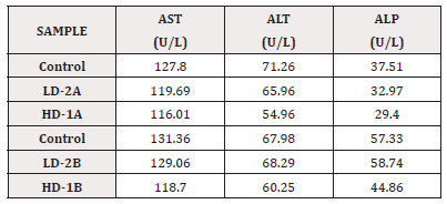
Table 2 Shows the serum and enzymes results.
Discussion
Since ancient times, people have attempted to treat a variety of illnesses and alleviate physical pain by using medicinal herbs. Everyone with whom they were acquainted knew something about the uses of therapeutic herbs [16] (Nazim, et al., 2017). The plants are multifunctional and a rich source of therapeutic compounds that can be exploited to create human-use drugs. Curiously, 80% of people worldwide, particularly in developing nations, still rely on plant-based medicines for disease prevention and treatment (World Health Organization, 2010) [21]. Various studies have shown that Musa paradisiacal has a variety of beneficial effects in a number of disease conditions, including diabetes mellitus, hypertension, hyperlipidemia, thyroid dysfunction, and body weight (Mallick, et al., 2016, Parmar & Kar, 2017).
Musa paradisiaca has a variety of nutrients, including minerals and bioactive compounds, which are why almost all of the plant's parts have been utilized to treat a variety of illnesses. For instance, an aqueous extract of fermented unripe Musa paradisiacal fruits and peels has anti-microbial [12] (Kapadia, et al., 2015) and anti-ulcer genic properties (Ezekwesili, et al., 2014) [10]. Research has also demonstrated that M. paradisiaca fruit pulp aqueous extract has wound-healing and antioxidant qualities [2] (Shodehinde and Oboh, 2018). It may also be used as a treatment for hepatic dysfunction and diabetes [6,17].
Discussion on the Morphological Observation
According to the morphological investigation, there is a noticeable and substantial rise (p<0.05) in the final body weight compared to the initial body weight in the cases of control vs low dose, high dosage versus low dose, and high dose versus control. The animal's final weight was a considerable increase above its starting weight. On the other hand, the low dosage group and high dose group had mean final body weights that were considerably higher than their initial body weights (p<0.05), respectively. As a result, the reason for the increase in body weight could be attributed to its moisturizing and diuretic qualities, while unripe Musa paradisiaca peels and fruits have been shown to have appetite-boosting [8] and anti-hepatic, anticancer, and fungicidal qualities [5].
This is consistent with the study conducted by Patrick, et al. (2020), which found that, in comparison to the diabetic control group, the Musa paradisiaca diabetic treated rats showed a significant increase in body weight and HDL- cholesterol (p<0.05) but a significant decrease in blood glucose (p<0.05), serum total cholesterol, triglyceride, LDL- cholesterol, and VLDL cholesterol concentrations.
Discussion on the Histological Studies
The liver's typical cellular and tissue architecture was demonstrated by the study's findings. The liver is made up of parenchymal cells, the portal triad (P), the central vein (C), and numerous other non-parenchymal cell types, all of which are important. In addition to being the target of autoimmune liver disorders, biliary epithelial cells also play a crucial role in the coordination of many immune cells involved in both innate and acquired immunity (Plate 1). Our current study's outcome is consistent with the conclusions of [9].
The study's histology findings for the control animals display a central vein (C) and portal triad (P) with many hepatocytes with sinusoids surrounding them. As can be seen in (Plate 1), the tissue looks normal. However, in the low dose group, the central vein (C) was visible, along with many surrounding hepatocytes with sinusoids, and the parenchyma displayed pseudorosettes tissue, as can be seen in (Plate 2) above. These results are consistent with research by Olagunju, et al. (2020) [18], which found that giving plantain weed extract reduced several markers of inflammation brought on by liver injury. On the other hand, the results of administering a high dose of aqueous extract from Musa Paradiasica reveal a central vein (C) surrounded by many hepatocytes with sinusoids. Additionally, as Plate 3 illustrates, the tissue looks normal. This finding is consistent with Redoy, et al.'s [18] research from 2021, which found that plantain weed extract significantly reduced inflammation and liver enzymes to prevent liver damage.
Our current study's results are consistent with those of Agrawal, et al. (2018), who also looked into the hepatoprotective properties of Musa paradisiaca stem in rats treated with CCl4 and paracetamol-induced hepatotoxicity models. In the experimental animals, the administration of hepatotoxins (CCl4 and paracetamol) resulted in significant biochemical and histological deteriorations in the liver. However, pretreatment with alcoholic extract (500 mg/kg) significantly reduced the elevated levels of serum enzymes such as serum glutamic-oxaloacetic transaminase (SGOT), serum glutamic pyruvic transaminase (SGPT), alkaline phosphatase (ALP), and bilirubin levels. Furthermore, the alcoholic and aqueous extracts reversed the hepatic damage towards the normal level, providing further evidence of the hepatoprotective activity of Musa paradisiaca stem. On the liver of CCl4 and paracetamol-induced hepatotoxicity animal models, the alcoholic extract (250 and 500 mg/kg, p.o.) and the aqueous extract (500 mg/kg, p.o.) of the stem of Musa paradisiaca produce notable effects.
The veins at the core of each lobule in the liver are known as the central venules, or the liver's central veins. They take in blood that has been mingled in the liver sinusoids and put it back into the hepatic veins. This is in line with a study by Logan, et al. (2007), which used masson lichun red acid reddish staining (Masson staining), hematoxylin and eosin staining (H&E), and periodic acid and Schiff staining (PAS) to investigate structural alterations of the liver. The model mice showed focal necrosis, mussy hepatic cords, and fibrosis in the H&E staining, while the T2DM mice showed significant hepatocellular damage in the masson staining.
Discussion on the effect of aqueous extract of Musa Paradisiaca on the serum electrolyte and Liver enzymes
When group A (control=122.24) was compared to group B (LD-2A=127.18), there was a significant difference that increased. Comparing group A, there was a similarly substantial rise (LD-2A=127.18) and (HD-1A=131.81), respectively. In contrast, group B has greater values for low dose sodium (LD-2B=137.11) compared to control (=125.51) and HD-1B (128.90).
Table 2 illustrates the considerable increase in the relationship between electrolyte abnormalities and hepatocellular damage in low dosage A & B group (LD-2A=127.18 mmol/L and LD-2B=137.11 mmol/L) when blood Na+ concentration was low (hyponatraemia). The deficiency in Na concentration decreased at a value of less than 130 mmol/L (asymptotic characteristic), despite an observable continuous increase in group A high dose (HD-1A=131.81 mmol/L) and a significant decrease in group B high dose (HD-1B= 128.90 mmol/L) [19]. This is related to the loss in arterial blood volume effectiveness [4] and the predominance of antidiuretic hormone secretion caused by impaired renal solute-free water perfusion [11]. Most cirrhotic patients develop ascites, which is explained by the fluid route changing into splanchnic circulation [13].
Additionally, Table 2 shows a non-significant decrease in K+ serum concentration when compared with A group high dose (HD–1A=1.54 mmol/L) and a significant rise in K+ serum concentration in the A group when comparing the low dose (LD-2A=1.56 mmol/L) with the control group (control=1.42 mmol/L) (P value < 0.05). The majority of liver disease patients experience abnormal K+ levels; this is consistent with the findings of [3], which show that a similar event occurs during treatment by medication intake or extracellular shift, impairing glomerular filtration due to kidney injury in ESLD and leading to hyperalkalism.
Additionally, there was a discernible rise in Cl and AST in groups A and B as compared to the control group and the high dose group. While there was a decrease in group B's high dose ALT and ALP when compared to high dose (HD-2B) with low dose (LD-1B), there was also an increase in group A's ALT and ALP when compared to control to high dose.
Conclusion
Herbs and medicinal plants serve an important role in maintaining general health, they also have therapeutic effects as well as antioxidant agent. These plants even though proven beneficial for health can be life threatening if not taken with caution. The flavonoid constituent of Musa Paradiasica shows substantial amount of antioxidants properties, which indicates their ability to fight cancerous cells, also the dietary fiber has been reported to have beneficial effects on conditions related to liver disease and liver cancer, including blood glucose [22], insulin sensitivity [7], liver fat content [14], and metabolic syndrome.
Electrolytes system disturbance is very common and associated with hepatocellular injury, decreasing in sodium concentration (hyponatremia) especially in cirrhotic patient and increasing in chloride concentration (hyperchloraemia) in patients who have end stage of liver disease. Liver enzymes are considered as an indicator to monitor the condition of the liver that increasing in GOT and GPT pointing to hepatocellular injury.
Acknoledgments
The authors wish to acknowledge the huge contributions of the staff of the Department of Anatomy and Forensic Anthropology, Faculty of Basic Medical Sciences, University of Cross River State, Nigeria towards this research work
Disclosure
We the authors report no conflict of interest.
References
- Adeolu A T, Enesi D O (2019) Assessment of proximate, mineral, vitamin and phytochemical compositions of plantain (Musa paradisiaca) bract-an agricultural waste. International Research Journal of plant science 4(7): 192-197.
- Agrawal A, Sharma M, Rai S K, Singh B, Tiwari M, et al. (2018) The effect of the aqueous extract of the roots of Asparagus racemosus on hepatocarcinogenesis initiated by diethylnitrosamine. Phytother Res 22(9): 1175-1182.
- Ahya S N, Soler M J, Levitsky J, Batlle D (2006) Acid-base and potassium disorders in liver disease. Semin Nephrol 26(6): 466-470.
- Asrani S K, Kamath P S (2013) Predictors of outcomes in patients with ascites, hyponatremia, and renal failure. Predictors of outcomes in patients with ascites, hyponatremia, and renal failure. Clinical Liver Disease 2(3): 132-135.
- Durazzo A, Denoeud F, Aury JM (2018) The banana (Musa acuminata) genome and the evolution of monocotyledonous plants. Nature 488: 213-217.
- Eleazu C O, Eleazu K C, Awa E, Chukwuma S C (2018) Comparative study of the phytochemical composition of the leaves of five Nigerian medicinal plants. Journal of Biotechnology and Pharmaceutical Research 3(2): 42-46.
- Hernández Aquino E, Muriel P (2018) Beneficial effects of naringenin in liver diseases: Molecular mechanisms. World J gastroenterol 24(16): 1679-1707.
- Islam M Z, Akter S (2019) Musa paradisiaca L. and Musa sapientum L.: A phytochemical and pharmacological review. Journal of Applied Pharmaceutical Science 1(5): 14-20.
- Ishibashi H, Nakamura M, Komori A, Migita K, Shimoda S (2019) Liver architecture, cell function, and disease. Semin Immunopathol 31(3): 399-409.
- Ikpeazu A O (2017) Chemical composition of unripe (green) and ripe plantain (Musa paradisiaca). J Sci Food Agric 24(6): 703-707.
- Kim J H, Lee J S, Lee S H, Bae W K, Kim N H, et al. (2009) The association between the serum sodium level and the severity of complications in liver cirrhosis. Korean J Intern Med 24(2): 106-112.
- Kapadia KPS, Bhowmik D, Duraivel S, Umadevi M (2015) Traditional and Medicinal Uses of Banana. Journal Pharmacology Phytochemistry 1: 51-63.
- Laragh J H, Ames R P (1963) Physiology of body water and electrolytes in hepatic disease. Medical Clinics of North America 47(3): 587-606.
- Liu X, Yang W, Petrick J L, Liao L M, Wang W, et al. (2021) Higher intake of whole grains and dietary fiber are associated with lower risk of liver cancer and chronic liver disease mortality. Nat Commun 12(1): 1-9.
- Nkendah S T, Rahmana M W, Sahaa R, Hasana M M, Deba A (2020) Jute stick powder as a potential low-cost adsorbent to uptake methylene blue from dye enriched wastewater. Desalin Water Treat 153: 279-287.
- Nazim M, Girija K, Lakshman K, Divya T (2017) Hepatoprotective activity of Musa paradisiaca on experimental animal models. Asian Pac J Trop Biomed 2(1): 11-15.
- Ojewole H A, Adewunmi O (2018) Anti-inflammatory medicinal plants: The effect of three species on serum antioxidant system and lipids levels in rats. NISEB Journal 11(2).
- Olagunju A I, Oluwajuyitan T D, Oyeleye S I (2020) Effect of plantain bulb’s extract-beverage blend on blood glucose levels, antioxidant status, and carbohydrate hydrolysing enzymes in streptozotocin-induced diabetic rats. Prev Nutr Food Sci 25(4): 362.
- Oldridge J, Karmarkar S (2015) Fluid and electrolyte problems in renal dysfunction. Anaesthesia & Intensive Care Medicine 16(6): 262-266.
- Redoy M R A, Rahman M A, Atikuzzaman M, Shuvo A A S, Hossain E, et al. (2021) Dose titration of plantain herb (Plantago lanceolata L.) supplementation on growth performance, serum antioxidants status, liver enzymatic activity and meat quality in broiler chickens. Italian Journal of Animal Science 20(1): 1244-1255.
- World Health Organization (2021) Increased resistance of SARS-CoV-2 variant P. 1 to antibody neutralization. Cell host & microbe 29(5): 747-751.
- Zelber Sagi S, Salomone F, Mlynarsky L (2017) The Mediterranean dietary pattern as the diet of choice for non‐alcoholic fatty liver disease: evidence and plausible mechanisms. Liver Int 37(7): 936-949.


 We use cookies to ensure you get the best experience on our website.