Research Article 
![]() Creative Commons, CC-BY
Creative Commons, CC-BY
Magnetic Resonance Therapy (MRT) and Relativity
*Corresponding author: Yin Rui, BeiHang University, China.
Received: November 08, 2024; Published: November 15, 2024
DOI: 10.34297/AJBSR.2024.24.003246
Abstract
Epoxide within molecular structures is recognized as hallmarks of potent carcinogenic cells. This paper proposes a novel approach, Magnetic Resonance Therapy (MRT), which selectively eliminates epoxide in cancerous tissues while preserving oxygen in healthy tissues, offering a potentially side-effect-free method for cancer treatment and prevention. The study presents a series of in vitro, in vivo, and clinical experiments to support this innovative therapeutic strategy. The underlying mechanism of MRT is grounded in the principles of relativity for rotational frames, and an experimental validation of this theoretical framework is provided. Furthermore, this research unveils previously undescribed relativistic phenomena, including space-time exchange and critical cylinder effects. Due to space constraints, the detailed mechanism of epoxide elimination will be elucidated in a subsequent publication. This groundbreaking approach to cancer therapy holds promise for significant advancements in oncological interventions and warrants further investigation.
Keywords: Cancer, Epoxide, Magnetic resonance therapy, Relativity for rotational frames
Part 1. The Practice of MRT Treating Cancer
It is well known that lots of diseases are caused by oxidation of cell molecules. As pointed by James D. Watson in his book “Molecular Biology of the Gene” [1]: “the procarcinogens become into powerful carcinogens, as the epoxide appears in their molecules.” An efficient and non-hazardous way to clean epoxide is proposed in this paper. We call it Magnetic Resonance Therapy (MRT). No medicine, no radioactivity, no scalpel, the MRT uses weak magnetic field (less than 10Gs) to clean epoxide. Its equipment consists of a signal generator (the plastic box in Figure 1) and a group of coils with air core (the white cylinder in Figure 1). The signal generator produces currents, those flow through the coils to generate both main and RF magnetic fields acting on the lesion. The coils are positioned 20cm away from the body surface, since the resonance happens beyond that place until 60cm. The treatment starts when the signal generator is turned on. The standard dosage is three times in one-hour per day. The device is very light (the total weight is 2Kgs only), easy-managed, so that it can be used not only in hospitals but also at home. And since it is no-hazard but benefit for body, it can be used to prevent cancer. Every year taking MRT for a month, even in sleep time, can keep the cancer away all life (Figure 1).
The Killed Process of Cancer Cells By MRT
Giving cultured squamous cancer cells MRT and observing the killed process of cells, we find that as MRT has been given for 5 minutes, lots of expanding bubbles bulge on the membrane of cells; as MRT has been given for 10 minutes, all bubbles expand to break, cytoplasm overflow, perforations appear at the place covered by bubbles before. The cell is dead currently. Continuing MRT, perforations become bigger and bigger, the cell is broken. Five batches of 100 rats were taken to experiments, following are the results of 2 batches.
Cell Experiment in Vivo
Five days later after inoculating the Ehrlich (liver ascites cancer) line into the abdomen of 20 mice (BALB/c), every mouse grew up ascites including lots of Ehrlich cancer cells. Take 10 of them as experimental group and give them 8 days MRT (90minutes/day). The microscope photo-pictures are shown as Figure 2(A), where (a) is the cancer cells in control mice, every cell is complete. (b) are the cells in experimental mice, perforations, overflowing cytoplasm, pieces of broken cells and dissolving into ascites can be observed (Figure 2).

Figure 1: Equipment and Usage.

Figure 2: (A)Cell experiment in Vivo (B) Animal experiment MRT treated Lewis’s lung cancer in black mice with 100% cure rate.
MRT Treated Lewis Lung Cancer in Black Mice With 100% Cure Rate
Four days after inoculating Lewis’s lung cancer line, eleven mice of C57BL/6J all grew cancer blocks with the size of about 3mm. Three of them were taken as the control group, four of them were taken as the experimental group with small dose (10 minutes/day), and the other four mice were taken as big dose group (70 minutes/ day). One week later we ended MRT, the cancer blocks in control mice all grew to the size of about 2cm, but the cancer blocks in six experimental mice (four of big dose group and two of small dose group) disappeared. However, there was a tuber in the size of about 1cm for two mice of small dose group. We dissected four mice. The cancer block in control mouse is shown as Figure 2(B)(a) (the left one), the middle one is the tuber in the mouse taken small dose. The picture of dissected mouse without cancer block is shown in Figure 2(B)(b).
Two weeks later the other two mice of control group dead. However, one month later the tuber in the left mouse of the small dose group naturally disappeared. Nine months later the left five mice were all alive with very good health, and we ended this experiment. Such cure rate (100%) was scarce for animal experiment of cancer.
Clinical Experiments
More than 50 volunteers have taken MRT, Follows are some cases.
Case 1: A 64-year-old man suffered from lung squamous cancer. The x-ray photo taken on 14.3.1994 is shown as Figure 3(a), where the white elliptic part in upper left lung is the original cancer block. Two weeks Later the metastasized cancer blocked the bronchus, air couldn’t get into left lung, the left lung atelectasis (so-called white lung) as shown in Figure 3(b). Before MRT, the patient could neither sit nor lay, four or five times of shock happened every day. We gave him a one-week MRT. On the second MRT day he could sit, after the third MRT day he could walk in the garden of the hospital. The x-ray photo taken on the last MRT day is shown as Figure 3(c). His left lung could expanse again. 40 days later another x-ray photo was taken and is shown in Fig.2 (d). The situation was much better (Figure 3).

Figure 3: MRT treated lung cancer.
Case 2: A fifteen-year-old boy suffered from a finger tumor as shown in Figure 4(a). Two months later after taking MRT three days (3×1hour/day), a piece of hard crust shell was taken off the tumor as shown in Figure 4(b). After taking another three days MRT, the tumor constricted month by month as shown in Figure 4(c)(d)(e) (Figure 4).

Figure 4: MRT treats finger tumor.
Case 3: The bile duct of an old man was obstructed by cancer block. Jaundice appeared on his face, hands and body as shown in Figure 5(a). He had no appetite and had a dregs-like stool. He tokes the MRT home. As the MRT had been given for three hours, a boundary line of jaundice appeared on his forehead, above this line the jaundice disappeared (Figure 5).
As the MRT was continuing, the boundary line was going down and down. As the MRT had been given for six hours, the jaundice on his face and hands disappeared. As the MRT had been given for nine hours, the old man said: ‘I am so hungry’. We ended that day’s MRT and gave him a bowl of gruel. Gobbling up the gruel, he said: ‘I am hungry too’. His daughter smiled and asked: ‘can he eat so much?’ We said: ‘doesn’t matter, give him dinner please’. On the second day his stool became normal. Nevertheless, we gave him another two days MRT again, the picture taken on the third MRT day is shown in Figure 5(b).

Figure 5: MRT treated bile duct cancer.
Above cases show that one or two weeks MRT can cure the cancer with small size. But the big size cancer should take MRT for long time. In this case, MRT can be taken at home and self-service by patients without the help of doctor. The following are some cases (Figure 6).

Figure 6: MRT treated lower jawbone cancer.
Case 4: A 78-year-old woman suffered from lower jawbone cancer, and metastasized. She couldn’t eat or sleep. She tokes the MRT home. The dose was 4×40 minutes/day. Since taking MRT, she could go to sleep every night, because the pain relaxed. Figure 6 (a) and (b) are the pictures taken before MRT, Figure 6 (c) and (d) are those taken after one-month MRT. Wemparing them, we see that the cheek swollen disappeared, large area of cancer-ulcer with liquid oozed on chin healed up.
Case 5: MRT treated breast small cell cancer: At the beginning the size of cancer block was 14cm×12cm as shown in (Figure 7). After one-month MRT (3×1hour/day), the mass in the upper left region disappeared, while the middle portion was charred and necrotic with a cavity being formed as shown in (Figure 7). After one-year MRT, the cancer tissue carbonized and necrotized into cavities as shown in Figure 7. But the skin around the cancer was without any change, although it suffered one-year MRT as well. This means the MRT can only kill cancer and no affect to normal tissue (Figure 7).

Figure 7: MRT treated breast small cell cancer.
Case 6: An 86-year-old man has suffered from prostate cancer for several years. His PSA was 100↑ (below 4 normal) on 25.5.2020. The PET-CT photo taken on 2020.6.5 is shown as Figure (8), where every black point was the cancer bone metastasis. Since 2020.7.19 he had taken MRT at home (3×1hour/day). After 5 months MRT the PSA was decreased to 2.1, then stop MRT. However, the PSA still decreased, two months later decreased to 1.1, then increased: five months later increased to 1.89. Then started MRT again, one month later PSA decreased to 0.431. The PET-CT photo taken in September of 2021 is shown in (Figure 8). In terms of this photo we know that 80% of bone metastasis disappeared (Figure 8).

Figure 8: MRT treated prostate cancer.
Prevention And Early-Treatment of Cancer
Case 7: A 43-year-old female teacher was tested squamous cancer cells in white deposits on her tongue in 1994. The doctor suggested her to cut part of tongue. She was a teacher; without a complete tongue how could she give lecture? She asked us for help. We gave her one-week MRT (90 minutes/day) at the very beginning, then 3 months MRT (60 minutes every week). Now, 30 years is over, she fulfilled work and retired with wonderful health (Figure 9).
Tumor markers in the blood have exceeded the standard before cancer forms solid tumors. This is the best time for early treatment. MRT is a simple device and easy to operate and has no side effect, so it can be used as “pre-cancer” treatment at home. Following is an example:
Case 8: A 79-year-old man in physical examination on 2014.12.29 was found that his Squamous Cell Carcinoma (SCC) cell antigen was 12.9 (below 1.5 normal). The doctor in no way helped him, only allowed him to take another test after 3 months. He asked us for help and toke 20 days MRT at home from 2015.1.10 to 2015.1.30. After that 3 examinations were performed on 2015.1.30: 2015.3.13. and 2015.4.10. The results were: 8.1;4.1 and 0.8. Then in 2016,17,18,19 every year he tokes one-month MRT. Till now, ten years are over, his SCC has always been in the normal region.

Figure 9: MRT treated cerebral hemorrhage.
The effect of MRT is clean epoxide, which is not only harmless, but beneficial to the body. The MRT can be used not only to treat cancer but also to prevent cancer. Put the device on the bed and turn on it before going to bed and turn it off after waking up. Doing it like this one month per year can
keep cancer away all life. On the other hand, it can be used to anti-oxidation, since it can clean epoxide. Following is an example:
Case 9: 67 years old women suffered from cerebral hemorrhage, after emergency operation, she kept life but left wet brain as shown in (Figure 9), so that she couldn’t walk, eat and speak. We gave her three months MRT (3×40 minutes/day). After one-month MRT, she could eat meal with chopsticks, go to bathroom freely, and talk fluently. However, there was blood in her brain as shown in (Figure 9). After three months MRT the blood in her brain disappeared. The place where the blood occupied before became empty as shown in (Figure 9). No need for more cases, the efficiency of MRT has been exhibited. Now let us turn to the mechanism of MRT, that is based on Relativistic Effect of Rotation (RER), which was proposed by us in references [2-4]. Since lots of natural laws revealed by RER are unknown for modern physics, we have to give an experiment to prove it first [5].
Part 2. The Physical Foundation of MRT
Experimental observation of Relativistic Effect of Rotation
It is well known that the tangential velocity v , angular velocity ω , and the radial distance r have the relation of v =rω . Therefore, any rotation has a corresponding radial distance rc, where v reaches the speed of light c:rc=c/ω. We call c r critical radius. All points with their radial distance form a cylindrical surface called critical cylinder. The scientists believe that the tangential velocity Outside Critical Cylinder (OCC) will surpass the speed of light, but superluminal is impossible, so they give-up research it. However, our experiments demonstrate that there are observable physical phenomena in OCC, which are opposite to those Inside Critical Cylinder (ICC). Now let us show the experiments.
The Experiment Setup and Result
A helical electrode is connected to the negative output terminal of a DC High Voltage Generator (HVG). When the HVG is turned on, an abundance of free electrons will accumulate on the electrode. The electrode is affixed to a resin plate, which is secured in a plastic dish. The two ends of two U-type ferrite cores clamp the electrode, resin plate and the plastic dish together, while the other two ends are inserted into the exciting coils, as shown in (Figure 10).
A Square Wave Current Generator (SWCG) feeds the coils, producing a magnetic field that is transferred to the electrode via two ferrite cores, causing the free electrons on the electrode to precession. The angular velocity of precession is given by Larmor equation: ω =γB. for electron, its gyromagnetic ratio γ =2.6667×10¹⁰ (Hz/Tesla), as B= 0.12 Tesla, the precession angular velocity is ω =2.01×10¹⁰ (r/s), the corresponding critical radius rcis 1.492 cm, approximately 1.5 cm. A 20% carbon ink containing a high concentration of anionic surfactants (negatively charged ions) was utilized as the test charge. To begin, warm water is added to the plastic dish, and a small quantity of ink is injected 1 to 1.3 cm to the right of the electrode. The HVG is then turned on to recharge electrons on the electrode. The ink is repelled by the electrons, showing a distinct rightward shift as illustrated in the 4 sequential screenshots from the video presented in Figure 11(b). Once the ink’s right edge is repelled to the black mark indicating the resin board’s center, the SWCG is activated to induce precession of the electrons. Subsequently, the ink on the left side of 1.5cm continues being repelled rightward, whereas the ink in right side of 1.5cm begins being attracted leftward by the precession electrons. Figure 11(c) sequentially presents 6 screenshots from the experimental video depicting this stage of the precession (Figure 11).

Figure 10: The experimental device.

Figure 11: Ten screenshots of an experimental video.
This experimental result demonstrates following conclusions
a) The electric force of precession electrons exists in OCC but has the opposite direction to that in ICC. This is a relativistic effect of rotation; we call it the Critical Cylinder Effect (CCE). CCE reveals the incompleteness of Coulomb’s law and Newton’s gravitational law.
b) The observed critical radius indicates the precession of electrons possesses definite axis and constant angular velocity, so it is real rotation. Since only a rotating object can create precession when an external torque is applied, the spin of electron must be real rotation, contrary to claims of “not being a real rotation.” in quantum spin theory.
c) Furthermore, quantum field theory believes that the electric force is transferred by virtual photons, which means that electrons emit virtual photons and hit the negative ions, causing the momentum change of the negative ions; the rate of momentum change is the force on the negative ions. However, this fails to explain why photon exchanges would exert opposing forces on either side of the critical radius. Electrical force must interact with a medium rotating in sync, i.e. its electric field but not virtual photons. In fact, the strong weak force, and gravity are the relativistic effect of electromagnetic force, that we will explain in another paper.
d) There is field force in OCC which means the tangential velocity of electric field of the processing electron does not exceed the speed of light. In OCC, the relation between v and of fixed axis rotation is no longer v = rω , but v = c2 / (rω), which has not yet been known for modern physics. Reference (4) has seriously proved it. Now let us give the simple theoretic proof.
Space-Time Exchange and Tangential Velocity of a Rotation
Suppose frame A’ is rotating with a constant angular velocity ω about a fixed axis z (z’) relative to frame A (Figure 12). It is well known that substituting the local reference frames of event point P for inertial frames, i.e. substituting (ds,dp,dz,dt) for (x,y,z,t)and ρω for ν into the (inverse) Lorentz transformation of inertial frames (1), the (inverse) Lorentz transformation for rotational frames can be got for the area of Inside Critical Cylinder (ICC) as shown in (2):
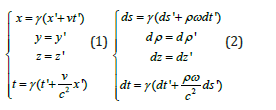

Figure 12: (s, z, ρ, ICT) coordinate system.
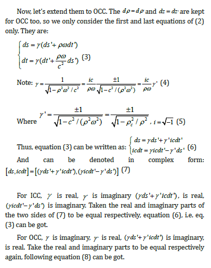
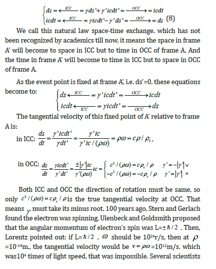
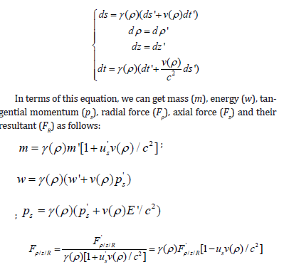
At OCC γ(ρ) taken minus root, so the mass, energy, tangential momentum and force have opposite sign against those at ICC. We call it Critical Cylinder Effect (CCE). The experiment given above proves the radial force in OCC is opposite against that in ICC. That means the relativistic effects of rotatopn, proposed by us, is correct.
Conclusion
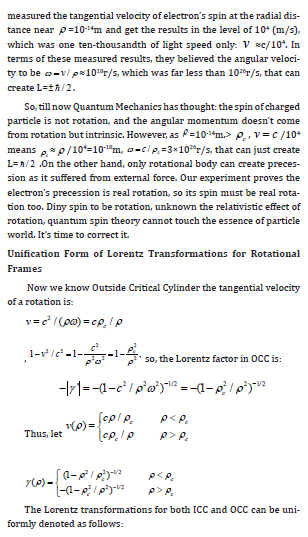
Cancer and cardio- and brain-vascular diseases are the top two killers of mankind. MRT can not only treat but also prevent them. Besides, MRT can treat lots of other diseases caused by oxidation. Once it is popularized into every family, the level of health will be increased. The RRF reveals lots of natural laws, which are unknown for modern physics, such as space time exchange, the tangential velocity for OCC is c2 / (ρω), the critical cylindrical effects, which indicate the force will change direction, as the acceptor is changed from ICC to OCC, and is verified by experiment given in this paper. Only based on RRF, the essence of particle world can be revealed, we will give them in another paper.

Figure 13: Video
Acknowledgement
None.
Conflict of Interest
None.
References
- James D Watson (1977) Molecular Biology of the Gene. Wa Benjamin Inc.
- Yin Rui (1997) Rotating Lorentz Transformation and Unification of Forces. BUAA Press.
- Yin Rui (2000) Unification of Forces According to Relativity for Rotations. Hadronic Journal 23(5): 487-549.
- C Møler (1972) The Theory of Relativity. Clarendon Press Oxford.
- Yin Rui, Yin Ming, Wang Yang (2024) Critical cylindrical Effect and space-time exchange in rotational reference frames of special relativity. World Academy of Science Engineering and technology Physical and Mathematical Sciences 18(10): 110.


 We use cookies to ensure you get the best experience on our website.