Research Article 
 Creative Commons, CC-BY
In Vitro Studies of Skin Burn Infections Using Casuarina Equisetifolia L. Fabricated Silver Nanoparticles
*Corresponding author: Shubhangi Moharekar, Savitribai Phule Pune University, Pune and Department of Biotechnology, New Arts, Commerce and Science College, Ahmednagar, India.
Received: January 11, 2025; Published: January 21, 2025
DOI: 10.34297/AJBSR.2025.25.003334
Abstract
In present work, Silver Nanoparticles (AgNPS) were synthesized by biological as well as chemical method. Biosynthesis of silver nanoparticles was done using dried and burned fruit powder of Casuarina equisetifolia. The characteristics of synthesized nanoparticles were confirmed by UVVisible spectroscopy, X- Ray Diffraction (XRD) and Scanning Electron Microscopy (SEM). The size of biologically synthesized nanoparticle was comparatively smaller than chemically synthesized one. The biologically synthesized AgNPs showed remarkable antibacterial activity against five pathogenic bacteria isolated from burn patient’s skin viz. Pseudomonas aeruginosa, Staphylococcus aureus, Escherichia coli, Acinetobacter sp. and Klebsiella pneumoniae. Based on these findings, it was concluded that the present study provides potential of biologically synthesized AgNPs as an effective antibacterial agent against skin pathogens and hence can be used for medical application to prevent skin burn infections. These results clearly indicate that AgNPS could provide alternative topical therapy in treatment of skin burn infection.
Keywords: Antibacterial agent, Casuarina equisetifolia, Silver nanoparticle, Skin Burn Infections
Introduction
Nanotechnology is the study of nano sized materials dealing with their synthesis and wide range applications. Various properties of a material depend on its size and shape, as size of material reduces to nano-scale its properties do change. Noble metal nanoparticles such as gold, silver, platinum and palladium have been most effectively studied [1-3]. These nanoparticles can be synthesized using physical, chemical and biological methods. Chemically synthesized nanoparticles are not favourable for biomedical application due to its toxicity and flammable nature of starting chemical compounds. The biological approach to synthesize metal nanoparticles is of great scientific interest and current need of nanotechnology research. Use of biological materials such as microorganisms, plant extract or biomass for nanoparticle synthesis is an eco-friendly and economic alternative to chemical and physical methods [4- 6]. The presence of diverse phytochemical constituents present in plant material may play role in biosynthesis of metal nanoparticles. In recent years, several plants have been reported for synthesis of silver, copper and gold nanoparticles such as extracts of neem, aloe vera, wheat, lemongrass, tamarind, ginger and garlic [7-12].
The emerging infections in burn patient represent a serious therapeutic challenge because bacteria leukocidin and tissue-destroying enzymes which leads to extensive skin loss and heavy contamination (Kang 2016). As a result, wound healing process get altered or delayed. Therefore, topical antimicrobial therapy to control microbial colonization and proliferation of microbial pathogens including multidrug- resistant organisms is most important to take care of burn wound. It has been estimated that in developing countries about 75% of the mortality associated with burn injuries is related to sepsis [13]. The reason behind this phenomenon is the increasing resistance to antibiotics in infectious bacteria due to its overuse; therefore, there is a need for an alternative therapy. Nanoparticles can serve as potent alternative to the antibiotics, because nanoparticles have antibacterial and antifungal activity [14]. Casuarina equisetifolia is traditionally used as a medicinal plant [15,16], uses like anti-inflammatory, antibacterial, anti-diabetic, hepatoprotective activity, antioxidant, antispasmatic etc [17]. The leaf of C. equisetifolia are used as folk medicine for treating nervous disorder, diarrhea, stmoch-ache, cough, ulcers, diabetes etc [8-10]. The fruit if C. equisetifolia is woody and oval structure made up of numerous carpuls, each with single seed and small wing. The fruits of C. equisetifolia plant were used in the given study because tribal people used it to treat skin burns because it is well known that plants- derived NPs are less harmful side effects than that of chemically synthesized NP [18]. To my knowledge for the first time, the present work reports green synthesis of AgNPs from medicinally valuable plant C. equisetifolia fruit extract as a reducing agent. In the present work, we investigated the biosynthesis of AgNPs using fruit of Casuarina equisetifolia. The properties of obtained AgNPs were characterized by UltraViolet-Visible (UV-Vis) spectroscopy, Scanning Electron Microscopy (SEM) and X-Ray Diffraction (XRD) techniques. This work provided a potent application of AgNPs to serve as an alternative to antibiotics against skin burn pathogens.
Materials And Methods
Materials
Fruits of Casuarina equisetifolia were collected from campus of NACAS College, Ahmednagar. Dried fruit powder and burned fruit powder of above plant were prepared and further used for stock preparation. Analytical grade silver nitrate (AgNO3) was purchased from Merck chemicals. Autoclaved distilled water was used for preparation of 100mM AgNO3 stock solution.
Preparation of Plant Extract
Collected dried fruits were washed to remove dust particle. Fine powder of both dried fruits as well as burned fruit, was prepared. 20 g of fine powder was suspended in 100 ml sterile distilled water, boiled and filtered through Whatman No.1 filter paper and stored at 4°C to be used for further experiments.
Chemical Synthesis of Silver Nanoparticles
The solution of 1mM aqueous AgNO3 was heated up to boiling. To this solution 5m of trisodium citrate (stabilizing agent) was added drop by drop and mixed vigorously. It was heated up to 2 hours and then cooled at room temperature. It was then centrifuged and pellet (nanoparticles) was dried at 72°C overnight.
Biological Synthesis of Silver Nanoparticles
For biological synthesis, prepared dried and burned fruit extract (10ml) was mixed with 90 ml of 1mM aqueous AgNO3 separately. Further it was kept at 72°C in water bath. The silver nanoparticle synthesis was identified by the appearance of dark brown color from yellow colored solution.
Characterization of Silver Nanoparticles
a. Ultraviolet-Visible Spectral Analysis
UltraViolet-Visible spectral analysis of reaction mixture was done by using UV-Vis double beam spectrophotometer of Systronics Ltd. within the range of 200-800 nm. Distilled water was used to adjust the baseline.
b. X-Ray Diffraction Analysis
Silver nanoparticle solution obtained was purified by repeated centrifugation at 10,000 rpm for 20 min followed by re-dispersion of the pellet into 10ml of sterile distilled water. After drying of purified silver nanoparticles, the structure and composition were analyzed by XRD. The XRD spectra was recorded using X-ray diffractometer (Bruker GXS D-8) operated at voltage of 40 kV and a current of 30 mA with CuKα radiation by using 2θ from 10-80°. The crystallite domain size was calculated from the width of the XRD peaks using the Scherrer formula [19].

Where, D is the average crystallite domain size perpendicular to the reflecting planes, λ is the X-ray wavelength, β is the Full Width at Half Maximum (FWHM), and θ is the diffraction angle.
c. Topographical Analysis by SEM
Scanning Electron Microscopic (SEM) analysis was done using Hitachi S-4500 SEM machine. Film of the sample were prepared on a carbon coated copper grid by dropping a very small amount of sample on the grid, extra solution was removed using a blotting paper and then the film on the SEM grid were allowed to dry by putting it under a mercury lamp for 5 min.
Isolation of Infectious Microorganisms from Burn Skin
The freshly discarded burned patient swab was collected from biomedical waste of Civil Hospital. Ahmednagar. Immediately swab was brought to the lab and Swab culture technique was used to isolate infectious microbes. Swabs were inoculated on Blood agar and MacConkey’s Agar plate and further incubated at 37°C for 24 hr. For further identification, isolated colonies were grown on selective media like Cetrimide agar, Mannitol Salt agar, Worfel-Ferguson agar, Leed’s Acinetobacter medium etc. The purity and identification of each isolate was confirmed in laboratory based on morphological and biochemical characterization according to Bergey’s manual of determinative bacteriology, 9th edition.
Antibacterial Activity of Silver Nanoparticle
Sterilized Mueller Hinton Agar (MHA) plates were prepared for antibacterial activity determination. Plates were then spreaded with isolated bacterial cultures (P. aeruginosa, S. aureus, E. coli, Acinetobacter sp. and K. pneumoniae). Sterilized 6 mm cork borer was used to prepare agar wells. Aliquote of 100 μl of chemically and biologically synthesized AgNPs were placed into the wells followed by 10 min incubation in refrigerator. The plates were incubated at 37°C for 24 hrs and zone of inhibition was measured. The results were compared with standard antibiotic Gentamycin (10μg /ml). Assay was performed in triplicates.
Statistical Analysis
The data was analysed using a normal probability plot to confirm that the data followed a normal distribution and the goodness of fit for the data was evaluated using adjusted R-squared. Tukey’s Post-hoc analysis was conducted using Tukey’s test to identify significant differences in antibacterial activity between the different bacterial isolates.
Results and Discussion
In the present study, biosynthesis of silver nanoparticles was successfully done using dried and burned fruit powder extract of Casuarina equisetifolia. Silver has been known for its antimicrobial capacity against many bacterial strains [18]. Green synthesis of Ag- NPs was done from aqueous silver ions by using the biomass reducing and capping agents of Casuarina equisetifolia fruit extract. After 24 hrs the colorless reaction was changed into yellowish brown colour, indicating that formation of AgNPs [16-4]. The plant was previously reported to have properties like antibacterial [20,21], antifungal [21], antioxidant [16], antihyperlipidemic, anti-diabetic [22] and antiulcerogenic [23]. C. equisetifolia was reported to have phyto-constituents like β-sitosterol, cholesterol, casuarine, catechin, citrulline, gallicin, gentisic acid, isoquercitrin, juglanin, kaempferol, rutin, trifolin taraxerol, lupenone, Iupeol, gallic acid, sitosterol [24,25]. Park, et al., [26] reported that polysaccharides and phytochemicals are usually responsible for green synthesis of nanoparticles.
During synthesis period of 24 hours, a characteristic yellowish- brown color was observed on addition of silver nitrate solution to the fruit powder extract, indicating synthesis of AgNPs in biological method (Figure 1).
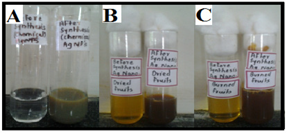
Figure 1: Yellowish brown colloidal solution of silver nanoparticles synthesized using A: chemical method B: Dried fruits and C: Burned fruits.
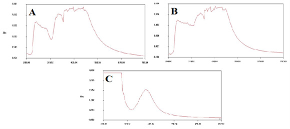
Figure 2: UV-Visible absorption spectra for silver nanoparticles synthesized A: chemically (Peak point-416nm) B: using dried fruits (Peak point- 434nm) C: using burned fruits (Peak point-412nm).
Confirmation of AgNP formation was done by taking UV-Vis spectrum of the solution, in the range of 200–800 nm. Result of UVVis spectroscopic analysis of these solutions revealed a characteristic absorption peak at 416 nm for chemically synthesized AgNPs, 434 nm and 412 nm for AgNPs synthesized using dried and burned fruits, respectively confirmed the synthesis of AgNPs [1] (Figure 2).
A result of Scanning Electron Microscopy (SEM) and X-Ray Diffraction (XRD) analysis gives an idea about size, shape and nature of synthesized nanoparticles. Scanning Electron Microscopy (SEM) showed presence of relatively cubic and triangle shaped nanoparticles with average size ranging 1-100 nm, it further confirmed the agglomeration of AgNPs (Figure 3).
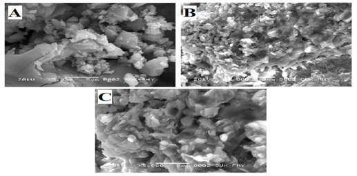
Figure 3: SEM micrograph for silver nanoparticles synthesized by A: Chemical method B: Dried fruits and C: Burned fruits.
X-Ray Diffraction (XRD) analysis revealed crystalline nature of chemically and biologically synthesized AgNPs. Sharp peak in the XRD patterns suggests very small size of nanoparticles. Size of the silver nanoparticles synthesized by chemical method, dried fruits and burned fruits was calculated using Scherrer’s formula [19] as 45 nm, 13 nm and 33 nm respectively (Figure 4). This gives resemblance with particle size range obtained from results of SEM image. It further indicated that biologically synthesized silver nanoparticles were significantly smaller than chemically synthesized (Figure 3 and 4). It has been reported that small NPs are able to inhibit or destroy microbial cells easily [27].
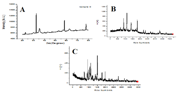
Figure 4: XRD pattern of silver nanoparticles synthesized by A: Chemical method (45nm), B: using dried fruits (13nm) C: using burned fruits (33nm).
Bacteria were isolated from infected burned skin sample on Blood Agar. The colonies giving hemolysis on Blood Agar media were further selected and grown on selective media like Cetrimide agar, Mannitol salt agar, MacConkeys agar, Leed’s acinetobacter medium and Worfel-Ferguson medium. Further biochemical characterization was performed for above five isolated colonies. After morphological and biochemical characterization (Table 1), all the five isolates were identified with the help of Bergey’s Manual as Isolate 1- Pseudomonas aeruginosa, isolate 2 - Staphylococcus aureus, isolate 3 - Escherichia coli, isolate 4 - Acinetobacter sp. and Isolate 5 - Klebsiella pneumonia. These above five isolates were previously reported for infections in burn patients and are found mostly resistant towards different antibiotics used in study [13].
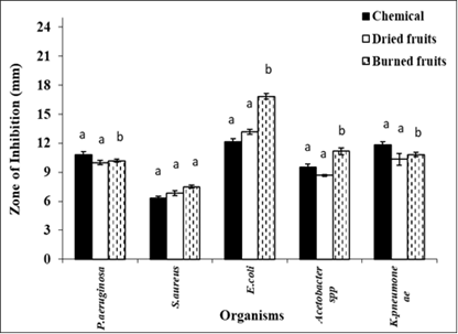
Bactericidal effect of chemically and biologically synthesized AgNPs was evaluated using agar well diffusion method against previously isolated five pathogenic bacteria. The data was analyzed using a normal probability plot to confirm that the data followed a normal distribution and adjusted R square value is 72.55, indicating a good fit of the model. Tukeys pair-wise comparison clearly indicates that AgNPs synthesised using burned fruit powder of C. equisetifolia demonstrated the significant antibacterial activity against all the five isolates (except for Staphylococcus aureus) (Figure 5). Nanopartical size of burned fruit powder was significantly smaller which suggest that tiny NPs may penetrates microbial cell wall easily and destroy various organelles. It has been observed that AgNPs synthesized from leaves and fruits are effective antimicrobial agent [28]. It was observed that, E. coli, Acinetobacter spp. is highly susceptible to nanoparticles synthesised using burned fruits of C. equisetifolia, whereas S. aureus is less susceptible to both chemically and biologically synthesised nanoparticles. In general, AgNPs are nontoxic [29] and used for wound dressing in clinics for many years. The AGNPs are used in wound healing because its takes lesser healing time [30] and show better collagen alignment and greater mechanical strength.
Conclusion
In present work, we have developed a method for synthesis of AgNPs using fruit of C. equisetifolia. To the best of our knowledge, nanoparticles synthesis using burned fruit powder of C. equisetifolia has not yet been reported. Biological method gives production of smaller sized silver nanoparticles compare to chemical method. The results of study emphasized on application of synthesized nanoparticle in treatment of skin burn infection. These results clearly indicate that AgNPS could provide alternative topical therapy in treatment of skin burn infection.
Acknowledgement
None.
Conflict of Interest
None.
References
- Prathna TC, Mathew L, Chanrasekaran N, Raichur AM, Mukarjee A, et al. (2010) Biomimetic Synthesis of Nanoparticles: Science, Technology & Applicability. Biomimetics Learning from Nature edited by A. Mukherjee.
- Moharekar S, Bora P, Daithankar V and Uplane (2014) Exploitation of Aspergillus niger for synthesis of silver nanoparticles and their use to improve shelf life of fruit and toxic dye degradation. IJIPSR 2(9): 2106-2118.
- Arvizo RR, Bhattacharyya S, Kudgus R, Giri K, Bhattacharya R, et al. (2012) Intrinsic therapeutic applications of noble metal nanoparticles: past, present and future. Chemical Society Reviews 41(7): 2943-2970.
- Sastry M, Ahmad A, Khan MI, Kumar R (2004) Microbial nanoparticle production, in Nanobiotechnology, ed. by Niemeyer CM and Mirkin CA. Wiley-VCH, Weinheim: 126-135.
- Bhattacharya D, Rajinder G (2005) Nanotechnology and potential of microorganisms. Crit Rev Biotechnol 25(4): 199-204.
- Mohanpuria P, Rana K, Yadav K (2008) Biosynthesis of nanoparticles: technological concepts and future applications. J Nanopart Res 10: 507-517.
- Shivshanka S, Rai A, Ahmad A, Sastry M (2004) Rapid synthesis of Au, Ag, and bimetallic Au core-Ag shell nanoparticles using Neem (Azadirachta indica) leaf broth. J Colloid Interface Sci 275(2): 496-502.
- Prathap SC, Chaudhary M, Pasricha R, Ahmad A, Sastry M, et al. (2006) Synthesis of gold nanotriangles and silver nanoparticles using Aloe vera plant extract. Biotechnol. Prog 22(2): 577-583.
- Armendariz V, Jose Yacaman M, Duarte Moller A, Peralta Videa JR, Troiani H, et al. (2004) HRTEM characterization of gold nanoparticles produced by wheat biomass. Rev Mex Fis 50(1): 7-11.
- Shivshankar S, Rai A, Ahma A, Sastry M (2005) Controlling the optical properties of lemongrass extract synthesized gold nanotriangles and potential application in infrared-absorbing optical coatings. Chem. Mater. 17: 566-572.
- Ankamwar B, Chaudhary M, Sastry M (2005) Gold Nanotriangles Biologically Synthesized using Tamarind Leaf Extract and Potential Application in Vapor Sensing. Metal-Organic and Nano-Metal Chemistry 35: 19-26.
- Lal SS, Nayak PL (2012) Green synthesis of gold nanoparticles using various extract of plants and spices. IJSID 2(3): 325-350.
- Shahzad MN, Ahmed N, Khan IH, Mirza AB, waheed F, et al. (2012) Bacterial Profile of Burn Wound Infections in Burn Patients. Ann Pak Inst Med Sci 8(1): 54-57.
- Wright GD (2005) Bacterial resistance to antibiotics: Enzymatic degradation and modification. Advanced Drug Delivery Reviews 57: 1451-1470.
- Chen HY (1997) Flora of Hainan; Science Press: Beijing, China.
- Dhan P, Suri S, Upadhyay G, Singh BN (2007) Total phenol, antioxidant and free radical scavenging activities of some medicinal plants. Int. J Food Sci Nutr 58(1): 18-28.
- Tiwari1 S, Talreja S (2023) A Critical Overview on Casuarina equisetifolia. Pharmacognosy Reviews (17)34: 255-261.
- Hano C, Abbasi BH (2022) Plant-Based Green Synthesis of Nanoparticles: Production, Characterization and Applications. Biomolecules 12(1): 31.
- Parmar R, Mangrola MH, Parmar BH, Joshi VG (2012) A software to calculate crystalline size by Debey-Scherrer Formula using VB.NET. Multi-Disciplinary Edu Global Quest.
- Parekh J, Jadeja D, Chanda S (2005) Efficacy of aqueous and methanol extracts of some medicinal plants for potential antibacterial activity. Turk jrnl Biol 29: 203-210.
- Gumgumjee NM, Hajar AS (2012) Antimicrobial efficacy of Casuarina equisetifolia extracts against some pathogenic microorganisms. J Med Plants Res 6(47): 5819-5825.
- Sriram N (2011) Antidiabetic and antihyperlipidemic activity of bark of Casuarina equisetifolia on streptozotocin induced diabetic rats. Inter J Pharm Rev Res 1(1): 4-8.
- Shalini S, Kumar AS (2011) Study on phytochemical profile and antiulcerogenic effect of Casuarina equisetifolia L. Asian J Pharm Sci Technol 1(1): 7-12.
- Cambie RC, Ash J (1994) Fijian medicinal plants. Australia: CSIRO
- Rastogi RP, Mehrotra BN, Sinha S, Pant P, Seth R, et al. (1998) Compendium of Indian medicinal plants.
- Park Y, Hong YN, Weyers A, Kim YS and Linhardt RJ (2011) Polysaccharides and phytochemicals: a natural reservoir for the green synthesis of gold and silver nanoparticles. IET Nanobiotechnology 5(3): 69-78.
- Singh RS, Singh T, Pandey A (2019) Chapter 1 - Microbial Enzymes—An Overview. Advances in Enzyme Technology: 1-40.
- Moustafal M, Al Emam A, Sayed M, Alamri S, Alghamdii H, et al. (2021) Green Synthesis of Ag Nanoparticles from Aqueous Extracts of Leaves and Fruit of Casuarina equisetifolia against Candida albicans and other Clinical Isolates. International Journal of Agriculture & Biology: 1-6.
- Kwan KHL, Kelvin WK, Yeung KWK, Liu X, Wong KKY, et al. (2014) Silver nanoparticles alter proteoglycan expression in the promotion of tendon repair. Nanomedicine: Nanotechnology, Biology and Medicine 10(7): 1375-1383.
- Chauhan N, Tyagi AK, Kumar P, Malik A (2015) Antibacterial Potential of Jatropha curcas Synthesized Silver Nanoparticles against Food Borne Pathogens. Front Microbiol 7: 1748




 We use cookies to ensure you get the best experience on our website.