
Review Article 
 Creative Commons, CC-BY
Therapeutic Strategies for Early Recurrent Exposure
in the Treatment of Giant Scalp Masses with Scalp
Expanders: Systematic Review and Case Report/h1>
*Corresponding author: Yongyan Cui, MD, Xiangwen Xu, MD, Department of Plastic and Reconstructive Surgery, Peking university Shenzhen
hospital, 1120 Lianghua Road, Shenzhen, Guangdong, 518036, P.R. China.
Received:April 10, 2025; Published:April 14, 2025
DOI: 10.34297/AJBSR.2022.26.003470
Abstract
Background: The cosmetic treatment of large scalp masses is usually performed with a tissue dilator two-stage surgery. Dilator exposure is the most
common complication during dilation, with repeated exposure being the biggest challenge.
Methods: The literature on exposure management of expander under different conditions was systematically reviewed. Screening consists of searching
for keywords (exposure, squeeze, management, skin extension, skin extension, and complications) in a format suitable for PubMed, Medline,
and Embase database syntax. The risk of bias in included studies was assessed using the Joanna Briggs Institute (JBI) Critical Assessment Checklist.
Results: Eleven studies were included, with a total of 29 extender exposed patients. Seven exposures from four studies reported a success rate of
85.7% (6/7) of removing the expander at late exposure and performing a second stage of surgery. Twenty-one exposures from seven studies reported
an early sustained expansion success rate of 81.0% (17/21). Four studies reported five failures, but only two of them analyzed the factors that
led to the failure.
Conclusion: This paper initially conducts a systematic review of the existing literature, examining the management strategies for dilator exposure
at both early and late. Subsequently, the article presents a case report, where the patient experienced early repeated exposure of the expander and
refused its removal. In this condition, a detailed treatment plan was formulated, which included ultra-rapid dilation without suturing, combined with
antibiotic therapy, and rapid tissue reconstruction with the aid of flap rearrangement.
Keywords:Tissue Expansion Devices, Scalp Expander, Expander Exposure, Nevus, Systemic Review, Case Report
Introduction
A two-stage procedure involving the use of tissue dilators is
often used to treat large lumps for cosmetic purposes [1, 2], especially
those located in the scalp area. During the first stage of the
procedure, a tissue dilator is inserted under the aponeurotic cap to
prevent harmful loss of scalp vascularization [3] and pressure on
the hair follicles [4]. The presence of distinct anatomical features in
the scalp, such as a hard skull, lack of elasticity in scalp tissue, and
considerable tension at the wound edge caused by the aponeurotic
flap, may lead to an increased likelihood of exposure of the dilator
after surgery [5, 6]. When the tissue dilator is exposed, removing
the dilator immediately or continuing the dilation and then performing
a second stage surgery are two common strategies. While
earlier studies have documented the management of extender exposure
in a variety of situations, there is still a lack of research that
specifically addresses detailed aspects of this management.
This paper systematically reviewed the literature on the treatment
of expander rupture at different stages. At the same time, we
also presented a case report involving repeated exposure to a dilator
during treatment of a large scalp sebaceous nevus. In addition,
through the exposure management of the expander in this case, a
thorough plan of repeated exposure of the expander is proposed.
Methods
This study was conducted using the 2020 Preferred Reporting
Project Statement for Systematic Review and Meta-Analysis (PRISMA).
Acceptance Standard
A comprehensive systematic review of the published literature
describing extended exposure management was conducted under
the preferred reporting item of the guidelines for systematic reviews
and meta-analyses. Those that did not discuss skin dilators,
where the management plan was unclear, non-English studies, conference
abstracts, review articles, letters and reviews were excluded.
Medical Literature Search
The Medline and Embase databases were searched from their
creation until June 6, 2024. Filtering consists of searching for the
following keywords in a format that fits the syntax of each database:
exposure, squeeze, management, skin extender, skin extender,
and complications. For example, PubMed’s search strategy is “scalp
and (dilation or dilation) and complications and (management or
repair or salvage).” After deleting duplicate references, the remaining
215 references were screened according to title and abstract, of
which 147 were excluded. We obtained the full text of 68 articles,
and 11 studies were selected for inclusion in this review based on
the application of inclusion and exclusion criteria. Includes articles
detailing the risk management of expansion machines.
Study Selection and Data Collection
The authors initially screened the full text of these studies by
title/abstract respectively. The data included in the study were extracted
into Microsoft Excel sheets. Information on eligible studies,
including the first author’s name, year of publication, study background,
patient demographics, cause of exposure, complications,
treatment, and end results, was extracted and cross-checked by the
two authors. Differences between authors are resolved by a third
author.
Biased Risk Assessment
The potential risk of bias in this study was assessed by the Joanna
Briggs Institute (JBI) Case Series Critical Assessment Tool. The
validity and reliability of the condition assessment, continuous recruitment
of participants, reporting of patient demographics and
clinical data, clinical management, and follow-up are sources of
bias in the assessment of this checklist.
Results
Search Procedure
The filtering process is shown in Figure 1. Ten case serials and
one case report published between 1987 and 2017 were included.

Figure 1:
Research Characteristics
Table1 summarizes data including authors, number of patients
reported, causes of exposure, complications, corresponding management,
and outcomes. And depending on the time of exposure
during dilation, cases were classified as either “early” (stage 2 surgery
with insufficient skin dilation) or “late” (Stage 2 surgery with
sufficient skin dilation). A total of 29 patients in 11 studies experienced
dilator exposure (Table 1). Be included. Seven exposures
from four studies reported a success rate of 85.7% (6/7) when the
expander was removed at a later stage and a second stage surgery
was performed. Twenty-one exposure cases from seven studies
reported early sustained expansion, with a success rate of 81.0%
(17/21). Of the included studies, four reported five failures, but
only two of them analyzed factors that contributed to the failure,
including early exposure to self-aerating osmotic dilators and lack
of systemic antibiotics.

Table 1:Summary of characteristics of included studies.
Risk of Bias in Research
Figure 2 is a summary of the risk of bias assessment based on
the key assessment tools of the JBI case series. Details are set out
in Supplement 1. According to our assessment, the most common
sources of possible bias in the included studies were continuous
and complete participant inclusion and statistical analyses, which
were not reported in an explicit form to make conclusive judgments.
Inappropriate demographic reporting is also another source
of bias in these studies.
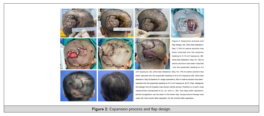
Figure 2:
Case Report
A 49-year-old woman was admitted on June 2, 2023, complaining
of progressive enlargement of the occipital tubercles over 40
years. Nodules were found at birth, up to about 10×5cm, accompanied
by progressive pruritus and occasional secretions, which had
a certain impact on sleep. On June 5, 2023, a 400 cm3 rectangular
tissue dilator was implanted. Start dilating gradually 1 week after
surgery with 10-20mL saline twice a week. Over a period of 1.5
months, the dilator expanded by a total of 150mL, and the patient
complained of extreme discomfort during the expansion. A week
later, the patient returned to the hospital reporting a 2cm crack at
the incision site (the left edge of the dilator) and yellow transparent
exudate. The skin covering the expander redness and temperature
rise. Since then, she has suffered from 3 times of expander exposure
(Table 2).

Table 2:Strategies and corresponding outcome of the expander exposure in case report.
After the 3rd exposure of expander, since the patient was adamant
about keeping the dilator, we decided to go for ultra-rapid
dilation without closing the wound (Figure 2A to 2C). Here is the
volume of saline injected into the expander: 120mL in the first
week, 50mL in the second week, and 35 mL in the third week, for a
total exposed area of 21.2cm2. Fewer complaints of pain are heard
from patients due to the de-tensioning effect caused by the wound
opening. Over the course of 17 days, 205mL of normal saline was
injected into the expander. Given that a quarter of the dilator is
exposed to the outside of the wound, continuing the injection will
reduce skin expansion and may make the infection worse. Therefore,
dilation surgery is suspended and the patient is prepared for
surgical intervention.
When the patient returned to the clinic on September 8, 2023,
there was a laceration of approximately 3.5×6cm between the dilator
and the mass, as shown in Figure 2D. The folds of the expander
protruded from the wound, and a yellow, cloudy liquid flowed out
at the same time. The temperature near the edge of the wound increases
slightly, with a reddish tinge. After a thorough evaluation
of the enlarged skin, we decided to perform reconstructive surgery
with Z-plasty (Figures 2E and 2F). After surgery, acupuncture was
used to relieve congestion at the edges of the flap (Figure 2G). The
patient returned to the clinic one month and six months after the
operation without any complications (Figures 2H and 2I).
Discussion
Causes of exposure include scalp expansion, inadequate skin
closure, incorrect placement of dilators, pre-irradiated areas, simultaneous
insertion of more tissue dilators, use of two-stage dilators,
refusal to use dilators, and sharp folds of dilators [7-11]. In
this case, scalp expansion and sharpening of the dilated pleats may
be the cause of exposure. As shown in Figure 2, with the help of high
tension and these folds, the expander can be pushed out from the
incision site, which is similar to Vergnes ‘s findings [12]. As Onishi
has found, repeated failures of wound closure are inevitable despite
debridement and antibiotic treatment [13]. This can be explained
by the fact that after the first exposure, the synergistic effect of infection
at the wound edge and further expansion may lead to repeated
failure of wound healing.
Referring to the previous article, the treatment of exposure can
be divided into two types (Table 1). If the dilator exposure occurs at
an advanced stage and it is considered that there is sufficient skin
for reconstruction or that it can be obtained using an auxiliary flap,
the dilator can be removed and then a second stage of surgery can
be performed [8,11,12,14,15]. If exposure occurs early, resulting
in insufficient reconstructed skin, the implementation of extended
repair becomes more complicated. In the case of infection-induced
exposure, the dilator must be removed promptly, along with pocket
irrigation using an antibacterial solution. After reinserting the
dilator, care should be taken to close the wound without straining
it. After debridement, systemic antibiotic therapy is also required
under the guidance of pathogen examination and drug sensitivity
test [13]. When exposure is not caused by infection, debridement
of the wound is recommended without replacing the dilator [16-
20]. In addition, reducing the volume of tissue dilators and suturing
with minimal tension have been shown to be effective in preventing
wound rupture [17]. Unlike exposure due to infection, systemic
antibiotic use is limited to cases where there is clinical or laboratory
evidence of infection [18,19]. In both conditions, dilation can
begin after the lesion has healed. Another notable advantage of an
extender is that multiple extenders are plugged in and the exposed
extender cannot be retained after evaluation. In this case, the expander
can be removed by overexpansion of the remaining expander
[21,22].
While many studies have reported various rescue options, most
lack detail. In this case, we provide an exhaustive plan to repeatedly
expose the expander. First, we used an approach similar to the one
described earlier. Studies have shown up to three exposures in the
same location. As soon as the first dilator exposure was detected,
we performed dilator drainage and debridement, followed by sutures
and intravenous sequential oral antibiotics. After the wound
heals, dilation surgery is performed.
Unfortunately, recurrent cracking occurs at the insertion site
during dilation. Since the patient showed no signs of serious infection
at the time of treatment, and strongly rejected dilation replacement
due to financial and time constraints, we optimized the rescue
plan from the following aspects while continuing the dilation [1].
(1) The schedule of saline injection after exposure: Compared to a
traditional injection schedule, we give relatively large amounts of
saline solution in a short period of time to achieve the required volume
(ultra-rapid expansion). Although it may aggravate the tearing
of the incision, this can reduce the risk of infection from prolonged
exposure. (2) Evaluation of skin dilation: In the previous study,
there is a formula describing the relationship between the injection
solution and the expanded skin: at this time, when 150mL of solution
was injected, the area of the expanded skin should be 38.2cm2
[2,23]. In the absence of exposure, when the volume of the solution
in the expander reaches 205mL, the area of the expansion skin
should be 60.82cm2. But in this case, a quarter of the expander is
exposed, that is, the area of the expansion skin should be 45.62cm2.
Since the sebaceous nevus is about 50cm2 in size, the enlarged skin
area is almost sufficient for reconstruction. As expected, after the
dilator was removed, the actual measured area of expanded skin
during reconstruction surgery was 47.5cm2, which was very close
to 45.62 cm2. The successful results of this case show that, in the
case of preventing infection, even if the expansion caused by wound
tearing is slightly reduced, sustained expansion under exposure can
reach the target volume with the help of wound edge structures.
(3) Flap design: When faced with a large defect caused by a wound,
direct suturing can cause excessive tension, which can lead to necrosis
of the skin edges, resulting in skin loss after expansion [3].
Therefore, aileron flaps is a widely used technique in the field of
tissue expansion and reconstruction [8,24]. In this particular case,
a Z-shaped flap was used. On the one hand, the amount of stretched
skin required to effectively close the defect is less compared to the
rotary flap technique. On the other hand, it has the potential to
significantly reduce regional tensions, thereby reducing recurrent
problems and the opening of wounds [25]. Without the need to
remove the dilator after repeated exposure, this protocol provides
another option to achieve the desired results through ultra-fast dilation
techniques and flap rearrangement.
Conclusion
This paper initially conducts a systematic review of the existing
literature, examining the management strategies for dilator exposure
at various stages-both early and late. Subsequently, the article
presents a case report involving the treatment of a large scalp mass
with a scalp expander, where the patient experienced early repeated
exposure of the dilator and refused its removal. In response to
this situation, we formulated a detailed treatment plan, which included
ultra-rapid dilation without suturing, combined with antibiotic
therapy, and rapid tissue reconstruction with the aid of flap
rearrangement.
Acknowledgements
This study was supported by Shenzhen Key Medical Discipline
Construction Fund (SZXK026).
References
- Marcus J, Horan DB, Robinson JK (1990) Tissue expansion: past, present, and future. J Am Acad Dermatol 23(5 Pt 1): 813-825.
- Romo Muñoz MI, Sánchez Galán A, Miguel M, Vanesa Nuñez Cerezo, Bryant Noriega Rebolledo, et al. (2018) Using Tissue Expanders as a Choice for the Treatment of Congenital Facial Nevus in the Young Child. Eur J Pediatr Surg 28(1): 105-108.
- Raposio E, Raposio G (2022) Scalp reconstruction by tissue expansion: tips and tricks. Acta Biomed 93(5): e2022313.
- Radwan AM, Zide MF (2019) Tissue Expansion in the Head and Neck. Atlas Oral Maxillofac Surg Clin North Am 27(2): 167-173.
- El Sakka DM (2015) Tissue expanders in post-burn alopecia: with or without galeotomies? Ann Burns Fire Disasters 28(3): 210-214.
- Azzi JL, Thabet C, Azzi AJ, Mirko S Gilardino (2020) Complications of tissue expansion in the head and neck. Head Neck 42(4): 747-762.
- Wang HD, Ibrahim Z, Quan A, Jennifer Bai, Benjamin T Ostrander, et al. (2020) Pediatric Tissue Expansion: Predictors of Premature Expander Removal in a Single Surgeon's Experience with 472 Expanders. Plast Reconstr Surg 145(3): 755-762.
- Gosain AK, Turin SY, Chim H, John A LoGiudice (2018) Salvaging the Unavoidable: A Review of Complications in Pediatric Tissue Expansion. Plast Reconstr Surg 142(3): 759-768.
- Rivera R, LoGiudice J, Gosain AK (2005) Tissue expansion in pediatric patients. Clin Plast Surg 32(1): 35-44, viii.
- (2021) Association, C.o.A.M.o.C.B.Compilation of Annual Meeting of Chinese Burn Association 2012: 286-288.
- Cho JY, Jang YC, Hur GY, Jang Hyu Koh, Dong Kook Seo, et al. (2012) One stage reconstruction of skull exposed by burn injury using a tissue expansion technique. Arch Plast Surg 39(2): 118-123.
- Vergnes P, Taieb A, Maleville J, J M Bondonny (1993) Repeated skin expansion for excision of congenital giant nevi in infancy and childhood. Plast Reconstr Surg 91(3): 450-455.
- Onishi K, Nakajima T, Nakanishi Y (1990) Infection and exposure in scalp expansion-A successful outcome. Eur J Plast Surg 13: 89-92.
- Yeşilada AK, Akçal A, Dağdelen D, Deniz Özgür Sucu, Leyla Kılınç, et al. (2013) The feasibility of tissue expansion in reconstruction of congenital and aquired deformities of pediatric patients. Int J Burns Trauma 3(3): 144-150.
- Paletta C, Campbell E, Shehadi SI (1991) Tissue expanders in children. J Pediatr Surg 26(1): 22-25.
- Dogra BB, Sharma A, Singh M, Bijitesh Chakravarty (2007) Tissue expansion: A simple technique for complex traumatic soft tissue defects. Int Surg 92(1): 37-45.
- Harving S, Ravnsbaek J, Johansen LV (1989) Salvage of exposed implants. Scand J Plast Reconstr Surg Hand Surg 23(2): 143-144.
- Neligan PC, Peters WJ (1987) The use of tissue expansion in burn scar reconstruction. J Burn Care Rehabil 8(2): 107-110.
- De Agustin JC, Morris SF, Zuker RM (1993) Tissue expansion in pediatric burn reconstruction. J Burn Care Rehabil 14(1): 43-50.
- Chun JT, Rohrich RJ (1998) Versatility of tissue expansion in head and neck burn reconstruction. Ann Plast Surg 41(1): 11-16.
- Karagergou E, Papas A, Foroglou P, Euterpi Demiri (2012) Management of the complications of tissue expansion during a 5-year period (2005-2010). J Plast Surg Hand Surg 46(3-4): 167-171.
- Kim SW, Kim YH, Kim JT (2011) Successful treatment of large forehead defect after the failure of tissue expansion: changing plan and strategy. J Craniofac Surg 22(6): 2129-2131.
- Pamplona DC, Weber HI, Leta FR (2014) Optimization of the use of skin expanders. Skin Res Technol 20(4): 463-472.
- Gaxiola García MA, Sánchez AP (2019) Salvaging the Unavoidable: A Review of Complications in Pediatric Tissue Expansion. Plast Reconstr Surg 144(3): 517e-518e.
- Tayyaba FU, Amin MM, Attaur Rasool S, Uzma Naseer, Akashah Ambar, et al. (2015) Reconstruction of post burn scalp alopecia by using expanded hair-bearing scalp flaps. Pak J Med Sci 31(6): 1405-1410.
*Corresponding author: Yongyan Cui, MD, Xiangwen Xu, MD, Department of Plastic and Reconstructive Surgery, Peking university Shenzhen hospital, 1120 Lianghua Road, Shenzhen, Guangdong, 518036, P.R. China.
Received:April 10, 2025; Published:April 14, 2025
DOI: 10.34297/AJBSR.2022.26.003470
Abstract
Background: The cosmetic treatment of large scalp masses is usually performed with a tissue dilator two-stage surgery. Dilator exposure is the most
common complication during dilation, with repeated exposure being the biggest challenge.
Methods: The literature on exposure management of expander under different conditions was systematically reviewed. Screening consists of searching
for keywords (exposure, squeeze, management, skin extension, skin extension, and complications) in a format suitable for PubMed, Medline,
and Embase database syntax. The risk of bias in included studies was assessed using the Joanna Briggs Institute (JBI) Critical Assessment Checklist.
Results: Eleven studies were included, with a total of 29 extender exposed patients. Seven exposures from four studies reported a success rate of
85.7% (6/7) of removing the expander at late exposure and performing a second stage of surgery. Twenty-one exposures from seven studies reported
an early sustained expansion success rate of 81.0% (17/21). Four studies reported five failures, but only two of them analyzed the factors that
led to the failure.
Conclusion: This paper initially conducts a systematic review of the existing literature, examining the management strategies for dilator exposure
at both early and late. Subsequently, the article presents a case report, where the patient experienced early repeated exposure of the expander and
refused its removal. In this condition, a detailed treatment plan was formulated, which included ultra-rapid dilation without suturing, combined with
antibiotic therapy, and rapid tissue reconstruction with the aid of flap rearrangement.
Keywords:Tissue Expansion Devices, Scalp Expander, Expander Exposure, Nevus, Systemic Review, Case Report
Introduction
A two-stage procedure involving the use of tissue dilators is often used to treat large lumps for cosmetic purposes [1, 2], especially those located in the scalp area. During the first stage of the procedure, a tissue dilator is inserted under the aponeurotic cap to prevent harmful loss of scalp vascularization [3] and pressure on the hair follicles [4]. The presence of distinct anatomical features in the scalp, such as a hard skull, lack of elasticity in scalp tissue, and considerable tension at the wound edge caused by the aponeurotic flap, may lead to an increased likelihood of exposure of the dilator after surgery [5, 6]. When the tissue dilator is exposed, removing the dilator immediately or continuing the dilation and then performing a second stage surgery are two common strategies. While earlier studies have documented the management of extender exposure in a variety of situations, there is still a lack of research that specifically addresses detailed aspects of this management.
This paper systematically reviewed the literature on the treatment of expander rupture at different stages. At the same time, we also presented a case report involving repeated exposure to a dilator during treatment of a large scalp sebaceous nevus. In addition, through the exposure management of the expander in this case, a thorough plan of repeated exposure of the expander is proposed.
Methods
This study was conducted using the 2020 Preferred Reporting Project Statement for Systematic Review and Meta-Analysis (PRISMA).
Acceptance Standard
A comprehensive systematic review of the published literature describing extended exposure management was conducted under the preferred reporting item of the guidelines for systematic reviews and meta-analyses. Those that did not discuss skin dilators, where the management plan was unclear, non-English studies, conference abstracts, review articles, letters and reviews were excluded.
Medical Literature Search
The Medline and Embase databases were searched from their creation until June 6, 2024. Filtering consists of searching for the following keywords in a format that fits the syntax of each database: exposure, squeeze, management, skin extender, skin extender, and complications. For example, PubMed’s search strategy is “scalp and (dilation or dilation) and complications and (management or repair or salvage).” After deleting duplicate references, the remaining 215 references were screened according to title and abstract, of which 147 were excluded. We obtained the full text of 68 articles, and 11 studies were selected for inclusion in this review based on the application of inclusion and exclusion criteria. Includes articles detailing the risk management of expansion machines.
Study Selection and Data Collection
The authors initially screened the full text of these studies by title/abstract respectively. The data included in the study were extracted into Microsoft Excel sheets. Information on eligible studies, including the first author’s name, year of publication, study background, patient demographics, cause of exposure, complications, treatment, and end results, was extracted and cross-checked by the two authors. Differences between authors are resolved by a third author.
Biased Risk Assessment
The potential risk of bias in this study was assessed by the Joanna Briggs Institute (JBI) Case Series Critical Assessment Tool. The validity and reliability of the condition assessment, continuous recruitment of participants, reporting of patient demographics and clinical data, clinical management, and follow-up are sources of bias in the assessment of this checklist.
Results
Search Procedure
The filtering process is shown in Figure 1. Ten case serials and one case report published between 1987 and 2017 were included.
Figure 1:
Research Characteristics
Table1 summarizes data including authors, number of patients reported, causes of exposure, complications, corresponding management, and outcomes. And depending on the time of exposure during dilation, cases were classified as either “early” (stage 2 surgery with insufficient skin dilation) or “late” (Stage 2 surgery with sufficient skin dilation). A total of 29 patients in 11 studies experienced dilator exposure (Table 1). Be included. Seven exposures from four studies reported a success rate of 85.7% (6/7) when the expander was removed at a later stage and a second stage surgery was performed. Twenty-one exposure cases from seven studies reported early sustained expansion, with a success rate of 81.0% (17/21). Of the included studies, four reported five failures, but only two of them analyzed factors that contributed to the failure, including early exposure to self-aerating osmotic dilators and lack of systemic antibiotics.
Table 1:Summary of characteristics of included studies.
Risk of Bias in Research
Figure 2 is a summary of the risk of bias assessment based on the key assessment tools of the JBI case series. Details are set out in Supplement 1. According to our assessment, the most common sources of possible bias in the included studies were continuous and complete participant inclusion and statistical analyses, which were not reported in an explicit form to make conclusive judgments. Inappropriate demographic reporting is also another source of bias in these studies.
Figure 2:
Case Report
A 49-year-old woman was admitted on June 2, 2023, complaining of progressive enlargement of the occipital tubercles over 40 years. Nodules were found at birth, up to about 10×5cm, accompanied by progressive pruritus and occasional secretions, which had a certain impact on sleep. On June 5, 2023, a 400 cm3 rectangular tissue dilator was implanted. Start dilating gradually 1 week after surgery with 10-20mL saline twice a week. Over a period of 1.5 months, the dilator expanded by a total of 150mL, and the patient complained of extreme discomfort during the expansion. A week later, the patient returned to the hospital reporting a 2cm crack at the incision site (the left edge of the dilator) and yellow transparent exudate. The skin covering the expander redness and temperature rise. Since then, she has suffered from 3 times of expander exposure (Table 2).
Table 2:Strategies and corresponding outcome of the expander exposure in case report.
After the 3rd exposure of expander, since the patient was adamant about keeping the dilator, we decided to go for ultra-rapid dilation without closing the wound (Figure 2A to 2C). Here is the volume of saline injected into the expander: 120mL in the first week, 50mL in the second week, and 35 mL in the third week, for a total exposed area of 21.2cm2. Fewer complaints of pain are heard from patients due to the de-tensioning effect caused by the wound opening. Over the course of 17 days, 205mL of normal saline was injected into the expander. Given that a quarter of the dilator is exposed to the outside of the wound, continuing the injection will reduce skin expansion and may make the infection worse. Therefore, dilation surgery is suspended and the patient is prepared for surgical intervention.
When the patient returned to the clinic on September 8, 2023, there was a laceration of approximately 3.5×6cm between the dilator and the mass, as shown in Figure 2D. The folds of the expander protruded from the wound, and a yellow, cloudy liquid flowed out at the same time. The temperature near the edge of the wound increases slightly, with a reddish tinge. After a thorough evaluation of the enlarged skin, we decided to perform reconstructive surgery with Z-plasty (Figures 2E and 2F). After surgery, acupuncture was used to relieve congestion at the edges of the flap (Figure 2G). The patient returned to the clinic one month and six months after the operation without any complications (Figures 2H and 2I).
Discussion
Causes of exposure include scalp expansion, inadequate skin closure, incorrect placement of dilators, pre-irradiated areas, simultaneous insertion of more tissue dilators, use of two-stage dilators, refusal to use dilators, and sharp folds of dilators [7-11]. In this case, scalp expansion and sharpening of the dilated pleats may be the cause of exposure. As shown in Figure 2, with the help of high tension and these folds, the expander can be pushed out from the incision site, which is similar to Vergnes ‘s findings [12]. As Onishi has found, repeated failures of wound closure are inevitable despite debridement and antibiotic treatment [13]. This can be explained by the fact that after the first exposure, the synergistic effect of infection at the wound edge and further expansion may lead to repeated failure of wound healing.
Referring to the previous article, the treatment of exposure can be divided into two types (Table 1). If the dilator exposure occurs at an advanced stage and it is considered that there is sufficient skin for reconstruction or that it can be obtained using an auxiliary flap, the dilator can be removed and then a second stage of surgery can be performed [8,11,12,14,15]. If exposure occurs early, resulting in insufficient reconstructed skin, the implementation of extended repair becomes more complicated. In the case of infection-induced exposure, the dilator must be removed promptly, along with pocket irrigation using an antibacterial solution. After reinserting the dilator, care should be taken to close the wound without straining it. After debridement, systemic antibiotic therapy is also required under the guidance of pathogen examination and drug sensitivity test [13]. When exposure is not caused by infection, debridement of the wound is recommended without replacing the dilator [16- 20]. In addition, reducing the volume of tissue dilators and suturing with minimal tension have been shown to be effective in preventing wound rupture [17]. Unlike exposure due to infection, systemic antibiotic use is limited to cases where there is clinical or laboratory evidence of infection [18,19]. In both conditions, dilation can begin after the lesion has healed. Another notable advantage of an extender is that multiple extenders are plugged in and the exposed extender cannot be retained after evaluation. In this case, the expander can be removed by overexpansion of the remaining expander [21,22].
While many studies have reported various rescue options, most lack detail. In this case, we provide an exhaustive plan to repeatedly expose the expander. First, we used an approach similar to the one described earlier. Studies have shown up to three exposures in the same location. As soon as the first dilator exposure was detected, we performed dilator drainage and debridement, followed by sutures and intravenous sequential oral antibiotics. After the wound heals, dilation surgery is performed.
Unfortunately, recurrent cracking occurs at the insertion site during dilation. Since the patient showed no signs of serious infection at the time of treatment, and strongly rejected dilation replacement due to financial and time constraints, we optimized the rescue plan from the following aspects while continuing the dilation [1]. (1) The schedule of saline injection after exposure: Compared to a traditional injection schedule, we give relatively large amounts of saline solution in a short period of time to achieve the required volume (ultra-rapid expansion). Although it may aggravate the tearing of the incision, this can reduce the risk of infection from prolonged exposure. (2) Evaluation of skin dilation: In the previous study, there is a formula describing the relationship between the injection solution and the expanded skin: at this time, when 150mL of solution was injected, the area of the expanded skin should be 38.2cm2 [2,23]. In the absence of exposure, when the volume of the solution in the expander reaches 205mL, the area of the expansion skin should be 60.82cm2. But in this case, a quarter of the expander is exposed, that is, the area of the expansion skin should be 45.62cm2. Since the sebaceous nevus is about 50cm2 in size, the enlarged skin area is almost sufficient for reconstruction. As expected, after the dilator was removed, the actual measured area of expanded skin during reconstruction surgery was 47.5cm2, which was very close to 45.62 cm2. The successful results of this case show that, in the case of preventing infection, even if the expansion caused by wound tearing is slightly reduced, sustained expansion under exposure can reach the target volume with the help of wound edge structures. (3) Flap design: When faced with a large defect caused by a wound, direct suturing can cause excessive tension, which can lead to necrosis of the skin edges, resulting in skin loss after expansion [3]. Therefore, aileron flaps is a widely used technique in the field of tissue expansion and reconstruction [8,24]. In this particular case, a Z-shaped flap was used. On the one hand, the amount of stretched skin required to effectively close the defect is less compared to the rotary flap technique. On the other hand, it has the potential to significantly reduce regional tensions, thereby reducing recurrent problems and the opening of wounds [25]. Without the need to remove the dilator after repeated exposure, this protocol provides another option to achieve the desired results through ultra-fast dilation techniques and flap rearrangement.
Conclusion
This paper initially conducts a systematic review of the existing literature, examining the management strategies for dilator exposure at various stages-both early and late. Subsequently, the article presents a case report involving the treatment of a large scalp mass with a scalp expander, where the patient experienced early repeated exposure of the dilator and refused its removal. In response to this situation, we formulated a detailed treatment plan, which included ultra-rapid dilation without suturing, combined with antibiotic therapy, and rapid tissue reconstruction with the aid of flap rearrangement.
Acknowledgements
This study was supported by Shenzhen Key Medical Discipline Construction Fund (SZXK026).
References
- Marcus J, Horan DB, Robinson JK (1990) Tissue expansion: past, present, and future. J Am Acad Dermatol 23(5 Pt 1): 813-825.
- Romo Muñoz MI, Sánchez Galán A, Miguel M, Vanesa Nuñez Cerezo, Bryant Noriega Rebolledo, et al. (2018) Using Tissue Expanders as a Choice for the Treatment of Congenital Facial Nevus in the Young Child. Eur J Pediatr Surg 28(1): 105-108.
- Raposio E, Raposio G (2022) Scalp reconstruction by tissue expansion: tips and tricks. Acta Biomed 93(5): e2022313.
- Radwan AM, Zide MF (2019) Tissue Expansion in the Head and Neck. Atlas Oral Maxillofac Surg Clin North Am 27(2): 167-173.
- El Sakka DM (2015) Tissue expanders in post-burn alopecia: with or without galeotomies? Ann Burns Fire Disasters 28(3): 210-214.
- Azzi JL, Thabet C, Azzi AJ, Mirko S Gilardino (2020) Complications of tissue expansion in the head and neck. Head Neck 42(4): 747-762.
- Wang HD, Ibrahim Z, Quan A, Jennifer Bai, Benjamin T Ostrander, et al. (2020) Pediatric Tissue Expansion: Predictors of Premature Expander Removal in a Single Surgeon's Experience with 472 Expanders. Plast Reconstr Surg 145(3): 755-762.
- Gosain AK, Turin SY, Chim H, John A LoGiudice (2018) Salvaging the Unavoidable: A Review of Complications in Pediatric Tissue Expansion. Plast Reconstr Surg 142(3): 759-768.
- Rivera R, LoGiudice J, Gosain AK (2005) Tissue expansion in pediatric patients. Clin Plast Surg 32(1): 35-44, viii.
- (2021) Association, C.o.A.M.o.C.B.Compilation of Annual Meeting of Chinese Burn Association 2012: 286-288.
- Cho JY, Jang YC, Hur GY, Jang Hyu Koh, Dong Kook Seo, et al. (2012) One stage reconstruction of skull exposed by burn injury using a tissue expansion technique. Arch Plast Surg 39(2): 118-123.
- Vergnes P, Taieb A, Maleville J, J M Bondonny (1993) Repeated skin expansion for excision of congenital giant nevi in infancy and childhood. Plast Reconstr Surg 91(3): 450-455.
- Onishi K, Nakajima T, Nakanishi Y (1990) Infection and exposure in scalp expansion-A successful outcome. Eur J Plast Surg 13: 89-92.
- Yeşilada AK, Akçal A, Dağdelen D, Deniz Özgür Sucu, Leyla Kılınç, et al. (2013) The feasibility of tissue expansion in reconstruction of congenital and aquired deformities of pediatric patients. Int J Burns Trauma 3(3): 144-150.
- Paletta C, Campbell E, Shehadi SI (1991) Tissue expanders in children. J Pediatr Surg 26(1): 22-25.
- Dogra BB, Sharma A, Singh M, Bijitesh Chakravarty (2007) Tissue expansion: A simple technique for complex traumatic soft tissue defects. Int Surg 92(1): 37-45.
- Harving S, Ravnsbaek J, Johansen LV (1989) Salvage of exposed implants. Scand J Plast Reconstr Surg Hand Surg 23(2): 143-144.
- Neligan PC, Peters WJ (1987) The use of tissue expansion in burn scar reconstruction. J Burn Care Rehabil 8(2): 107-110.
- De Agustin JC, Morris SF, Zuker RM (1993) Tissue expansion in pediatric burn reconstruction. J Burn Care Rehabil 14(1): 43-50.
- Chun JT, Rohrich RJ (1998) Versatility of tissue expansion in head and neck burn reconstruction. Ann Plast Surg 41(1): 11-16.
- Karagergou E, Papas A, Foroglou P, Euterpi Demiri (2012) Management of the complications of tissue expansion during a 5-year period (2005-2010). J Plast Surg Hand Surg 46(3-4): 167-171.
- Kim SW, Kim YH, Kim JT (2011) Successful treatment of large forehead defect after the failure of tissue expansion: changing plan and strategy. J Craniofac Surg 22(6): 2129-2131.
- Pamplona DC, Weber HI, Leta FR (2014) Optimization of the use of skin expanders. Skin Res Technol 20(4): 463-472.
- Gaxiola García MA, Sánchez AP (2019) Salvaging the Unavoidable: A Review of Complications in Pediatric Tissue Expansion. Plast Reconstr Surg 144(3): 517e-518e.
- Tayyaba FU, Amin MM, Attaur Rasool S, Uzma Naseer, Akashah Ambar, et al. (2015) Reconstruction of post burn scalp alopecia by using expanded hair-bearing scalp flaps. Pak J Med Sci 31(6): 1405-1410.




 We use cookies to ensure you get the best experience on our website.