
Research Article 
 Creative Commons, CC-BY
Discovery of Hyaluronic Acid in the Heart as a Lubricating Mechanism
*Corresponding author:Jorge Carlos Trainini MD, PhD. Presidente Perón Hospital. National University of Avellaneda, Argentina.
Received:July 28, 2025; Published:August 05, 2025
DOI: 10.34297/AJBSR.2025.27.003628
Abstract
Objective: The sliding between myocardial segments, produced by torsion-detorsion, theoretically implies the existence of an antifriction mechanism
to avoid dissipating the energy used by the heart. So, is there a specific histology that explains this fact? Do the venous ductules of Thebesius
and Langer play a role in this mechanism? Is there an organic lubricant that can facilitate the sliding of overlapping cardiac segments?
Material and Methods: Seventy-one hearts from the morgue and slaughterhouses were used: a) 54 two-year-old cattle weighing 1300-1900g
(average 1650g); b) 17 humans (three at 8, 16 and 23 weeks of gestation respectively; four infants at 30 days, 36 days, 10 weeks and 27 weeks; one
4-year-old child; one 10-year-old child and eight adults with an average weight of 300g).
Results: In all the hearts in this study, we found hyaluronic acid in the cleavage planes between the myocardial bundles, in addition to the venous
ducts of Thebesius and Langer.
Conclusions: This functional association between the Thebesian and Langer ducts, together with the appreciable amount of hyaluronic acid found in
our research on human and animal hearts, and its lubricating role in the rest of the body, could be crucial in understanding the dynamics of the helical
heart. Ventricular torsion is thus correlated with a lubrication mechanism that facilitates the sliding of myocardial segments to prevent energy loss.
Keywords:Hyaluronic acid, Myocardial friction, Cardiac anatomy, Fulcrum
Introduction
The segments that constitute the continuous myocardium when folded in a helix must overlap. The modeling achieved in our three-dimensional study [1,2] clearly shows this concept of superposition of the segments with the consequence of constituting the helical myocardium (Figure 1). The sliding between internal and external segments of the myocardium assumes opposite directions in its movements, during the systolic phase and protodiastolic phase of myocardial contraction of the heart, generating friction. It is understandable that the friction of the segments also implies, from a physical perspective, an opposition to movement.
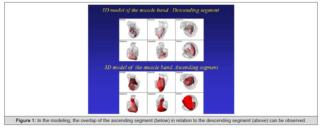
Figure 1:In the modeling, the overlap of the ascending segment (below) in relation to the descending segment (above) can be observed.
However, to fulfill this dynamic, it has been crucial to interpret that there is a support point for the myocardium, as we have demonstrated and given the name of cardiac fulcrum through anatomical and histological investigations (Figures 2 and 3) [3,4].
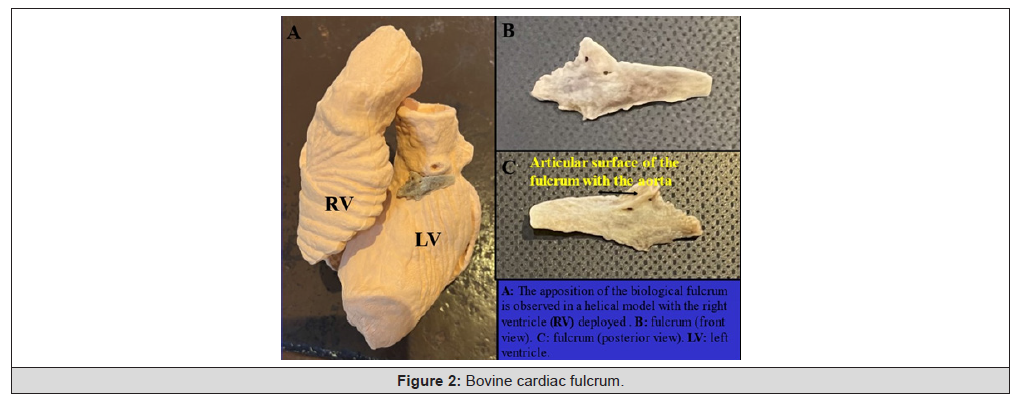
Figure 2:Bovine cardiac fulcrum.
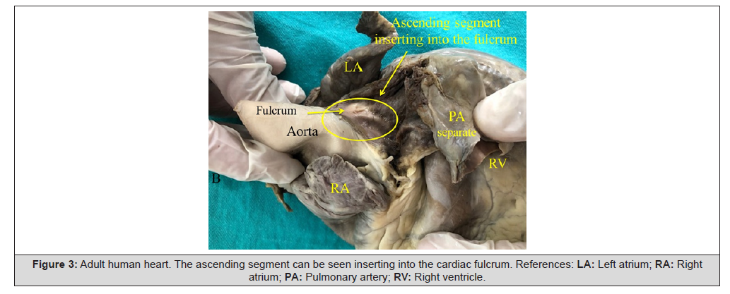
Figure 3:Adult human heart. The ascending segment can be seen inserting into the cardiac fulcrum. References: LA: Left atrium; RA: Right atrium; PA: Pulmonary artery; RV: Right ventricle.
We know that the helical structure of myocardial continuity allows fulfilling the dynamics of ventricular torsion-detorsion. Anatomical studies, the progress in the understanding of cardiac dynamics through the propagation of the electrical impulse, and ultrasound studies with ventricular rotation confirm the structure- function relationship of the heart [5]. Ventricular torsion represents the functional need that makes the heart fibers adopt an 8-shaped configuration. This is manifested as myocardial in its three axes: longitudinal, circumferential and radial, depending on the helical fiber orientation. The study of this rotation is a potential marker of myocardial disease severity when its twists deviate from normal values. Ventricular torsion may become a more adequate marker of the heart’s condition, even superior to functional class and fraction eyection in the understanding of heart failure [6-8]. During systole, the three cardiac axes contract generating torsion in the muscle mass which is “wrung as a towel” to achieve its ejective action. To analyze this activity, it has been essential to understand the path followed by the stimulus in the continuous myocardium, which explains cardiac movements. In the protodiastolic phase of myocardial contraction, during the first 100ms of diastole, the longitudinal axis lengthens and then with ventricular filling it expands and untwists, increasing its circumferential and radial axes. The helical myocardial anatomy, ventricular torsion, friction, and intraventricular ejective blood vortex correlate in the sequential explanation of cardiac dynamics. The sliding between myocardial segments, produced by torsion-detorsion, theoretically implies the existence of an antifriction mechanism to avoid dissipating the energy used by the heart. So, is there a specific histology that explains this fact? Do the venous ductules of Thebesius and Langer play a role in this mechanism? Is there an organic lubricant that can facilitate the sliding of overlapping cardiac segments?
Material and Methods
In an attempt clarify the discussion on the structural conformation of the myocardium, imaging procedures have been used improving its resolution. However, by not acting directly on its anatomy and histology, they have been unable to define essential details of its organizational pattern. In this research, fresh bovine and human hearts were used to obtain detailed descriptions aimed at elucidating the true spatial myocardial architecture. Seventy-one hearts from the morgue and slaughterhouses were used: a) 54 twoyear- old bovines with a weight between 1300 and 1900g (average 1650g); b) 17 humans (three at 8, 16 and 23 weeks of gestation respectively; four infants at 30 days, 36 days, 10 weeks and 27 weeks; one 4-year-old child; one 10-year-old child and eight adults with an average weight of 300g). Anatomical, histological and histochemical were carried out. The heart was fixed in 10% buffered formalin, and histology was performed using hematoxylin-eosin and Masson’s trichrome staining technique in four-micron sections. Ten percent formalin was used as buffer and immunohistochemistry staining was also implemented [9]. All samples were subjected to histochemical analysis with Alcian blue staining, a reliable marker to identify the presence of hyaluronic acid as an antifriction mechanism, and even provide a semi-quantitative assessment
Results
In all the hearts in this study, we found hyaluronic acid in the cleavage planes between the myocardial bundles, in addition to the venous ducts of Thebesius and Langer, which were first described by Vieussens in 1706 [10]. These venous ducts originate from intramyocardial veins (Thebesius) or tributaries of the major coronary vein (Langer), cross the myocardium and drain into the cardiac cavities (Figure 4). We have even observed it in human pregnancy at 23 weeks (Figure 5).
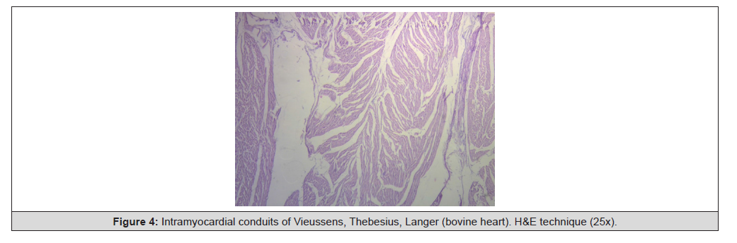
Figure 4:Intramyocardial conduits of Vieussens, Thebesius, Langer (bovine heart). H&E technique (25x).
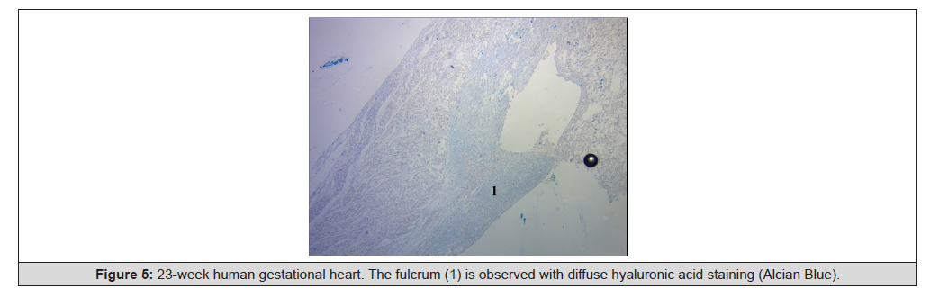
Figure 5:23-week human gestational heart. The fulcrum (1) is observed with diffuse hyaluronic acid staining (Alcian Blue).
A friction plane for cardiac pathophysiology occurs in the left ventricular wall. The fibers of the descending segment on the anterior surface of the left ventricle run deep through the mesocardium, crossing obliquely and internally with those of the ascending segment, which are located externally. This important friction plane is located between the two segments. Furthermore, between the cardiomyocytes, we found lacunar spaces with a capillary network and a plasma fluid rich in hyaluronic acid, which was stained light blue with Alcian blue. We found no differences between bovine and human hearts.
Discussion
It is logical to consider that there must be an antifriction mechanism that prevents dissipation of energy during myocardial segment sliding throughout the generation of torsion-detorsion movements. Is there a histological basis to explain this fact? Do the Thebesian and Langer venous conduits play a role in this mechanism? Can the myocardium have an organic lubricating source? Francisco Torrent Guasp believed that an elastic membrane could be found that would limit friction, avoiding excessive energy expenditure, but he ended up accepting that neither macroscopically nor with the microscope could he find any septum. [11] On the other hand, Luis Becú in a personal letter to Torrent Guasp in 1996, reported an appealing idea: “[The] muscle bundles, one on top of the other but in discordant orientations have been objected in several ways, but a basic objection is that if this were true, the friction between them would be great, generating heat and wasting energy… making necessary an antifriction lubricating mechanism… [there is] a vast system of flat lacunar blood spaces usefully arranged in spaces between layers and bands of muscle tissue… that have ample communication with the coronary veins… and what is much more significant and interesting, with the lumen of both ventricles through the ghostly Thebesian veins” [1,11,12].
During our investigation, we considered that the antifriction mechanism could be generated by the loose “spongy” myocardial content with transmural blood currents (sinusoids), rudiments of the evolutionary and embryonic heart, in which Adam Thebesius´ veins (German anatomist, 1686-1732) [13] and those described by Karl Langer (Vienna, 1819-1887) [14] would play a fundamental role (hydraulic system). These coronary-ventricular connections, also studied by Vieussenius (1715), Minot (1900), Lewis (1904), Grant (1926), and Wearn (1928), would reduce the shearing stress between the muscle surfaces in the ventricular torsion-detorsion movements [15]. In fact, a study of historical chronology indicates that the first to describe these ducts was Julius Vieussens (1635- 1715) in 1706. [10] Later, in 1708, it was certified by Thebesius. To reach these conclusions, Vieussens tied both vena cavae and the pulmonary veins. He then injected saffron dye into the coronary arteries. After its normal course through the capillaries, veins and coronary sinus, he observed with surprise that the cardiac cavities were also stained. He called these ducts “ducti carnosi.” For his part, Thebesius provided a more detailed description of these ducts, which came to be named after him [10]. These small venous conduits that originate from coronary arteries (Vieussens) intramyocardial veins (Thebesian) or from branching vessels of the major coronary vein (Langer), run across the myocardium and drain into the cardiac chambers (Figure 6). Obviously, the ones that drain into the left ventricular chamber deoxygenate the blood coming from the lungs. In fact, this results in greater oxygen content in the left atrium than in the corresponding ventricle. Some essential questions arise here: Why would these venous conduits play a counterproductive function for the organism by deoxygenating arterial blood? What is their true role? Initial studies on these conduits considered that they could oxygenate the myocardium in case of coronary artery occlusion.
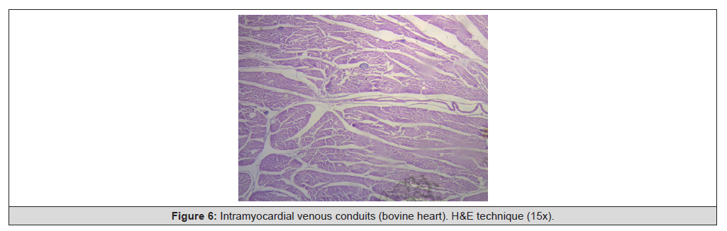
Figure 6:Intramyocardial venous conduits (bovine heart). H&E technique (15x).
In this regard, in all the investigated hearts, we have found hyaluronic acid in the cleavage planes between the myocardial bundles associated with Thebesian and Langer venous conduits. We found hyaluronic acid stained with “Alcian blue” (Figures 6-9) in the lacunar structure between the myocardial bundles in bovine and human hearts, which together with Thebesian and Langer venous conduits as branches for the necessary hydration of this element, could clarify this lubricating effect, and thus counteract the friction between surfaces as an antifriction mechanism. Actually, between the different segments there is no specific sliding membrane, but rather friction surfaces intersected by a spongy system that covers the entire myocardium and where hyaluronic acid with its lubricating capacity facilitates the sliding movement between them (Figures 10-12) [15,16]. In our study, hyaluronic acid was very scarce in the trigones, aorta and pulmonary artery, but abundant in the myocardium and papillary muscles. No differences were encountered between bovine and human hearts.
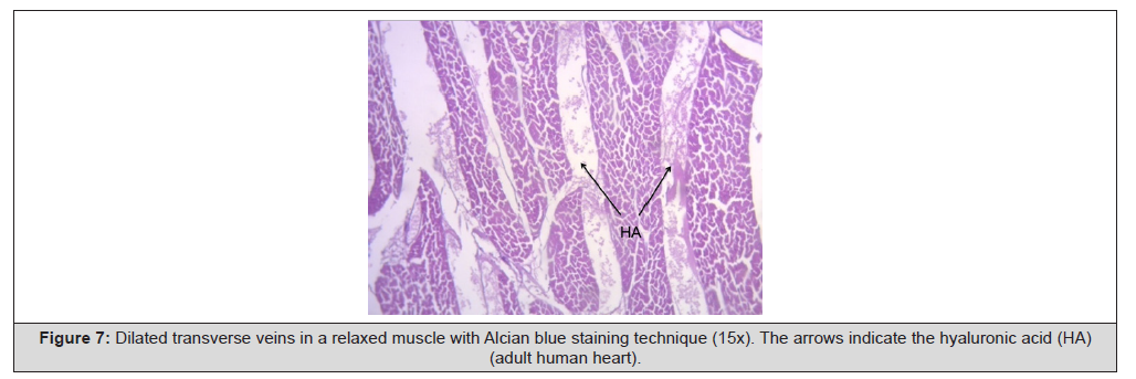
Figure 7:Dilated transverse veins in a relaxed muscle with Alcian blue staining technique (15x). The arrows indicate the hyaluronic acid (HA) (adult human heart).
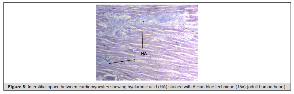
Figure 8:Interstitial space between cardiomyocytes showing hyaluronic acid (HA) stained with Alcian blue technique (15x) (adult human heart).
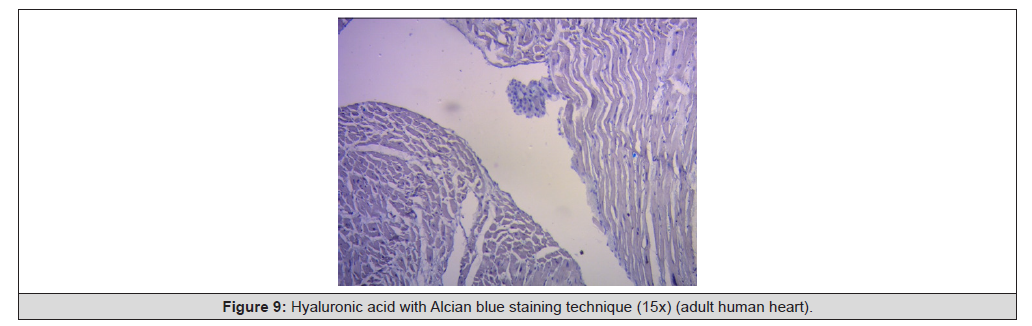
Figure 9:Hyaluronic acid with Alcian blue staining technique (15x) (adult human heart).
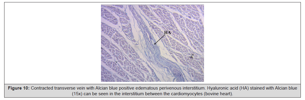
Figure 10:Contracted transverse vein with Alcian blue positive edematous perivenous interstitium. Hyaluronic acid (HA) stained with Alcian blue (15x) can be seen in the interstitium between the cardiomyocytes (bovine heart).
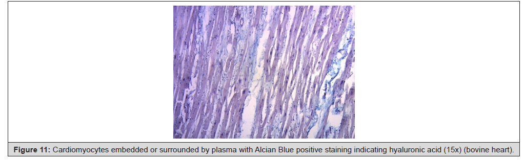
Figure 11:Cardiomyocytes embedded or surrounded by plasma with Alcian Blue positive staining indicating hyaluronic acid (15x) (bovine heart).
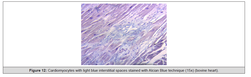
Figure 12:Cardiomyocytes with light blue interstitial spaces stained with Alcian Blue technique (15x) (bovine heart).
Hyaluronic acid, which appeared in the first chordate animals, is abundant in connective tissues [17]. Half of hyaluronic acid is found in the skin and 25% in the bones and joints. Due to its capacity to bind water it is an excellent lubricant and provides great resistance to mechanical pressures [18]. Hyaluronic acid helps to create the fabric of the extracellular matrix through its interaction with proteoglycans or collagen. When it is very concentrated it may interact with itself creating networks or meshes that bestow viscoelastic properties to tissues. During development it helps the morphogenesis of embryonic structures. For example, it may be released from basal epithelial layers, curving their shape due to the great amount of water it attracts [19] (Figure 10-12).
Conclusions
This functional association between the Thebesian and Langer venous conduits and the substantial amount of hyaluronic acid found in our investigations in bovine and human hearts, added to its lubricating role in the rest of the body, could be crucial to understand the helical heart dynamics. Thus, ventricular torsion is correlated with a lubricating mechanism that facilitates myocardial segment sliding to avoid energy loss. Across these venous conduits and by the helical contraction, plasmatic fluid with hyaluronic acid would be continuously driven through a rich capillary network. In our investigation, we have found between the cardiomyocytes, spaces with a capillary network containing a plasmatic fluid rich in hyaluronic acid.
Acknowledgement
None.
Conflict of Interest
None.
References
- Trainini Jorge, Beraudo Mario, Wernicke Mario, Trainini Alejandro, Lowenstein Jorge, et al. (2024) Fundamental Conclusions on Research into the Anatomy and Organization of the Helical Heart. Am J Biomed Sci & Res 23(5).
- Valle J, Herreros J, Trainini J, García Jimenez E, Talents M, et al. (2017) Three-dimensional definition of Torrent Guasp´s ventricular muscle band and its correlation with electric activation of the left ventricle (abstract). XXIII Congreso SEIQ, Madrid, España BJS 105(s2).
- Trainini JC, Lowenstein J, Beraudo M, Wernicke M, Trainini A, et al. (2021) Myocardial torsion and cardiac fulcrum (Torsion myocardique et pivot cardiaque). Morphologie 105(348): 15-23.
- Trainini JC, Beraudo M, Wernicke M, Carreras Costa F, Trainini A, et al. (2022) Evidence that the myocardium is a continuous helical muscle with one insertion. REC: CardioClinics 57: 194-202.
- Trainini JC, Elencwajg B, López Cabanillas N, Herreros J, Lowenstein J, Bustamante Munguira J, et al. (2017) Ventricular torsion and cardiac suction effect: The electrophysiological analysis of the cardiac band muscle. Interventional Cardiol 9: 45-51.
- Eriksson T, Prassl A, Plank G, Holzapfel G. (2013) Influence of myocardial fiber/sheet orientations on left ventricular mechanical contraction. Mathematics and Mechanics of Solids 18: 592-606.
- Töger J, Arvidsson PM, Bock J, Kanski M, Pedrizzetti G, et al. (2018) Hemodynamic forces in the left and right ventricles of the human heart using 4D flow magnetic resonance imaging: Phantom validation, reproducibility, sensitivity to respiratory gating and free analysis software. PLoS One 13(4): e0195597.
- Torrent Guasp F, Buckberg G, Carmine C, Cox J, Coghlan H, et al. (2001) The structure and function of the helical heart and its buttress wrapping. I. The normal macroscopic structure of the heart. Seminars in Thorac and Cardiovasc Surg 13(4): 301-319.
- Karamchandani JR, Nielsen TO, van de Rijn M, West RB (2012) Sox10 and S100 in the diagnosis of soft-tissue neoplasms. Appl Immunohistochem Mol Morphol 20(5): 445-450.
- Vieussens R (1706) Nouvelle decouvertes sur le coeur. Toulouse.
- Trainini JC, Herreros J (2019) El explorador del corazó Biografía de Francisco Torrent Guasp. Ed Biblos, Buenos Aires.
- Trainini JC (2017) Letters between Pedro Cossio (1900-1986), Luis Becú (1927-1997) and Francisco Torrent Guasp (1931-2005). Rev Argent Cardiol 85: 253-256.
- Thebesius AC (1708) Medical dissertation on the circle of blood in the heart. Lugduul Batavorum.
- Langer K (1880) Die foramina Thebesii in Herzen des Menschen, Sitzungsber. h. Akad. Wissensch., Math. -naturw. Cl., Wien, 3 Abt., lxxxii, 25.
- Trainini J, Beraudo M, Wernicke M, Carreras Costa F, Trainini A, et al. (2022) The hyaluronic acid in intramyocardial sliding. REC: CardioClinics 58(2): 106-111.
- Trainini JC, Wernicke M, Beraudo M, Lowenstein J, Trainini A, et al. (2021) Hyaluronic acid in the heart. J Trends Biomedical Research 4: 1-5.
- Lorén C, Dahl C, Do L, Almaas V, Geiran O, Mörner, et al. (2019) Low molecular mass myocardial hyaluronan in human hypertrophic cardiomyopathy. Cells 8(2): 97.
- Hong yan Ding, Ya nan Xie, Qiang Dong, Koji Kimata, Yoshihiro Nishida, Naoki Ishiguro, et al. (2019) Roles of hyaluronan in cardiovascular and nervous system disorders. J Zhejiang Univ-Sci B Biomed&Biotechnol 20: 428-436.
- Bignami A, Asher R (1992) Some observations on the localization of hyaluronic acid in adult, newborn and embryonal rat brain. Int J Dev Neurosci 10(1): 45-57.




 We use cookies to ensure you get the best experience on our website.