
Research Article 
 Creative Commons, CC-BY
Evaluation of the Selective Stem Cell Enrichment Protocol (SCEP): A Personalized, Minimally Invasive Approach to Enhance Cellular Yield and Therapeutic Potential in Infertility-Related Conditions
*Corresponding author:VASANTHI PALANIVEL, Seragen Biotherapeutics Pvt Ltd, Helix Biotech Park, Bangalore Bioinnovation Centre, Electronic City Phase-1, Bangalore, 560100, Karnataka, India.
Received:June 06, 2025; Published:June 12, 2025
DOI: 10.34297/AJBSR.2025.27.003557
Abstract
Background: Autologous progenitor cell therapy holds promise for the treatment of infertility-related conditions including thin
endometrium, Asherman’s syndrome, poor ovarian reserve, premature ovarian failure, and male factor infertility. Traditional sources
of progenitors, such as bone marrow and adipose tissue, pose challenges in invasiveness, standardization, and cost. This study
introduces the proprietary Stem Cell Enrichment Protocol (SCEP) by Seragen, which isolates and enriches progenitor cells from
peripheral blood using a personalized, minimally invasive strategy.
Methods: Seventy-eight patients undergoing Seragen’s personalized fertility protocols (Endosera, Ovasera, Ashersera, and Semqualsera)
were retrospectively analyzed. Peripheral blood was aspirated following indication-specific SCEP protocol followed by
personalized G-CSF based progenitor cell mobilization. Mononuclear cells (MNC), CD34+ cell counts, and colony-forming units
(CFUs) were quantified. Correlations between yield and patient factors (age, comorbidities, aspirate volume) were assessed using
Pearson’s and chi-square tests.
Results: SCEP demonstrated efficient progenitor cell recovery across infertility indications. Mean MNC count was approximately
13.83x10^6 (per 10mL of mobilized blood) and mean CFU count was approximately 6.23 per 10 mL. The median MNC and CD34+
cell yields were approximately 2.64x10^8/kg and 0.67x10^6/kg, respectively, with cell viability exceeding 94%. Age and aspirate
volume showed significant negative correlations with yield (p< 0.001). Comorbidities reduced both MNC and CFU yields significantly
(p< 0.005). Personalized G-CSF regimens and minimal aspirate volumes optimized outcomes.
Conclusion: SCEP enables standardized, personalized enrichment of CD34+ progenitor cells from peripheral blood. It offers a compliant,
patient-friendly alternative to invasive harvesting methods, improving access and therapeutic utility in reproductive medicine.
Introduction
Infertility is increasingly recognized as a global health challenge. Epidemiological studies estimate that roughly 10–15% of couples of reproductive age are affected worldwide [1]. For example, an analysis found ~48.5 million couples (about 15%) experiencing infertility, with comparable rates in high-, middle- and low-income regions. These figures translate to about one in seven couples in developed countries and even higher in some developing region. The impact of infertility extendsfar beyond medical diagnoses, imposing profound psychological stress, social stigma and financial burden – standard ART interventions (e.g. IVF) often cost thousands of dollars per cycle [2]. Despite advances, many couples lack access to effective treatment due to cost and availability, and refractory infertility remains common hindering the success rate of IVF.
Certain forms of infertility are particularly intractable. Women with a persistently thin or nonresponsive
endometrium are notoriously difficult to treat. An endometrial thickness below ~7mm occurs in a minority (2–3%) of IVF cases (3) and is strongly associated with implantation failure. Causes include prior uterine injury (e.g. Asherman’s syndrome) or age-related vascular decline [3]. Likewise, diminished ovarian reserve (DOR) – marked by prematurely reduced follicle pool – affects an estimated 8–10% of women globally [4]. DOR sharply reduces oocyte yield and embryo quality and is a recognized poor-prognosis factor in ART. Severe male-factor infertility likewise contributes to this burden: studies suggest that abnormal semen parameters (oligo-/astheno-/ teratozoospermia or azoospermia) account for roughly 20–30% of infertility cases [1]. In sum, thin endometrium, poor ovarian reserve and severe semen deficits represent “problem cases” for current fertility treatments, often with no well-established therapies to restore function.
Regenerative medicine and stem/progenitor cell therapies have emerged as promising strategies for these challenging infertility conditions. In preclinical models, mesenchymal stromal cells (MSCs) and other progenitors have been shown to promote tissue repair in the uterus and gonads [5]. Crucially, the benefit derives largely from paracrine mechanisms: MSCs secrete a cocktail of cytokines, chemokines, growth factors and extracellular vesicles that modulate the tissue environment. For example, MSC-secreted VEGF and HGF activate endothelial and survival pathways (e.g. PI3K/Akt) to stimulate angiogenesis and cell proliferation. They also release immunomodulatory factors (e.g. TGF-β, IL-10) that temper inflammation and support regeneration [6]. In reproductive models, MSCs (and their secretome) have improved endometrial thickness, uterine blood flow and ovarian follicle counts after injury or chemotherapy. These findings suggest that stem/progenitor cells might repair or rejuvenate damaged reproductive tissues via angiogenesis, immunomodulation and niche restoration.
However, conventional stem-cell protocols have notable limitations. Many current approaches use bone marrow or adipose tissue as MSC sources, which entail invasive harvesting procedures (e.g. iliac crest aspiration, surgical liposuction) and require ex vivo cell isolation or expansion. Adiposederived MSCs, though more abundant, still require a liposuction step and laboratory processing. Culture expansion and processing can be costly and time-consuming, and none of these methods are truly tailored to individual patient biology. In practice, protocol variability, donor differences (age/ health) and lack of personalized dosing have hampered consistency. Moreover, use of donor (allogeneic) cells introduces immunologic and regulatory concerns. In short, conventional MSC therapies tend to be invasive, expensive, and non–patient-specific [6].
Peripheral blood offers an appealing alternative as an autologous progenitor source. In steady-state circulation, very few hematopoietic stem/progenitor cells (CD34”) are present, so simple phlebotomy yields minimal CD34” cells. However, for baseline yield of progenitor cells from peripheral blood the volume of blood needed is well above the regulatory guidelines hence proving to be hindrance. Further a selective enrichment process is needed to identify and isolate the progenitor cells from the circulating blood, where mobilization of cells in the circulating blood is undertaken.
In fact, growth-factor mobilization is well known in transplantation to increase circulating CD34”counts using stimuli such as G-CSF and other modalities.
Seragen’s Stem Cell Enrichment Protocol (SCEP) is a proprietary personalized, same-day procedure in which patients receive limited G-CSF injections based on their respective indications and responder type for mobilization, followed by peripheral blood collection and further selective enrichment processing to concentrate CD34+ progenitor cells and platelet-derived growth factors. Because SCEP uses only autologous blood draws (no tissue surgery or culture), it is minimally invasive and offers “zero downtime” for patients. The procedure integrates platelet lysate enrichment and defined separation steps to maximize CD34” and MNC yield in a single outpatient session. In essence, SCEP aims to deliver a personalized patient centric high-potency autologous cell cocktail tailored standardized to the standardization needs of the specific indication, without the delays or discomfort of conventional cell therapy protocols.
In this study we report the development and initial evaluation of the SCEP workflow. We measure cell-yield metrics (total nucleated cells, CD34” counts, viability) and characterize the final product’s composition. We also assess the protocol’s feasibility and patient-centric features (safety, convenience, time commitment) in our clinical setting. Our aim is to establish how SCEP can reliably produce a potent, autologous stem/progenitor cell preparation suitable for regenerative treatment of infertility.
Materials and Methods
Study Design and Population
This retrospective observational study included 78 patients treatedbetween January 2020 and December 2024 across Seragen- affiliated fertility centers. Patientsreceived SCEP-enriched preparations under the protocols Endosera (n=32), Ovasera (n=28), Semqualsera (n=4), and Ashersera (n=14). The inclusion criterion was availability of complete cellular yield data. Patients lacking complete records were excluded. The study received institutional ethical committee clearance and followed ICMR-CDSCO minimal manipulation guidelines. Written informed consent was obtained from all participants.
G-CSF Administration and Blood Collection
Patients received G-CSF doses (1–10 μg/kg/day) for 2–4 days based on age, comorbidities, and indication severity based on responder category. 40–80 mL of peripheral blood was aspirated 24 hours after the final dose as per SCEP protocol based personalized recommendations. Processing and Quantification Peripheral blood was processed using Seragen’s proprietary Selective enrichment- based isolation system. The yield was measured using: MNC count: via automated hematology analyzer (10^6 cells/L), CD34+ count: via flow cytometry (10^3 cells/μL) CFU assay: quantified by plating CD34+ cells in methylcellulose medium and counting colonies after 14 days. Statistical analysis was performed, employing Pearson’s correlation test to assess the relationships between continuous variables such as age, aspirate volume, and key outcome parameters including mononuclear cell count and colony-forming unit numbers. Furthermore, the chi-square test was strategically employed to evaluate the potential associations between categorical variables like sex and the presence of comorbidities, and their effects on cellular yield, allowing for a rigorous assessment of the influence of these factors on stem cell mobilization and collection outcomes. For continuous variables, Pearson’s correlation was utilized to find the relationships. A p-value <0.05 was considered statistically significant.
Results
Demographics and Indications
Our cohort (N=78) was predominantly female (60 women, 18 men; 77% female) with a median age of 39 years. This reflects typical demographics for autologous regenerative therapies in infertility, often affecting women in their late 30s. For context, clinical studies have applied autologous bone marrow#MSC therapies to women (aged ~24–42) with refractory thin endometrium or primary ovarian insufficiency (POI), sometimes achieving restored endometrial function and even pregnancy [6]. In our series, the leading indications were refractory thin endometrium (41%) and ovarian insufficiency (36%), with a small fraction for male-factor infertility (semen quality disorders, 5%) – the remainder had other diagnoses ($18%). Despite the female bias, prior stem cell mobilization studies have noted that female donors tend to mobilize fewer CD34^+ cells than males [9]; however, in this cohort sex did not significantly affect yield (consistent with recent findings) [10]. In summary, our patients were mostly women of childbearing age with poor endometrial or ovarian reserve.
Comorbidity Profile
Approximately one-quarter of patients were comorbidity-free (23.1%). The most common comorbidities were hypertension (34.6%), diabetes mellitus (11.5%), and rheumatoid/autoimmune disorders (5.1%). These conditions are known to impair stem#cell niche function. For example, diabetes has been shown to correlate with reduced peripheral CD34^+ mobilization [11]. In our cohort, comorbidity influenced yield negatively: patients with comorbidities had significantly lower MNC and CFU counts (p=0.001–0.005) than those without. In fact, multivariate analysis (ROC–Chi-square/ OR) confirmed that absence of comorbidity conferred dramatically higher odds of a successful harvest (≈9.85× greater odds of ≥10×10^6 MNCs, ≈14.7× for ≥3 CFUs). We also noted that individual comorbidities each carried risk: diabetes was associated with a ~9.2-fold risk of poor yield (for both MNC and CFU), consistent with prior observations of diabetic poor mobilizers. Rheumatoid disease similarly predicted a severe drop (OR≈11.5 for MNC <10×10^6 and OR≈27 for CFU <3). Hypertension showed the same pattern (OR≈13.5 for low MNC, ≈27 for CFU <3), suggesting that vascular comorbidity also hinders mobilization. In brief, metabolic or inflammatory diseases markedly reduced the efficiency of cell harvest.
Yield Metrics
Following personalized G-CSF administration (1-10 μg/kg/ day for 2-4 days) as part of a detailed study to optimize stem cell collection, and adherence to the SCEP protocol (40-80 mL aspirate volume), peripheral blood stem cell collection resulted in a median peripheral blood CD34+ cell count of 22.6/μL. Cell-cycle analysis revealed that 80% of CD34+ cells were in the G0 stage, with 70% co-expressing CD133, a marker for more immature progenitors, and high VEGF-A gene expression was observed. With modest volumes (median aspirated blood 60 mL, range 40-80 mL) and relatively high progenitor counts, mean MNC count was approximately 13.83 x 10^6 (per 10 mL of mobilized blood) and mean CFU count was approximately 6.23 per 10 mL. The median MNC and CD34+ cell yields were approximately 2.64 x 10^8/kg and 0.67 x 10^6/kg, respectively, with cell viability exceeding 94%. These values align with prior bone marrow aspirate studies (10) and demonstrate G-CSF mobilized peripheral blood yields comparable MNCs, CFUs, and CD34+ cells to BMAC, indicating unlike large-volume bone marrow aspirates, our relatively small aspirate volumes (50–80 mL) may have helped preserve progenitor concentration. This volume- effect is well known: smaller aliquots yield higher progenitor densities because larger marrow draws become diluted with peripheral blood.
Statistical Correlations
We evaluated how patient factors influenced yield. Age was inversely correlated with both MNC and CFU. The correlation coefficients were approximately r ≈ -0.54 for MNC and -0.55 for CFUs (p<0.001 for both). (For comparison, allogeneic donor studies have similarly found that older age predicts significantly lower CD34^+ mobilization.) Aspirate volume also inversely impacted yield: larger volumes were associated with fewer cells (r ≈ -0.59 for MNC, -0.50 for CFUs, p<0.001). This suggests the presence of a “dilution effect” – where bigger aspirates draw in more peripheral blood. Consistent with expectation, higher MNC counts paralleled higher CFU yields. We observed a strong positive correlation (r ≈ 0.76, p<0.001) between MNC count and CFU number. In other words, grafts with more mononuclear cells almost invariably produced more progenitor colonies. This suggests that total MNC count can serve as a surrogate marker for progenitor activity.
Comorbidity Impact on Mobilization Odds
Finally, we translated cellular yields observed in our peripheral blood stem cell collection study into clinical benchmarks and assessed how comorbidities shifted the odds of achieving “adequate” mobilization. Consistent with findings in similar studies [10,15], patients without any chronic disease had dramatically higher odds of reaching common success thresholds. In our data, being comorbidity-free was associated with ~9.85-fold greater odds of achieving ≥10×10^6 MNCs and ~14.7-fold greater odds of ≥3 CFUs (p≈0.005) compared to patients with any comorbidity. Conversely, each comorbidity was linked to greatly increased odds of failing those thresholds. For example, diabetes conferred about a 9-fold higher odds of an MNC count <10×10^6 (and similarly for CFU<3). Rheumatoid disease was even more severe: it carried roughly 11.5- fold higher odds of a sub%10×10^6 MNC and ~27-fold higher odds of <3 CFUs. We observed a similar detrimental effect of hypertension (OR ~13.5 for low MNC; ~27 for low CFU), underscoring that vascular comorbidity strongly impairs mobilization. This aligns with the general understanding that comorbidities can negatively affect treatment tolerability and outcomes [11,12]. Across various medical contexts, chronic inflammation has been tied to lower regenerative capacity and suppressed stem cell functionality. [13,14] In summary, absence of illness was the strongest predictor of a robust harvest, whereas common conditions (especially diabetes or chronic inflammation) markedly decreased stem cell yield, highlighting the importance of considering patient-specific factors in optimizing stem cell collection strategies [15]. Given these individual, procedural, and patientspecific factors, we were able to fine tune SCEP based personalized stem cell enumeration.
Discussion
This pilot study establishes the Selective Cell Enrichment Protocol (SCEP) as a reproducible, autologous, and minimally invasive strategy for harvesting progenitor cells in patients undergoing fertility treatments. The data affirm the protocol’s ability to achieve meaningful mononuclear and CFU yields across a heterogeneous population with varied infertility indications—namely refractory endometrium, diminished ovarian reserve, and compromised semen parameters. Importantly, the study also elucidates the influence of patient-intrinsic factors such as age and comorbidity burden on stem cell mobilization efficiency.
Consistent with prior findings in hematopoietic stem cell biology, both chronological aging and systemic illness, particularly diabetes mellitus, rheumatoid conditions, and hypertension, were associated with significantly diminished yields of mobilized CD34” cells and regenerative CFUs [1–3]. This aligns with prior observations that inflammatory cytokine profiles and endothelial dysfunction, prevalent in chronic disease states, impair both progenitor cell mobilization and regenerative function [4,5]. These insights underscore the critical need for personalized enrichment regimens, as implemented in SCEP, which tailors G-CSF dosing and blood aspiration parameters based on patient-specific predictors of response.
One of the most revealing findings was the inverse correlation between aspirated blood volume and cellular yield, suggesting that aggressive harvesting may paradoxically reduce progenitor concentration, possibly due to dilutional effects or procedural inefficiencies. This observation supports the emerging principle that regenerative efficacy is not linearly dose-dependent but instead optimized through personalized calibration of biological and procedural variables [6,7].
The application of G-CSF for peripheral blood mobilization is well-supported in the literature. Originally validated in the context of bone marrow transplantation, G-CSF has since been extended to regenerative gynecology. Studies by Zhao, et al. and later by Palanivel and colleagues demonstrated that SCEP based G-CSF usage to concentrate and use progenitor cells to deeply personalize patient centric protocol significantly enhances endometrial vascularity and EMT, translating into improved implantation and pregnancy rates among women with refractory thin endometrium [16-18].
A pilot study by Narmada, et al., (2024) have similarly observed that using SCEP for mobilized progenitor cells for male factor infertility due to non-obstructive azoospermia and severe oligoasthenoteratozoospermia (OATS) had improved sperm output and quality in men with poor sperm parameters Self-controlled pilot study done by Shyam, et al. (2025) using SCEP for mobilized progenitor cells and platelet derived growth factors i.e. Ovasera prior to STIM, to restore ovarian function and fertility in POR women, revealed significant improvements in ovarian metrics, including FSH (12.44 ± 3.25 to 10.22 ± 2.89 IU/L, p = 0.0319) and AFC (4.78 ± 1.92 to 7.22 ± 1.56, p = 0.0032), with stable AMH levels (0.34 ± 0.09 to 0.37 ± 0.08 ng/mL, p = 0.6022). Enhanced AFC-AMH correlation (r=0.62) suggested improved follicular dynamics. Among nine (9/14) patients, four 44.4% (4/9) achieved PGT-A-tested euploid embryos, with two ongoing pregnancies in first trimester. Two participants conceived and two awaiting transfer, and menopausal symptoms improved in 45%, with 50% regaining menses.
SCEP overcomes the limitations inherent in conventional stem cell harvesting approaches. Procedures relying on bone marrow aspiration or liposuction carry significant invasiveness, procedural morbidity, and logistical challenges, including cell culture requirements, cryopreservation, and compliance with complex regulatory standards [6,8,9,19-21]. SCEP adheres to CDSCO and ICMR criteria for less than “minimal manipulation,” thereby streamlining ethical approvals and facilitating deployment within routine ART practice [14,15]. Its outpatient, phlebotomy-based design and same-day processing reduce cost, eliminate hospitalization, and increase patient acceptance, which are critical for widespread clinical translation.
Conclusion
In summary, the SCEP framework represents a paradigm shift in the application of regenerative cell therapies for reproductive medicine. Through its patient-specific calibration of mobilization and processing parameters, SCEP reliably yields progenitor-rich, autologous infusates suitable for intrauterine or gonadal application. The data presented not only validate its feasibility and reproducibilitys but also highlight key personalization metrics—age, comorbidity, and aspirate volume—that influence its efficacy.
Importantly, SCEP achieves these outcomes while addressing longstanding barriers of cost, invasiveness, and regulatory complexity. As such, it holds the potential to standardize and scale autologous cell therapy across ART centers without reliance on culture facilities or surgical procedures. While these results are promising, randomized controlled trials comparing SCEP to traditional MSC or Allogenic MSC approaches are warranted to further substantiate its impact on reproductive outcomes and live birth rates.
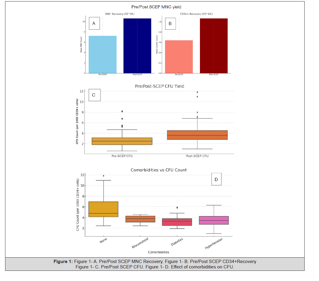
Figure 1:Figure 1- A. Pre/Post SCEP MNC Recovery. Figure 1- B. Pre/Post SCEP CD34+Recovery Figure 1- C. Pre/Post SCEP CFU. Figure 1- D. Effect of comorbidities on CFU.
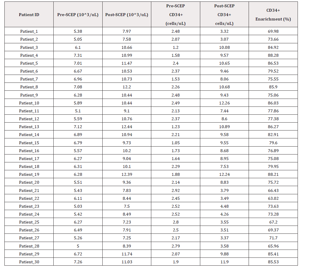
Table 1:Pre-SCEP/Post-SCEP MNC and CD34 yield.
References
- Agarwal A, Mulgund A, Hamada A, Michelle Renee Chyatte (2015) A unique view on male infertility around the globe. Reprod Biol Endocrinol 13: 37.
- Liu J, Qin Y, Liu H, Yonglin Liu, Yi Yang, et al. (2025) Global, regional, and national burden of female infertility and trends from 1990 to 2021 with projections to 2050 based on the GBD 2021 analysis. Sci Rep 15(1): 17559.
- Saad Naguib MH, Kenfack Y, Sherman LS, Chafitz OB, Morelli SS (2024) Impaired receptivity of thin endometrium: therapeutic potential of mesenchymal stem cells. Front. Endocrinol 14: 1268990.
- Kesharwani DK, Mohammad S, Acharya N, Joshi KS (2022) Fertility With Early Reduction of Ovarian Reserve. Cureus 14(10): e30326.
- Maacha S, Sidahmed H, Jacob S, Gentilcore G, Calzone R, et al. (2020) Paracrine Mechanisms of Mesenchymal Stromal Cells in Angiogenesis. Stem Cells Int 2020: 4356359.
- Chatzianagnosti S, Dermitzakis I, Theotokis P; Kousta E; Mastorakos G; et al. (2024) Application of Mesenchymal Stem Cells in Female Infertility Treatment: Protocols and Preliminary Results. Life 14(9): 1161.
- Karpova D, Rettig MP, DiPersio JF (2019) Mobilized peripheral blood: an updated perspective. F1000Res. 8: F1000 Faculty Rev-2125.
- Bansode M, Kore SA, Lee Y, Ghodake V, Verma R (2022) Clinical Approach of Novel Autologous Stem Cell Therapy for Primary Ovarian Insufficiency and Endometrial Regeneration. J Stem Cell Res Dev Ther 8: 102.
- Vasu S, Leitman SF, Tisdale JF, Hsieh MM, Childs RW, et al. (2008) Donor demographic and laboratory predictors of allogeneic peripheral blood stem cell mobilization in an ethnically diverse population. Blood 112(5): 2092-100.
- Martino M, Gori M, Pitino A, Massimo Gentile, Antonia Dattola, et al. (2017) Basal CD34+ cell count predicts peripheral blood stem cell mobilization in healthy donors after administration of granulocyte colony-stimulating factor: a longitudinal, prospective, observational, single-center, cohort study. Biol Blood Marrow Transplant 23(7): 1215–1220.
- Ferraro F, Lymperi S, Méndez Ferrer S, Saez B, Spencer JA, et al. (2011) Diabetes impairs hematopoietic stem cell mobilization by altering niche function. Sci Transl Med 3(104): 104ra101.
- Flannery M, Culakova E, Canin B, Peppone LJ, Ramsdale E, et al. (2021) Understanding Treatment Tolerability in Older Adults With Cancer [Review of Understanding Treatment Tolerability in Older Adults With Cancer]. J Clin Oncol 39(19): 2150-2163.
- Selimoglu Buet D, Wagner Ballon O, Saada V, Bardet V, Itzykson R, et al. (2015) Characteristic repartition of monocyte subsets as a diagnostic signature of chronic myelomonocyt i c leukemia. Blood 125(23): 3618-3626.
- Chang EL, Parker JD, Groshen S, Watkins K, Douer D (1998) Comparison of Progenitor Cell Content in Sequential Peripheral Blood Progenitor Collections after Mobilization with Chemotherapy and Granulocyte Macrophage Colony- Stimulating Factor. Acta Haematologica 99(2): 69-75.
- Fiala MA, Slade M, Wang S, Park S, DiPersio JF, et al. (2016) The impact of diabetes mellitus and other comorbidities on hematopoietic stem cell collection and hematologic recovery post-transplantation. In Leukemia & lymphoma/Leukemia and lymphoma 58(1): 241.
- Rajanna DB, Asha L, Vasanthi P, Srinivas C (2024) Endometrial preparation using CONCEIVF algorithm. Hum Reprod 39(Suppl 1): deae108.170.
- Anitha D, Vasanthi P, Srinivas C (2024) Evaluating Implansera in frozen embryo transfer. Hum Reprod 39(Suppl 1): deae108.693.
- Katakam DN, Sneha G, Srinivas C, Vasanthi P (2024) NOA-SERA for male infertility: case series.Hum Reprod 39(Suppl 1): deae108.433.
- Martino Massimo, Mercedes Gori, Tiziana Moscato, Virginia Naso, Anna Ferreri, et al. (2019) Challenge to Predict Mobilized Peripheral Blood Stem Cells on the Fourth Day of Granulocyte Colony-Stimulating Factor Treatment in Healthy Donors: Predictive Value of Basal CD34+ Cell and Platelet Counts. Biol Blood Marrow Transplant 25(8): 1586-1591.
- Tran MH, Cai Y, Stroncek D (2024) Liquid Storage of Peripheral Blood Stem Cell Products: Effects of Time and Temperature on Product Quality. Transplant Cell Ther 30(1): 120.e1-120.e10.
- Elshoury A, Maguire O, Conway A, Tario J, Soh KT, et al. (2023) Short-Term Storage of Mobilized Peripheral Blood Stem Cells in a Closed System Changes the Microenvironment and May Affect the Quantity of CD34+ and CD34+CD38-CD45RA-CD90+ Cells. Transplant Cell Ther 29(2): 112.e1-112.e9.




 We use cookies to ensure you get the best experience on our website.