
Research Article 
 Creative Commons, CC-BY
Fulcrum and Function of the Helical Heart: State of Research
*Corresponding author: Jorge Carlos Trainini MD PhD, Presidente Perón Hospital, Avellaneda, and National University of Avellaneda, Argentina.
Received:June 02, 2025; Published:June 09, 2025
DOI: 10.34297/AJBSR.2025.27.003550
Abstract
The pathway leading from cardiac structure to function induced to research on topics poorly explained by their mechanical organization
(1-3), but which should be considered complementary among them and essential for the physiology of the heart, to know:
1) Anatomical and histological investigation of the segmental sequence of the myocardial band.
2) Support and insertion of the myocardial band.
3) How is ventricular torsion produced?
4) Is there a histology that explains the phenomenon of friction between the layers of the myocardium? Is there an organic lubricating
source?
5) How is active protodiastolic ventricular suction produced?
6) Is it possible to consider in the heart a coupling phase between systole and diastole where cardiac suction takes place? Which
is the energy mechanism in the active suction phase?
7) Is the mechanical consequence of the cardiac structure the initiation of stimulation in the anatomical-functional unit formed
between the AV node and the cardiac fulcrum, and its continuity with the torsion of the myocardium, by opposite rotation between
the base and the apex with simultaneous shortening of both ventricles?
In conclusions: 1. We consider there is enough evidence that the fiber orientation and the opposite base –apex rotational movement of the heart, justifies the muscle band model; 2. The finding of the cardiac fulcrum gives support to the spiral myocardial band being the point of fixation that allows the helicoidal torsion; 3. The hyaluronic acid would act as a lubricant and provide great resistance to mechanical pressures in a functional association with the venous ducts of Thebesius and Langer, that would intervene as tributaries of the necessary hydration to said element and thus could counteract the friction of the surfaces by exercising an anti-friction mechanism; 4. This study explains the ventricular twist and the active suction mechanism during the isovolumic diastolic and early ventricular filling phases, in contrast with the traditional concept of passive relaxation during the diastolic isovolumic phase and the vis a tergo as mechanism for ventricular filling; 5. The deep anatomical, histológical, electrofisiological and functional investigations that our group have carried out at the level of the different segments of the muscular band allows a better interpretation of cardiac physiology and its clinical application is unquestionable in light of new cardiac images techniques.
Keywords:Cardiac function, Cardiac Fulcrum, Myocardial torsion
Introduction
The function of the heart corresponds to a mechanical dimension that should be addressed in terms of its structure, which is where we find the origin of the idea that led our research to explain its organic-functional integrity. If we stop in classical descriptions of the heart, we realize that anatomical attention was focused on its external and internal surfaces, granting scant importance to the intimate muscle conformation [4]. This was believed to be of a homogeneous solid nature with global uniform contraction, not considering that its mechanical capacity demanded a reinterpretation of its spatial anatomy and motions, leading us into other topics of its functioning that were completely disregarded by cardiology [5,6]. The anatomy of the heart was traditionally thought to be formed by spiralling muscle bundles, but these were never described in association with their physiology. Was Torrent Guasp who in 1970 [7,8] initiated the description and interpretation of the myocardial muscle band, starting point to understand its motions. This was demonstrated in multiple dissections showing that the ventricular myocardium is made up of a group of muscle fibers coiled unto themselves, resembling a rope, flattened laterally as a band, which by giving two spiral twists describes a helix that limits the two ventricles and defines their performance (Figure 1).
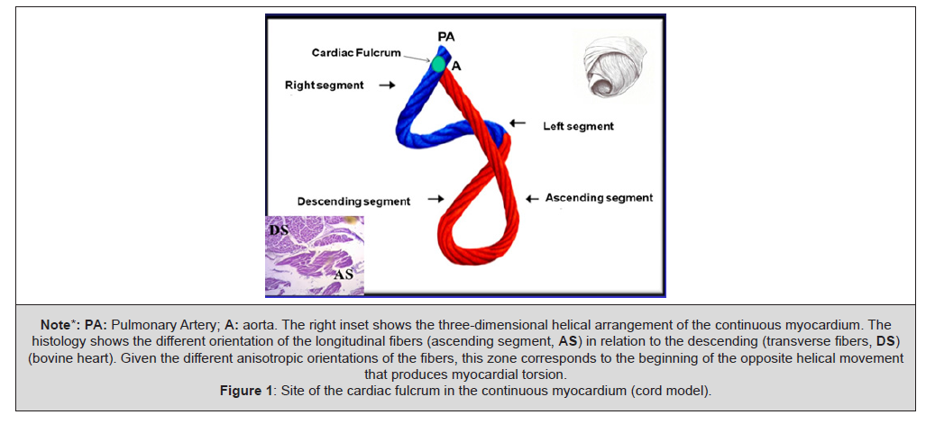
Figure 1:Site of the cardiac fulcrum in the continuous myocardium (cord model).
An explanation for this muscle homogenization with an intricate anatomical arrangement that hides the myocardial band, implies considering that its structural solidity is required in birds and mammals so that blood is ejected at high speed in a limited time span by an organ that must serve two circulations (systemic and pulmonary) [9]. The anatomical evolutionary state of the heart agrees with ventricular mechanics but lacked the understanding of an electrical propagation that could accurately explain the physiology. The studies in our investigations on this topic aim to show the integrity of an essential cardiac structure-function [1-10]. The left ventricular endocardial and epicardial electrical activation performed in patients with Three-dimensional Electro anatomical Mapping (TEM) allowed considering this fundamental topic to analyse it. This pathway leading from structure to function induced to research on topics poorly explained by their mechanical organization (1-3), but which should be considered complementary among them and essential for the physiology of the heart, to know:
1) Anatomical and histological investigation of the segmental sequence
of the myocardial band.
2) Support and insertion of the myocardial band.
3) How is ventricular torsion produced?
4) Is there a histology that explains the phenomenon of friction
between the layers of the myocardium? Is there an organic lubricating
source?
5) How is active protodiastolic ventricular suction produced?
6) Is it possible to consider in the heart a coupling phase between
systole and diastole where cardiac suction takes place? Which
is the energy mechanism in the active suction phase?
7) Is the mechanical consequence of the cardiac structure the initiation
of stimulation in the anatomical-functional unit formed
between the AV node and the cardiac fulcrum, and its continuity
with the torsion of the myocardium, by opposite rotation
between the base and the apex with simultaneous shortening
of both ventricles?
Material and Methods
The methods used in this study to explain the hypothesis of the anatomy-functional integrity of the heart were:
1) Eighty-four hearts from the morgue, slaughterhouses, and breeding farms (for anurans) were used: a) 54 two-year-old cattle weighing between 1300 and 1900 g (average 1650 g); b) 17 humans (three at 8, 16, and 23 weeks of gestation, respectively; four infants at 30 days, 36 days, 10 weeks, and 27 weeks; one 4-year-old child; one 10-year-old child; and eight adults with an average weight of 300 g); c) three porcine hearts (400 g); d) 10 anuran hearts.
2) Anatomical, histological, histochemical, and radiological studies were performed. The hearts were fixed in 10% buffered formalin. Histology was performed with haematoxylin and eosin, Masson’s trichrome staining, and 4-micron sections. 10% formalin was used as a buffer. Immunostaining (s100-neurofilaments) was also performed. All samples underwent histochemical analysis using Alcian blue staining, a reliable marker for identifying the presence of hyaluronic acid, as an anti-friction mechanism, and also providing a semiquantitative assessment
3) Left ventricular endocardial and epicardial electrical activation in humans (five adult patients). The left ventricular endo and epicardial electrical activation sequence was studied using three-dimensional electroanatomic mapping (3D-EAM) with a navigation system and Carto mapping, enabling three-dimensional anatomical representation, with activation and electrical propagation maps. Isochronic and activation sequence maps were performed, correlating them with surface ECG. Since the left ventricular muscle structure is composed of an endocardial and an epicardial portion, or descending and ascending segments, respectively, in the anatomical classification (Figure1), two access routes were used to perform the mapping. The endocardial access was achieved by conventional atrial transeptal puncture and the epicardial access by percutaneous approach in the pericardial cavity with an ablation catheter. Endocardial and epicardial mapping were immediately and consecutively performed. In addition, the electrical activation propagation times of the muscle band were measured in milliseconds (ms) [13-15].
4) Echocardiography and RNM to corroborate previous studies and the usefulness of these knowledge in clinical practice.
Results
Myocadiac Architecture
Torrent Guasp demonstrated through multiple dissections in hearts of different species, including the human, that the ventricular myocardium is made up of a helical band (Figures 1 and 2) [3]. This study approach correlates with a cardiac structure presenting the remarkable characteristic of being a driving-suction pump with the size equivalent to a human fist and an average weight of 270grams, which ejects 4-6 litres/minute at a speed of up to 300cm/s, consuming only 10watts, and working without interruption for 80 years without maintenance, almost without noise, and no smoke. Its work is equivalent to the daily withdrawal of 1 ton of water 1m deep with a mechanical efficiency (work/energy relationship) of 50%, not achieved by man-made machines which only attain 30% efficacy. This allows ejecting 70% of the left ventricular volume with only 12% shortening of its contractile unit, the sarcomere (Figure 2) [16].
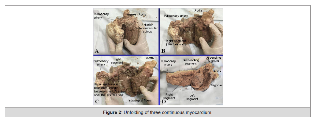
Figure 2:Unfolding of three continuous myocardium.
In view of the criticism or indifference met by the helical myocardial band, its dissection finds a structure with defined planes where the successive and related physiological heart motions of narrowing, shortening-twisting, lengthening-untwisting and expansion take place depending on the propagation of the electrical stimulus along its muscle pathways. It should be recalled that the myocardium constitutes a spiralling continuum in its fibers responding to the helical pattern in its muscle bundles. This arrangement indicates the need of generating a mechanical work that dissipates little energy. Therefore, the fiber layers very gradually shift their orientation, with more or less acute angles, to avoid that abrupt changes in the spatial organization dissipate the necessary work for cardiac function [17]. This situation generates a tangle of fibers that allows the band to behave as a continuous transmission chain with the epicardial fibers taking an oblique direction, the intermediate fibers a transverse course and the endocardial fibers also an oblique direction, but contrary to that of the epicardial plane. The endocardial and epicardial plane access angle is approximately 60 degrees in relation to the transverse fibers. Fiber orientation defines function and thus the ejection fraction is 60% when the normal helical fibers contract and falls to nearly 30% if only the transverse fibers shorten, reaching 15-20% if only the subendocardial longitudinal fibers are deformed. This occurs when the left ventricle dilates in cardiac remodeling and the fibers miss their oblique orientation, loosing muscular and mechanical efficiency [18]. In the progression from the ventricular base to the apex, the number of horizontal fibers decreases in relation to the oblique fibers, showing that the heart is organized as a continuous muscle helix (Figure 3).
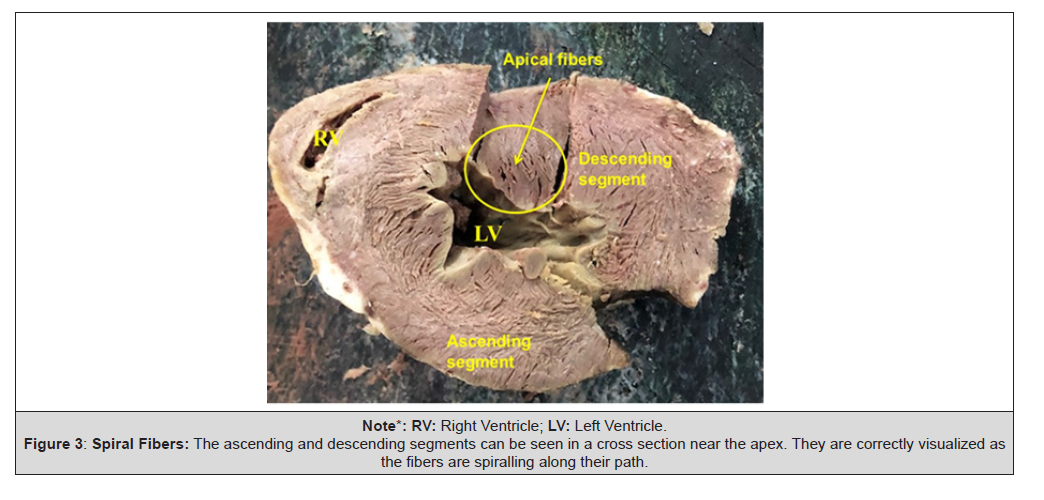
Figure 3:Spiral Fibers: The ascending and descending segments can be seen in a cross section near the apex. They are correctly visualized as the fibers are spiralling along their path.
Myocardial muscle bundles and bands, which derive from phylogenetic development, essentially shape a master axis of precise dynamic requirement. The spatial muscle structure adopted by the myocardial muscle band has a double function: a) to limit the ventricular chambers and b) to fulfil the suction and driving action in its role of cardiac pump.
Segmentation of the Myocardial Band
Figure 4 and 5 shows the unfolded muscle band. Being able to unfold the myocardium with a similar thickness in all its extension proves that the band is real and not a “heuristic” or biased construction. In its course, the myocardial band adopts a helical configuration that defines the two ventricular chambers. The myocardial band is characterized by a descending and an ascending band. The former includes the right, left and descending segments, while the latter is formed by the remaining ascending segment (Figures 1-5).
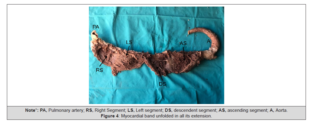
Figure 4:Myocardial band unfolded in all its extension.
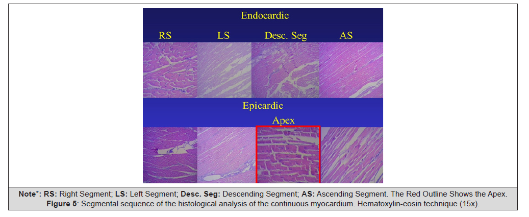
Figure 5:Segmental sequence of the histological analysis of the continuous myocardium. Hematoxylin-eosin technique (15x).
The myocardial band describes two spiral turns with the insertion of its initial end along the line extending from the pulmonary artery to the orifice of the tricuspid valve, called the palm-tricuspid cord, in front of the aorta, while its final end attaches below the aortic root. Both ends are fixed by an osseous, chondroid or tendinous nucleus, depending on the different species (animal or human) used in the studies. This nucleus, which we have called cardiac fulcrum [4,18-23], is the only perceptible edge where the muscle band fibers originate and end (Figures 1,6-10). These insertions must be understood as the supporting point of the myocardial band to fulfill its hemodynamic function. In our investigations we have not found insertion of cardiomyociytes in the collagen matrix of the trigones.
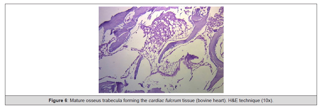
Figure 6:Mature osseus trabecula forming the cardiac fulcrum tissue (bovine heart). H&E technique (10x).
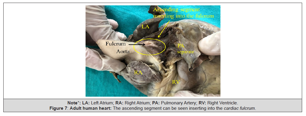
Figure 7:Adult human heart: The ascending segment can be seen inserting into the cardiac fulcrum.
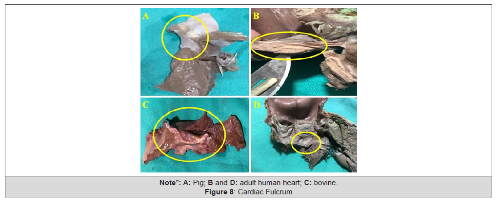
Figure 8:Cardiac Fulcrum.
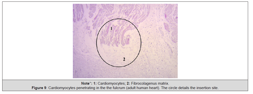
Figure 9:Cardiomyocytes penetrating in the the fulcrum (adult human heart). The circle details the insertion site.
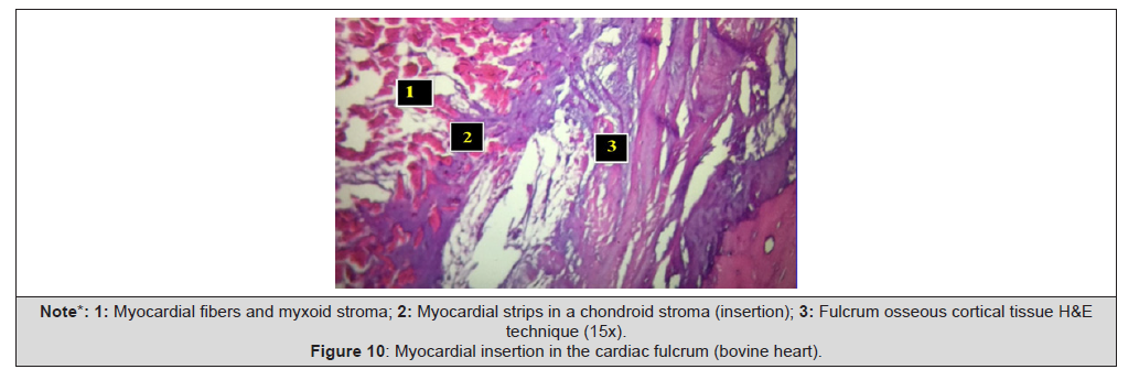
Figure 10:Myocardial insertion in the cardiac fulcrum (bovine heart).
Myocardial Support: The Cardiac Fulcrum
In anatomical investigations we have found in all bovine and human hearts a structure, whose osseus or chondroid histological composition is different, depending on the specimens analysed. Both the muscle fibers of the right segment (initial insertion) and those of the ascending segment (final insertion) are attached to this myocardial support. During the dissection of the myocardium, finding the cardiac fulcrum as the structure where the myocardium is inserted, led us to further enquiries: What are its characteristics? Where is it situated? What is its histology? Is its presence analogous in different species? How is the myocardial muscle inserted into this structure that we have called cardiac fulcrum? This site of insertion, which we have called cardiac fulcrum, is located in the vicinity of the tricuspid valve (right), the aorta (superior) and the pulmonary-tricuspid cord (anterior).To find it, it is necessary to move the pulmonary artery and the right segment to the left of the observer, baring the root of the aorta This maneuver reveals the fulcrum below the aorta and inferior to the right trigone, not continuous with it, below the origin of the right coronary artery, detached from the aortic continuity and constituting a complementary element between the aorta and the myocardium. An osseus formation, called “os cordis” in mammals (bovine, buffalo, sheep, antilope, deer, giraffe, camel, dog, cat pig, sea lion, horse, elephant, chimpanzee) is reported in the veterinary literature. Beyond its mere reference, no reason for its presence or function was ever assigned to this structure until our investigations, and was not described in humans.
The consistency of the cardiac fulcrum in bovine hearts, osseous on palpation, has been confirmed by histology. Its size, corroborated by dissection and computed tomography, is approximately 37mm long, 45mm wide and 15mm thick, with a triangular shape. The microscopic analysis of the bovine cardiac fulcrum shows a trabecular osteochondral matrix with segmental lines. Its general structure resembles the metaphyseal growth of long bones, and increased magnification reveals bone trabeculae with osteoblasts and segmental lines secondary to bone apposition. The same histological findings have been encountered in chimpanzees. The histological analysis of the fulcrum in adult human hearts (approximate size 25mm long and 15mm wide) evidenced a chondroid-tendinous matrix, which needs additional clarification. As a rule, there is similar consistency in the detection, location and morphology of the fulcrum in all the hearts analysed. They present myocardium insertion in the rigid fulcrum structure, forming a cardiomyocyte-matrix unit, independently of its osseous, cartilaginous or tendinous nature in the different specimens studied. As in any muscle, this point of attachment acts as a lever and also as a bearing, preventing the transfer of the ventricular rotation force, either by torque or torsion stress, to the great vessels, thus, dissipating the energy produced by the helical muscle movement.
Having found an osseous structure in the bovine os cordis and its relationship with the myxoid-chondroid texture in human hearts, even in embryos, is rational from the point of view of interpretation analysis. This disparity is associated with the different evolution given by age from chondroid to osseous material and with the greater power developed by bovine hearts, requiring a more rigid supporting point. Therefore, the histological analysis of the fulcrum in adult human hearts evidencing a chondroid-tendinous collagenous matrix, needs further clarification. The fact that it has been found in humans and different species implies that from a functional point of view its presence is synonym of myocardial insertion, as established in all histological analyses, becoming a solid point of reasoning to achieve its biomechanical function. And we find this demonstration when the histological examination is directed to the myocardial insertion in the cardiac fulcrum, be it of osseous, chondroid or tendinous nature. In all the hearts analyzed and according to the studies we have carried out in this investigation, this myocardial attachment was found to be as “ivy clinging to stone” in the rigid structure of the fulcrum, integrating an osseus, cartilaginous or tendinous cardiomyocyte-matrix unit. In this concept there is analogy between skeletal and myocardial muscle. The former contracts between a fixed and a mobile supporting point, and this situation is found in the continuous myocardium, as there is greater solidity in the insertion between the fulcrum and the ascending segment compared with the attachment of the right segment in this support.
At this point, fundamental questions arise: Why have we found that in adult human hearts the cardiac fulcrum has characteristics similar to a tendon despite it fulfils the same function of attaching the helical myocardium as in other species? Why does it not have the same structure as that found in the human embryo or child heart? Our interpretation is that perhaps the osseous-cartilaginous cardiac fulcrum is a vestigial organ specific of mammalian evolution. A vestigial structure must be understood as the preservation during the evolutionary process of genetically established attributes which have lost all or part of their ancestral function in a certain species. As a result, it is found in the initial process of human gestation, but later loses its osteo-cartilaginous histology, remaining as a tendinous matrix sufficient to achieve myocardial insertion and accomplish a muscle power which is much lower than that of larger mammals. Let us recall that in bovines the fulcrum found in this investigation is of osseus nature. To faithfully establish the identity of the cardiac fulcrum, a histological analysis has also been carried out on the trigones, trying to find cardiomyocytes as a probable cardiac muscle insertion in these structures. In our investigation, only collagenous tissue without cardiomyocytes was observed in the trigones, confirming that the fulcrum is the support of the myocardium, both at its origin and end (19,21-23) (Figure 6-10).
In the band we can distinguish the basal and apical loops. The basal loop extends from the base of the pulmonary artery to the central band twist. On the other hand, the apical loop courses from this point of inflexion to the base of the aorta. In turn, each loop consists of two segments. The basal loop consists of the right and left segments and the apical loop of the descending and ascending segments (Figure 1). In the general loop configuration, the basal loop envelops the apical loop, so that the right ventricular chamber presents as an open slit in the muscle mass thickness forming both ventricles). The segments, in turn, are defined by anatomical structures (Figure 4). These concepts indicate that the right ventricular free wall is formed by one loop (basal) and the left ventricular free wall by both loops (basal and apical). The fundamental point for cardiac mechanics is that the base and apical muscle fibers course in different directions. This disparity finds correlation with the fiber trajectories and the helical pattern of the muscle band limiting the ventricles.
Histological Analysis of the Myocardial Band.
The myocardium is a syncytial muscle with lateral bridges between its muscle fibers and a laminar histological arrangement of the muscle bundles. Due to its histological arrangement, the myocardium (syncythyum) behaves like an auxetic biological material; in other words, it contracts simultaneously in two directions (longitudinal and circumferential) and thickens in a third direction (radial, towards the ventricular centroid). The myocardium is made of incompressible and elastic muscle tissue, so if it contracts in two directions (longitudinal and circumferential) it must thicken in the third (radial). The histological analysis sequence of the unfolded myocardial band (Figure 5) demonstrates its linear orientation according to the segmental continuity of its spatial organization when the band is coiled, both in its internal and external surfaces. These orientations are identical in both surfaces (internal and external). It can be seen that the myocardial structure is not a lattice (crisscross of myocardial fibers) but a band. The lattice concept used was developed due to the band folding resulting in overlapping segments, which are functionally independent and with friction between their surfaces [20]. This arrangement is essential to achieve myocardial torsion, a fundamental action of cardiac mechanics that would be impossible with the lattice structure.
No segment of the band histological sequence explored in our investigations presents a lattice organization. As the external surface of the distal descending segment (Figure 5, lower panel) twists to become the ascending segment, the cardiomyocytes generate in the planimetric histological sections a different architecture in their orientation from that of the internal surface, only site (cardiac apex) where this situation occurs. The rest of the orientation is always parallel. In the apex, the spiral course of the myocardial fibers, which shift from the periphery towards the centre, determine a torsion where the subepicardial fibers become subendocardial, overlapping like the tiles of a roof, as evidenced in this image (Figure 3). As previously expressed, the opposite orientation and rotational movement of the left ventricular fibers, both at the basal and distal apical segment levels, explain myocardial band model. The interventricular septum consists of a ventral and a dorsal part. The first portion consists of the left descending segment, the intrarectal band (final segment of the muscle band) and the anterior septal band. The first two belong to the left ventricle and the remaining one to the right ventricle. The posterior region of the septum is composed of the left descending segment (corresponding to the left ventricle) and the posterior septal band (corresponding to the right ventricle). The spatial arrangement and the rotational motion of the ventricular fibers, conform the architectural plan of the muscle band. Up to this moment, a classic interpretation has been made of blood circulation through the different heart chambers, which does not correlate with its muscle dynamics. And this is, ultimately, the engine for the circulation established by the myocardial band, which also defines with its muscle structure, the limits of the chambers through which blood flows. This spatial arrangement of the muscle band, mainly at the level of descending and ascending segments, is the one that grants the ventricular chambers the shortening-twisting and lengthening-untwisting motions which are essential for cardiac function.
Muscle Friction
The opposing sliding motion of the left ventricular internal segments in relation to the external segments to achieve the mechanism of ventricular torsion, generates an inevitable friction between them (Figure 11). This would entail a high energy cost if the heart did not have a spongy system, with the participation of Thebesian and Langer venous conduits that act as a lubricating antifriction system: this is the hyaluronic acid, which flows across the myocardial thickness (Figure 12). Hyaluronic acid was one of our discoveries in this research [24-28].
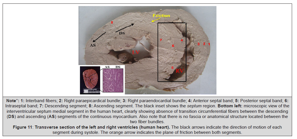
Figure 11:Transverse section of the left and right ventricles (human heart). The black arrows indicate the direction of motion of each segment during systole. The orange arrow indicates the plane of friction between both segments.
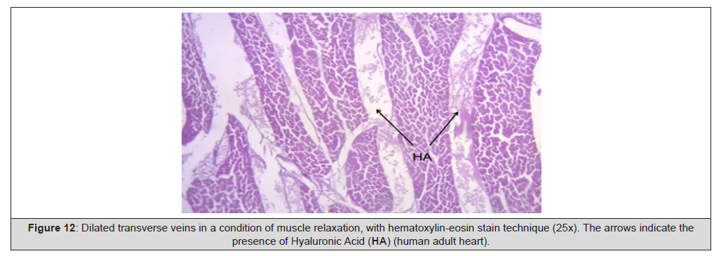
Figure 12:Dilated transverse veins in a condition of muscle relaxation, with hematoxylin-eosin stain technique (25x). The arrows indicate the presence of Hyaluronic Acid (HA) (human adult heart).
Cardiac Electric Activation
As EAM corresponded to the left ventricle, the activation wave previously generated in the right ventricle was not obtained. Electro anatomical mapping took an average of 20 minutes. There were no complications related to the procedure itself or any of the approaches. Figures 13-15 show the projection of the endocardial an epicardial electric activation. In all the Figures the right projection is observed in the left panel and the left anterior oblique projection is simultaneously observed in the right panel. The zones activated at each moment are detailed in red. On the lateral part, the activation of the descending and ascending muscle bands that make up the muscle structure of the ventricular band in the rope model is represented. In it, the area depolarized at that moment is represented in red and those that were previously activated and are in the refractory period are represented in blue. Below the rope model, the average electrical propagation time along the muscle band can be seen measured in milliseconds(ms) at the analyzed site. Activation of the left ventricle occurs 12.4ms±1.816ms after its onset in the interventricular septum (Figure 13 A). At that moment it also spreads to an epicardial area-ascending bands-evidencing a radial activation at a point we call “band crossover” that occurs on average 25.8ms±1.483ms after septal stimulation (Figure 13 B) and at 38.2ms±2.135 from cardiac activation onset. Synchronously, following the anatomical arrangement of the descending band, the activation moves axially towards the ventricular apex reaching it at an average of 58ms±2.0ms (Figures 14 A and B). At the band “crossover” the activation loses its unidirectional character and becomes slightly more complex. Three simultaneous wave fronts are generated: 1) the distal activation of the descending band towards the apical loop; 2) the depolarization of the ascending band from the crossover towards the apex and 3) the activation of this band from the crossover towards the end of the muscle band in the aorta. Figures 14 B, 15 A and B show the continuation and completion of this process. Intraventricular activation ends long before the termination of the QRS (Figure 15 A). The rest of the QRS corresponds to the late activation of the distal portion of the ascending band, which justifies the persistence of its contraction during the diastolic isovolumic phase, constituting the basis of the ventricular suction mechanism (Figure 15 B). A synthesis of the stimulation found in this study is shown with the rope model in Figure 16 (upper panel).
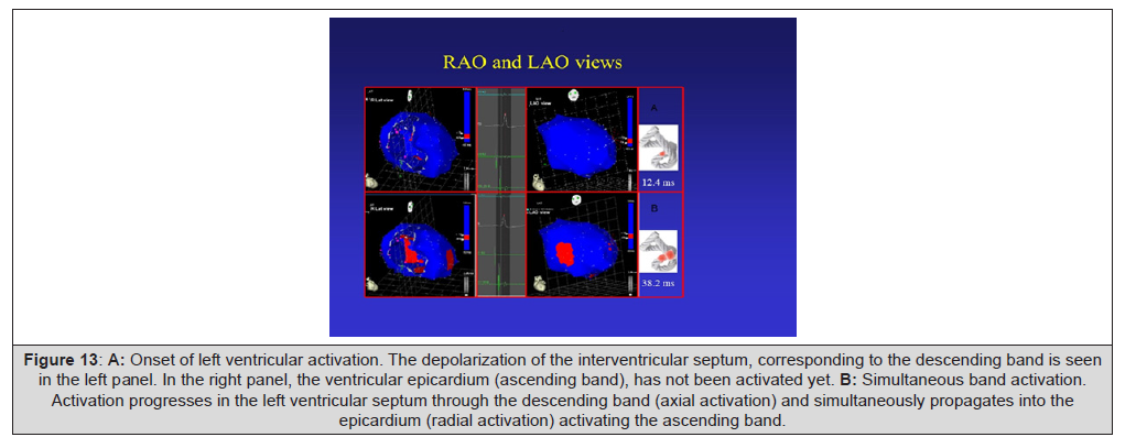
Figure 13:A: Onset of left ventricular activation. The depolarization of the interventricular septum, corresponding to the descending band is seen in the left panel. In the right panel, the ventricular epicardium (ascending band), has not been activated yet. B: Simultaneous band activation. Activation progresses in the left ventricular septum through the descending band (axial activation) and simultaneously propagates into the epicardium (radial activation) activating the ascending band.
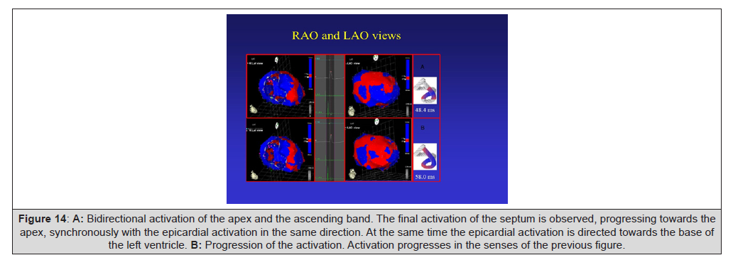
Figure 14:A: Bidirectional activation of the apex and the ascending band. The final activation of the septum is observed, progressing towards the apex, synchronously with the epicardial activation in the same direction. At the same time the epicardial activation is directed towards the base of the left ventricle. B: Progression of the activation. Activation progresses in the senses of the previous figure.
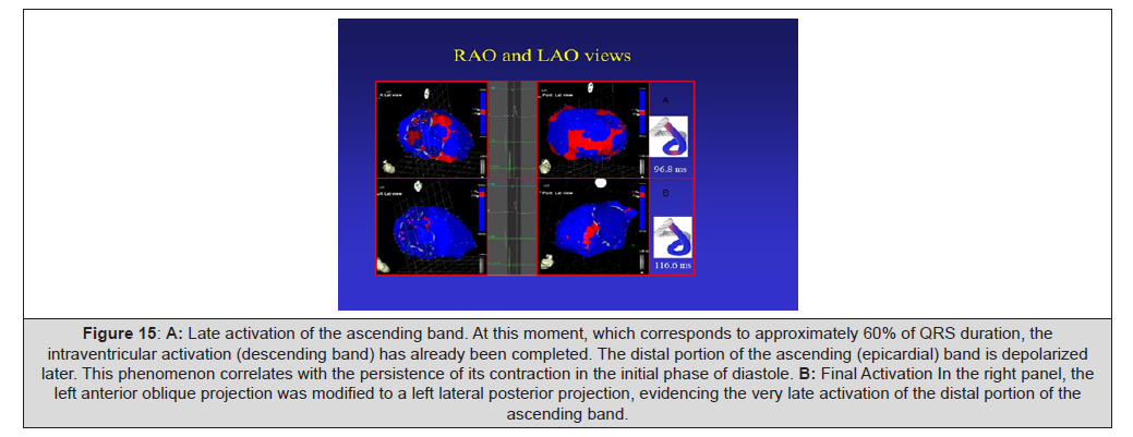
Figure 15:A: Late activation of the ascending band. At this moment, which corresponds to approximately 60% of QRS duration, the intraventricular activation (descending band) has already been completed. The distal portion of the ascending (epicardial) band is depolarized later. This phenomenon correlates with the persistence of its contraction in the initial phase of diastole. B: Final Activation In the right panel, the left anterior oblique projection was modified to a left lateral posterior projection, evidencing the very late activation of the distal portion of the ascending band.
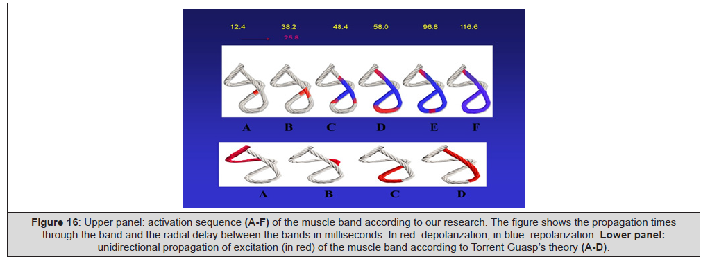
Figure 16:Upper panel: activation sequence (A-F) of the muscle band according to our research. The figure shows the propagation times through the band and the radial delay between the bands in milliseconds. In red: depolarization; in blue: repolarization. Lower panel: unidirectional propagation of excitation (in red) of the muscle band according to Torrent Guasp’s theory (A-D).
According to Torrent Guasp, longitudinal diffusion of stimuli along the ventricular myocardial band explained the performance of the heart (Figure 16, lower panel). However, this sequential “peristaltic” activation does not correlate with some currently wellknown fundamental phenomena, as clockwise and counterclockwise twisting at the left ventricular apex and base, which are mainly responsible for its mechanical efficiency (Figure 16, upper panel). Our research modifies these concepts since stimulus propagation is simultaneously axial and radial. A relationship must be found between activation and the mechanical result. The explanation is given by the simultaneous axial and radial electrical conduction pathways when they reach the band crossover, and is also supported by the spatial arrangement of the fibers, the subendocardial ones being located on the right side and the subepicardial ones on its left side (Figure 13-16).
Stimulus Propagation and Left Ventricular Twisting
As noted, left ventricular activation begins in the endocardial descending band, which is almost simultaneously depolarized axially and radially. At the crossover point of both bands, the activation spreads from the endocardium to the epicardium (radial propagation), that is, from the descending to the ascending band. From this point onwards, the ascending band depolarizes in two senses: towards the apex and towards the base, at the same time that the descending band completes its activation towards the apex (Figure 15). Thus, two essential phenomena occur:
1) As the apical loop depolarizes from the band crossover in two
simultaneous wave fronts (from the descending and from the
ascending bands) it generates their synchronized contraction.
2) The activation of the ascending band propagates from the
band crossover in two opposing directions: towards the apex
and towards the base (Figure 15). The resulting mechanical
contraction will also have a divergent direction, giving origin
to the apical and basal clockwise and counterclockwise rotations,
respectively.
The contractile behaviour like an auxetic biological material (contracts simultaneously in direction longitudinal and circumferential), together with the helical anatomical arrangement of the myocardial fibers around the ventricular cavity, results in thickening radial myocardial and left ventricular wringing, which is a combination of twisting and reduction of the longitudinal axis. This muscular action results in a squeezing of the left ventricle. Thickening occurs towards the ventricular centroid only, and is not accompanied by external thickening towards the pericardium. Furthermore, there is a decrease in cardiac diameter, as would be seen in a M-mode section of the LV in the parasternal long axis plane; in such circumstances, the external cardiac diameter would be smaller in Telesis tole than in tele diastole, despite myocardial thickening. This is due to the helical arrangement of the myocardial fibers, which contribute to the thickening of the myocardium.
Active Suction in the Diastolic Isovolumic Phase.
A fundamental issue we investigated, which did not have electrophysiological evidence, was to consider ventricular filling as an active phenomenon generated by a myocardial contraction that tends to lengthen the left ventricular apex-base distance after the ejective phase, thus producing a suction effect similar to a “suction cup”. This mechanism is explained by the persistence of the ascending segment contraction during the isovolumic diastolic phase [29- 35]. We have found that the endocardium is completely depolarized during the first part of the QRS. If according to our studies the depolarization of the ascending segment starts 25.8ms on average after that of the descending segment and its contraction persists for the same period of time, the condition of ventricular contraction will last approximately 400 ms. On the other hand, as ventricular systole lasts about 300ms, the remaining 100ms correspond to the diastolic isovolumic phase (erroneously called isovolumic relaxation, because as we see there is ventricular contraction). Briefly, during the initial part of this phase the ascending segment remains contracted as a result of the depolarization that occurred during the QRS [36,37]. The final part of the QRS corresponds in our investigation to the activation of the ascending segment. In this way, during the diastolic isovolumic phase, the contraction necessary to generate suction (“suction cup effect”) occurs. With the onset of untwisting during the diastolic isovolumic phase the ascending segment progressively lengthens, generating negative intraventricular pressure with this segment still contracted (active process) as an energy residue of the twisting process.
This suction phase between systole and diastole lasts around 100ms and is active during muscle contraction with an intraventricular pressure drop below zero (Figure 17). The difference in the duration of mean systolic strain between the systolic and postsystolic phase in the echocardiographic work of Mora Llabata, et al., is 88±7.1ms, a value analogous to those found in our research in relation to the duration of the ascending segment activation in the suction phase [38-40].
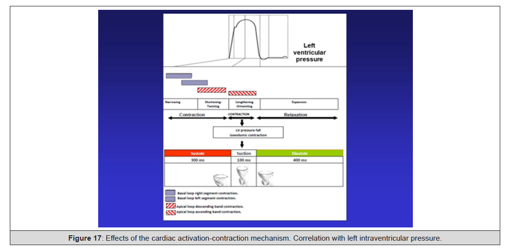
Figure 17:Effects of the cardiac activation-contraction mechanism. Correlation with left intraventricular pressure.
We have measured the left intraventricular pressure curve in patients who underwent cardiac resynchronization therapy in order to show the improvement at the level of the suction mechanism in the diastolic isovolumic phase. With the resynchronizer catheter positioned in the left ventricular endocardium (according to our research in the area of band “crossover”) normal electrical activation is restored, achieving normalization of the segments’ mechanical sequence and the restoration of the suction mechanism with the drop in left ventricular diastolic pressure [41]. In Figure 18 A, the curve corresponds to the left intraventricular pressure in a patient with LBBB in which the resynchronizer has been turned off. In Figure 18 B, in the same patient, the function of the device has been restored. It can be seen on the intraventricular pressure scale, to the right of each panel (in mmHg), that the diastolic pressure has decreased from 13 to 0mmHg with ventricular resynchronization. The increase in systolic pressure by 10mmHg has also been observed.
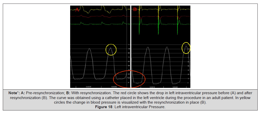
Figure 18:Left intraventricular Pressure
Echocardiography
Echocardiography has the ability to provide non-invasive understanding about the complex mechanism of myocardial contraction. For over 15 years the speckle tracking technique has been used to assess strain or myocardial deformation. This tool of two-dimensional (2D) ultrasound provides a regional qualitative analysis of left ventricular function, confirming the relationship between the cardiac anatomy of the helical muscle band and diverse echocardiographic findings. According to Ballester et al. the different segments of the basal and apical loops of Torrent Guasp’s myocardial band correlate with 2D ultrasound findings (10). The Left Ventricular Eyection Fraction (LVEF) results from the combined action of longitudinal and circumferential myocardic contraction. The distinctive helical structure of obliquely oriented myocardial fibers results in systolic rotation of the base and apex of the LV in opposite directions around its longitudinal axis (the algebraic subtraction of this rotation causes twisting of the heart muscle) and the simultaneous shortening of the LV as the base is displaced towards the apex, all of which results in ventricular wringing. The combination of these events determines ventricular radial thickening and LVEF. Speckle tracking echocardiography allows us to study the muscular behaviour of the myocardium by calculating Legendrian strain. Strain is negative when de original distance between 2 points decreases, as occurs in myocardial muscle contraction (longitudinal and circumferential contraction) and is positive when the distance between them increases, as occurs in muscle thickening (radial thickening). Its accuracy has been validated with sonomicrometry and especially with nuclear magnetic resonance, respectively.
The anatomical arrangement described by Torrent Guasp and the efficient myocardial function analyzed by our studies among others, demonstrate the following consequences [38,39]:
1) The myocardial muscle mass is more important at the mid-basal level where the basal and apical loop fibers join. This explains that the maximum strain amplitude obtained by radial strain occurs at this level (basal radial strain +44±18% vs. apical strain +23±16.5%).
2) The descending fibers adopt a progressively more oblique arrangement until they reach the apex. Effectively, as expected, longitudinal strain is markedly greater at the ventricular apex due to this oblique change in the spatial arrangement (apical longitudinal strain -21.9%±2.4 vs. basal longitudinal strain -19.6%±2.4).
3) Circumferential strain has greater amplitude at the apical level facilitating apical rotation (-25.6%±6.6); therefore, at the basal level the fibers are arranged more transversally, and consequently, basal strain results less important (-16.8±3.5).
4) To calculate the twist, the echocardiograph algorithm performs an algebraic sum (it adds the positive value of apical rotation to the negative one of basal rotation) In our experience, the value in normal subjects is around +19±9º, always with predominant apical rotation
5) Post-systolic longitudinal strain, which indicates the activity during relaxation, is basically produced in septal segments and in the anterior basal region corresponding to the anatomical location of the ascending segment [38].
The twist of an elastic and non-compressible material, such as the myocardium, involves a longitudinal shortening. In the case of the LV, the base and the apex rotate in opposite directions around the longitudinal axis of the LV at the same time as the base moves closer to the apex, which occurs due to the helical arrangement of the myocardial fibers. New parameters, possibly more adjusted to the current knowledge of physiology described, have been incorporated into our recent experience, including twist movements and the longitudinal shortening motions that are simultaneously developed during ventricular systole [39]. These are: a) the torsion index (twist/mitral annulus translational motion); and b) strain index (twist/left ventricular global longitudinal strain). Both parameters translate the active motions that are produced during systole “squeezing” the left ventricle, with a value of 13.1±4°/cm for the torsion index and 0.96±0.36°/% for the strain index in the healthy population studied. The “combined strain parameter” has also been postulated to assess left ventricular function. It includes the “strain product (twist × longitudinal strain)”, which is probably better to evaluate contractility, and the previously described “strain index (twist/global longitudinal strain), which would establish the type of predominant disease. Thus, a normal “strain product” (-387±137°x %) would translate into a preserved left ventricular global function, either by normality of both parameters or by the compensation provided between them (“pseudonormal” product). A reduced “strain product” could be due to the decrease of longitudinal strain, twist, or both, and would be characterized by the “strain index” (high, low or normal, respectively). Then, this “combined strain parameter” could help to better differentiate the disorder and in addition monitor the myocardial impairment derived from treatments or secondary to different cardiac diseases.
There are previous reports in the literature which have used the strain product (twist× longitudinal strain) as the best predictor of future cardiotoxicity by anthracyclines, due to a more global assessment of subendocardial and subepicardial fibers. The incorporation of strain parameters have allowed the recent classification of heart failure according to left ventricular mechanical function disorders. After observing specific anomalous models of ventricular mechanics in different subgroups of heart failure patients, an alternative approximation has been proposed to characterize it. Thus, cardiac failure can be classified into two big subgroups: 1) dominant longitudinal dysfunction; 2) transmural dysfunction (longitudinal and circumferential). This classification is based on the double helix oblique orientation of left ventricular myocardial fibers. Probably, the integration of pump and myocardial function parameters need to be jointly valued for a more accurate assessment of ventricular systolic function. Future studies should assess whether, during longitudinal follow-up of patients, values of myocardial torsion have prognostic value in terms of predicting ventricular failure. On the other hand, in processes that affect the entire myocardium from the early stages onwards, and not just the sub endocardium, myocardial torsion could provide extra information if both components are evaluated together.
In each different pathology, rotation, twisting and untwisting, are affected in different ways depending on myocardial torsion and the distribution of the most involved fibers, with clearly manifested characteristics in ventricular hypertrophy, diabetes, amyloidosis and heart valve and pericardial diseases. With a special technique known as Diffusion Tensor Magnetic Resonance Imaging with Multiresolution Tractography in a objective analysis, F Poveda and F. Carreras group in 2013 reveals a continuous helical myocardial fiber arrangement of both right and left ventricles, supporting the anatomical model of the helical ventricular myocardial band described by Torrent-Guasp and confirmed in our dissections [7].
In the multislice coronary Angio tomography studies, we have found that the analysis of the region described as cardiac fulcrum in the dissections performed, have an intensity in Hounsfield units above 110UH, while the adjacent muscle has units below 80UH. In Figure 19, the area described as fulcrum has an average of 132±4.5HU and the adjacent areas of myocardial muscle between 47.96±12.5 and 77.59±21.64. In echocardiographic image the cardiac fulcrum is shown in figure 20
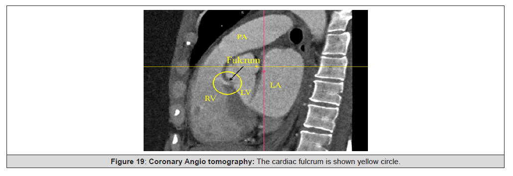
Figure 19:Coronary Angio tomography: The cardiac fulcrum is shown yellow circle.
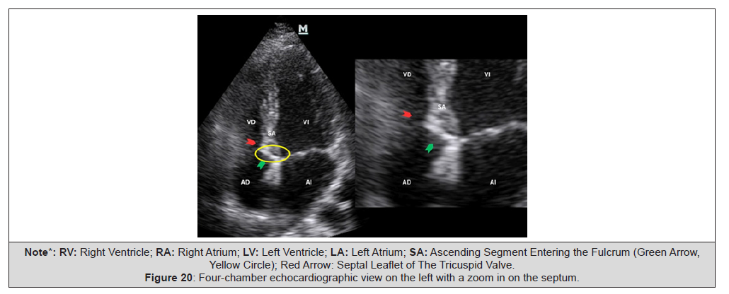
Figure 20:Four-chamber echocardiographic view on the left with a zoom in on the septum.
Relationship of the Cardiac Fulcrum to the AV Node
This analysis reveals, in both human and bovine hearts, an important issue for cardiac pacing therapy. Histological study found that the fulcrum is adjacent to the AV node, forming a rich cellular conglomerate of plexuses with neurofilaments. This contiguity between both structures is found in all specimens studied, both in bovine and human hearts. The fundamental finding of the research is that neurofilaments are also found inside the cardiac fulcrum. Fibroblasts and connective tissue are located in the thickness of the conduction system and between it and the working myocardium, and act as an insulating layer. It is also the connective tissue that constitutes the fibrous ring and the central fibrocartilaginous portion of the fulcrum, and thus allows the atrial and ventricular cavities to be electrically isolated (Figures 21 and 22).
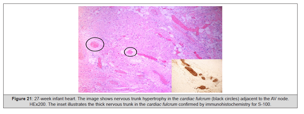
Figure 21:27-week infant heart. The image shows nervous trunk hypertrophy in the cardiac fulcrum (black circles) adjacent to the AV node. HEx200. The inset illustrates the thick nervous trunk in the cardiac fulcrum confirmed by immunohistochemistry for S-100.
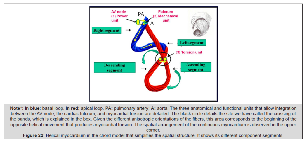
Figure 22:Helical myocardium in the chord model that simplifies the spatial structure. It shows its different component segments.
This investigation found a relationship between myocardial stimulation and its mechanical product. The mechanical consequence of the cardiac structure is the initiation of stimulation in the anatomical and functional unit between the AV node and the cardiac fulcrum, and its continuity in myocardial activation up to the almost simultaneous activation zone between the descending and ascending segments. This generates torsion of the left ventricular myocardium, due to opposite rotation between the base and the apex, with simultaneous shortening of both ventricles [42,43].
Discussion
Regarding the argued difficulty to dissect the myocardium, which is more apparent than real, we should consider that once the myocardial band originated as a loop in the arterial semicircle of amphibians and reptiles to adapt to the physiological demands of aerial life, the muscle bundles became firmly attached to their contact surfaces, hampering the necessary cleavage planes necessary for their anatomical dissection. The evolutionary goal was to develop a sufficiently solid hemodynamic structure with the strength to generate the suction and pumping of the blood volume that supplied the whole organism. Thus, every attempt to dissect an anatomical segment from the rest of the myocardium, avoiding the real cardiac arrangement, always turns into an obstacle due to the structural plan of the axes where the orientation of the myocardial band courses. Its spatial helical arrangement is in agreement with the mechanical function evidenced by the different segments that compose it. No segment of the band’s sequential histology, explored in our investigations, presents a mesh arrangement. The myocardial band cannot be anatomically suspended and free in the thoracic cavity because it would be impossible to eject blood at a speed of 200cm/s. Therefore, there must be a point of attachment, which was identified as the cardiac fulcrum (supporting point of leverage). In this supporting site, the muscle fibers are inevitably forced to “intertwine” with the connective, chondroid or osseous fulcrum, and our anatomical and histological investigations have shown that this insertion attaches both the origin and end of the myocardial band [31,44].
This structural composition keeps correspondence with the activation of the myocardial band. The stimulus runs by its muscle pathways, but in order to fulfill the function proposed by its helical arrangement, it is essential for it to simultaneously activate the left ventricular descending and ascending segments. The transmission of the stimulus between them generates the necessary ventricular torsion (a situation similar to “wringing a towel”) that enables the ejection of the blood content in a limited time span with the necessary force to adequately supply the whole body. The circulatory duct of annelids works with a peristaltic mechanism in its contractile progression. The impulse along its length preserves a pattern of axial transmission, but after the cardiac duct twists in birds and mammals, radial transmission of the impulse is added, allowing the helical motion indispensable to produce the successive twisting- shortening motions during systole and untwisting-lengthening in the subsequent suction phase. The ventricular narrowing phase at the beginning of systole is shaped by the contraction of the basal loop right and left segments. The overlapping shortening phase due to the descent of the base while twisting occurs, is produced longitudinally, as the ring contracts before the apex. The fact that the apex remains fixed is due to the movement of the base, descending in systole and ascending in diastole. This is better explained because the ascending band, rigid at the beginning of diastole, acts as a tight tutor keeping the apex immobile. The pressure generated to eject the highest amount of blood at the onset of ejection during an interval lasting 20% of the systolic phase is feasible due to the twisting motion. This action is achieved because the electrical stimulation propagates towards the descending band (axial propagation) and simultaneously to the ascending band (radial propagation).
Although the electrical conduction progresses along the myocardial band, radial propagation towards the ascending band plays an essential role in ventricular twisting by allowing opposing forces on its longitudinal axis to generate the necessary intraventricular pressure to achieve abrupt blood ejection. Thus, a twisting mechanism similar to “wringing a towel” would be produced. The following suction phase of the heart is not feasible due to the small difference with peripheral pressure. Neither can it be passive. The untwisting of the heart in the first 100ms of diastole (isovolumic diastolic phase) generates the negative intraventricular force to draw blood into the left ventricle, even in the absence of the right ventricle, as shown in experimental animals [35]. This suction phase (“suction cup” mechanism) is active with energy expenditure, and implies that the heart cycle consists of three stages: systole, suction and diastole.
Diastolic activation explains the insertion of a suction active coupling phase between systole and diastole, with muscle contraction, energy consumption, and a marked drop of intraventricular pressure. This effect draws blood into the ventricular chamber through a pressure difference with respect to the external pressure, and is responsible that in only 20% of the total filling time it should achieve 70% of the total filling volume. The new echocardiographic proposed parameters integrate values of longitudinal shortening and twisting and provided reference strain values obtained in a population of healthy subjects allowing a complete physiological assessment of cardiac systolic function, and could be used for the early detection and characterization of its alteration. There is a consensus that future clinical practice should adopt a combined approach in which changes in LV rotational mechanics and longitudinal shortening are considered and interpreted together. Likewise, the integration of complementary parameters of pumping and myocardial function should be considered for a more accurate evaluation of LV systolic function.
Limitations
It is very difficult in our environments to get human hearts for dissection. All this research is necessary to replicate with a greater number of hearts and patients.
Conclusions
1) We consider there is enough evidence that the fiber orientation
and the opposite base–apex rotational movement of the
heart, justifies the muscle band model
2) The finding of the fulcrum gives support to the spiral myocardial
band being the point of fixation that allows the helicoidal
torsion.
3) The hyaluronic acid would act as a lubricant and provide great
resistance to mechanical pressures in a functional association
with the venous ducts of Thebesius and Langer, that would intervene
as tributaries of the necessary hydration to said element
and thus could counteract the friction of the surfaces by
exercising an anti-friction mechanism.
4) This study explains the ventricular twist and the active suction
mechanism during the isovolumic diastolic and early ventricular
filling phases, in contrast with the traditional concept of
passive relaxation during the diastolic isovolumic phase and
the vis a tergo as mechanism for ventricular filling.
5) The deep anatomical, histológical, electrophysiological and
functional investigations that our group have carried out at
the level of the different segments of the muscular band allows
a better interpretation of cardiac physiology and its clinical
application is unquestionable in light of new cardiac images
techniques.
Acknowledgements
None.
Conflict of Interest
None.
References
- Ballester M, Ferreira A, Carreras F (2008) The myocardial band. Heart Fail Clin 4(3): 261-272.
- Trainini JC, Elencwajg B, López Cabanillas N, Herreros J, Lago N, et al. (2015) Stimuli propagation, muscle torsión and cardiac suction effect through electrophysiological research. In Basis of the New Cardiac Mechanics. The Suction Pump, Trainini JC and col. Lumen, Buenos Aires: 39-61.
- Torrent Guasp F, Buckberg G, Carmine C, Cox J, Coghlan H, Gharib M (2001) The structure and function of the helical heart and its buttress wrapping. I. The normal macroscopic structure of the heart. Seminars in Thorac and Cardiovasc Surg 13: 301-319.
- Trainini J, Lowenstein J, Beraudo M, Trainini A, Mora Llabata V, Wernicke M, et al. (2021) Myocardial torsion and cardiac fulcrum. Morphologie 105(348): 15-23.
- Trainini J, Wernicke M, Lowenstein J, Beraudo M, Wernicke M, et al. (2020) Cardiac helical function. Research Square.
- Trainini J, Beraudo M, Wernicke M, Lowenstein J, Carreras Costa F, et al. (2021) Physiology of the Helical Heart. Int J Anat Appl Physiol 7(5): 195-204.
- Torrent Guasp F (1998) The Structure and Function of The Heart. Rev Esp Cardiol 51: 91-102.
- Trainini JC, Lowenstein JA, Beraudo M, Mora Llabata V, Carreras Costa F, Cabezas Valle J, et al. (2022) Fulcrum and Torsion of the Helical Myocardium. Editorial Biblos Buenos Aires.
- Trainini J, Lowenstein J, Beraudo M, Valle Cabezas J, Wernicke M, et al. (2023) Anatomy and Organization of the Helical Heart. UNDAV Ed, Buenos Aires; Argentina.
- Poveda F, Gil D, Martí E, Andaluz A, Ballester M, et al. (2013) Helical structure of the cardiac ventricular anatomy assessed by diffusion tensor magnetic resonance imaging with multiresolution tractography. Rev Esp Cardiol 66:782-790.
- Carreras F, Ballester M, Pujadas S, Leta R, Pons Lladó G (2006) Morphological and functional evidences of the helical heart from non-invasive cardiac imaging. Eur J Cardiothoracic Surg 29(Suppl 1): S50-55.
- Trainini JC, Elencwajg B, López Cabanillas N, Herreros J, Lago N (2015) Ventricular torsion and cardiac suction effect. The electrophysiological analysis of the cardiac muscle. J Clin Exp Cardiolog 6: 10.
- Trainini JC, Beraudo M, Wernicke M, Carreras Costa F, Trainini A, et al. (2022) Evidence that the myocardium is a continuous helical muscle with one insertion. REC: Cardio Clinics 57: 194-202.
- Trainini JC, Beraudo M, Wernicke M, Carreras Costa F, Trainini A, et al. (2022) Anatomical investigation of the cardiac apex Rev Argent Cardiol 90: 125-130.
- Trainini J, Lowenstein J, Beraudo M, Wernicke M, Mora Llabata V, Lowenstein Haber D, et al. (2022) Cardiac Helical Function. Fulcrum and Torsion. Japan Journal of Clinical & Medical Research 2(3): 1-11.
- Trainini JC, Elencwajg B, López Cabanillas N, Herreros J, Lowenstein J Bustamante Munguira J, et al. (2017) Ventricular torsion and cardiac suction effect: The electrophysiological analysis of the cardiac band muscle. Interventional Cardiol 9 (1): 45-51.
- Biesiadecki B, Davis J, Ziolo M, Janssen P (2014) Tri-modal regulation of cardiac muscle relaxation; intracellular calcium decline, thin filament deactivation, and cross-bridge cycling kinetics. Biophys Rev 6(3-4): 273-289.
- Trainini J, Beraudo M, Mora Llabata V, Wernicke M, Lowenstein J, et al. (2020) The myocardial band theory. New Discoveries Supporting the Complex Mechanism of Myocardial Torsion. RETIC 3: 14-18.
- Trainini J, Beraudo M, Wernicke M, Trainini A, Lowenstein J, et al. (2021) Cardiac Fulcrum. Cardiovasc Surg Int 2(1): CSI-02-1011.
- Anderson R, Ho S, Redman K, Sanchez Quintana D, Punkenheimer P (2005) The anatomical arrangement of the myocardial cells making up the ventricular mass. Eur J Cardiothoracic Surg 28(4): 517-525.
- Trainini J, Beraudo M, Wernicke M, Trainini A, Haber Lowenstein D, et al. (2021) The myocardial support. Rev Argent Cardiol 89: 217-223.
- Trainini J, Beraudo M, Wernicke M, Trainini A, Haber Lowenstein D, et al. (2023) Myocardial support. Cardiac and Cardiovascular Research 4(2): 8.
- Trainini J, Beraudo M, Wernicke M, Herrero E, Trainini A, et al. (2025) Properties of the cardiac fulcrum. Rev Argent Cardiol 93: 41-47.
- Bignami A, Asher R (1992) Some observations on the localization of hyaluronic acid in adult, newborn and embryonal rat brain. Int J Dev. Neurosci 10(1): 45-57.
- Hong yan Ding, Ya nan Xie, Qiang Dong, Koji Kimata, Yoshihiro Nishida, et al. (2019) Roles of hyaluronan in cardiovascular and nervous system disorders. J Zhejiang Univ-Sci B (Biomed & Biotechnol) 20(5): 428-436.
- Lorén C, Dahl C, Do L, Almaas V, Geiran O, et al. (2019) Low molecular mass myocardial hyaluronan in human hypertrophic cardiomyopathy. Cells 8(2): 97.
- Trainini J, Werrnicke M, Beraudo M, Lowenstein J, Trainini A, et al. (2021) Hyaluronic acid in the heart. J Trends Biomedical Research 4: 1-5.
- Trainini J, Valle Cabezas J, Carreras Costa F, Beraudo M, Wernicke M, et al. (2022) Cardiac Energy. Clin Exp Invest 2022 Volume 3(1): 7-7.
- Cosín Aguilar J, Hernándiz Martínez A, Tuzón Segarra MT, Agüero Ramón Llin J, Torrent Guasp F (2009) Experimental study of the so-called isovolumetric relaxation phase of the left ventricle. Rev Esp Cardiol 62: 392-399.
- Buckberg GD, Coghlan HC, Torrent Guasp F (2001) The structure and function of the helical heart and its buttress wrapping. V. Anatomic and physiologic considerations in the healthy and failing heart. Semin Thorac Cardiovasc Surg 13(4): 358-385.
- Trainini J, Lowenstein J, Beraudo M, Wernicke M, Trainini A, et al. (2021) Myocardial torsion and cardiac fulcrum (Torsion myocardique et pivot cardiaque). Morphologie 105: 15-23.
- Arvidsson M, Töger J, Carlsson M, Steding Ehrenborg K, Pedrizzetti G, et al. (2017) Left and right ventricular hemodynamic forces in healthy volunteers and elite athletes assessed with 4D flow magnetic resonance imaging. Am J Physiol Heart Circ Physiol 312: H314-H328.
- Pedrizzetti G, Arvidsson PM, Töger J, Borgquist R, Domenichini F, et al. (2017) On estimating intraventricular hemodymamic forces from endocardial dynamics: A comparative study with 4D flow MRI. Journal of Biomechanics 60: 203-210.
- Maksuti E, Carlsson M, Arheden H, Kovács S, Broomé M, et al. (2017) Hydraulic forces contribute to left ventricular diastolic filling. Scientif reports 7: 43505.
- Trainini JC, Trainini A, Valle Cabezas J, Cabo J (2019) Left Ventricular Suction in Right Ventricular Dysfunction. EC Cardiology 6(6): 572-577.
- Trainini J, Valle Cabezas J, Beraudo M, Elencwajg B, Wernicke M, et al. (2024) Ventricular Complementarity. I J cardio & card diso 5(2).
- Trainini J, López MPH, Bastarrica ME, Trainini A, Cabezas JV, et al. (2024) Mechanisms of action of heart failure with preserved ejection fraction. Quan J Med Hea Sci 3(2): 73-83.
- Mora V, Roldán I, Romero E, Saurí A, Romero D, et al. (2018) Myocardial contraction during the diastolic isovolumetric period: analysis of longitudinal strain by means of speckle tracking echocardiography. J Cardiovasc Dev Dis 5(3): 41.
- Mora V, Roldán I, Romero E, Romero D, Bertolín J, et al. (2018) Comprehensive assessment of left ventricular myocardial function by two-dimensional speckle-tracking echocardiography, Cardiovascular Ultrasound 16(1): 16.
- Trainini J, Beraudo M, Wernicke M, Freire G, Trainini A, et al. (2025) Helical Heart Suction in Heart Failure with Preserved Ejection Fraction. AJBSR 25(6): 808-812.
- Elencwajg B, López Cabanillas N, Cardinali EL, Barisani JL, Trainini J, et al. (2012) The Jurdham procedure endocardial left ventricular lead insertion via a femoral transseptal sheath for cardiac resynchronization therapy pectoral device implantation. Heart Rhythm 9(11): 1798-1804.
- Trainini J, Wernicke M, Beraudo M, Cohen M, Trainini A, et al. (2023) Cardiac Fulcrum, its Relationship with the Atrioventricular Node. Rev Argent Cardiol 91: 429-435.
- Trainini J, Beraudo M, Wernicke M, Trainini A, Cohen M, et al. (2024) Relationship of the cardiac fulcrum and the AV node with ventricular torsion. AJBSR 24(2): 175-186.
- Trainini J (2024) In search of the cardiac fulcrum. The finding of the heart's support. Ed. Biblos, Buenos Aires, Argentina.




 We use cookies to ensure you get the best experience on our website.