
Case Report 
 Creative Commons, CC-BY
Integrating Augmented Reality and Image Fusion in Complex EVAR Using Low- Cost 3D Tools
*Corresponding author:Juan Carlos Gomez-Rodriguez, Vascular Surgery Department, Clínica Medellín, Medellín, Colombia. ORCID: https://orcid.org/0000-0002-2883-2181, Scopus Author ID: 57216120493
Received:July 17, 2025; Published:July 24, 2025
DOI: 10.34297/AJBSR.2025.27.003622
Abstract
We present the case of a complex infrarenal abdominal aortic aneurysm treated with Endovascular Aneurysm Repair (EVAR), involving
an accessory renal artery originating from the aneurysmal sac. The procedure integrated low-cost digital tools for preoperative
anatomical segmentation, 3D visualization through Augmented Reality (AR), and intraoperative image fusion for vascular
navigation.
The 3D reconstruction was performed using 3D Slicer (Boston, MA, USA), including the aorta, renal arteries, and lumbar spine. The
resulting STL model was visualized in an immersive AR environment using Meta Quest 3 (Meta Platforms, Inc., Menlo Park, CA, USA)
via SimLab VR Studio 14 (SimLab Soft, Amman, Jordan). This allowed the surgical team to familiarize themselves with the spatial
anatomy of the aneurysm and its relationship to visceral branches.
During the procedure, the Vessel Navigator system (Philips Healthcare, Best, Netherlands) enabled real-time fusion of the segmented
Computed Tomography Angiography (CTA) with live fluoroscopy. This generated a navigable vascular map projected onto
the patient’s anatomy, guiding selective catheterization of the accessory renal artery for preemptive embolization and stent graft
deployment.
The integration of AR and image fusion reduced the need for contrast media (34mL), minimized radiation exposure, and improved
catheterization accuracy. The intervention was completed successfully without intraoperative complications. The patient required
no additional support, was transferred to the general ward on postoperative day one, and was discharged on day two.
This case demonstrates the feasibility and clinical utility of combining AR and real- time image fusion with free or low-cost software
and consumer-grade devices in EVAR planning and execution. The approach enhances spatial understanding, procedural safety, and
technical precision, and is especially valuable in anatomically complex scenarios or resource-limited settings.
Keywords:EVAR, Abdominal Aortic Aneurysm, Augmented Reality, 3D Slicer, Image Fusion, Vessel Navigator, Accessory Renal Artery, Preoperative Planning, CTA Segmentation, Low-Cost Technology
Abbreviations:EVAR: Endovascular Aneurysm Repair; AR: Augmented Reality; CTA: Computed Tomography Angiography; STL: Standard Tessellation Language; 3D: Three-Dimensional; VR: Virtual Reality; IOPS: Intraoperative Positioning System; CT: Computed Tomography; mL: Milliliters; USD: United States Dollar
Introduction
Abdominal Aortic Aneurysms (AAA) represent a significant cause of morbidity and mortality, particularly in elderly patients. Among treatment options, Endovascular Aneurysm Repair (EVAR) has demonstrated lower perioperative risk compared to open surgery, especially in anatomically favorable cases. However, complex aneurysms—those involving visceral branches or anatomical variants— continue to pose technical challenges that require meticulous planning.
Detailed preoperative imaging is essential to minimize complications such as endoleaks, graft migration, and renal impairment due to excessive use of contrast agents. In cases with accessory renal arteries or tortuous anatomies, understanding the three-dimensional spatial relationship between the aneurysm and adjacent branches becomes critical.
Advances in 3D segmentation and Augmented Reality (AR) now offer enhanced visualization of vascular anatomy. These technologies allow clinicians to interact with patient-specific anatomy in an immersive way, improving orientation and decision- making. Nevertheless, implementation of such tools is often restricted by the high cost of dedicated workstations or proprietary platforms.
In this context, the integration of low-cost software and consumer- grade AR devices may offer a viable alternative for EVAR planning in complex scenarios. This case report describes the endovascular treatment of a complex infrarenal AAA using opensource 3D modelling, AR visualization, and intraoperative image fusion. The approach aims to enhance procedural precision, re duce contrast usage, and improve overall safety, particularly in resource- constrained environments.
Materials and Methods
A 78-year-old male patient was diagnosed with a complex infrarenal Abdominal Aortic Aneurysm (AAA), which included an accessory renal artery arising directly from the aneurysmal sac. An Endovascular Aneurysm Repair (EVAR) procedure was planned, with selective coil embolization of the accessory renal artery prior to endograft deployment, in order to prevent a type II endoleak.
Image Acquisition and 3D Modelling
A contrast-enhanced, submillimetric Computed Tomography Angiography (CTA) of the abdomen was acquired using a standard EVAR protocol. The arterial phase series, reconstructed at <1 mm slice thickness, was exported in DICOM (Digital Imaging and Communications in Medicine) format and imported into 3D Slicer (Boston, MA, USA), an open-source medical image analysis platform.
Segmentation was performed using the Segment Editor module within 3D Slicer. The abdominal aorta, both renal arteries (including the accessory renal artery), and lumbar vertebrae were manually identified (Figure 1). To improve segmentation efficiency and preserve anatomical precision, the “Grow from Seeds” algorithm was used. This semi-automatic method propagates label regions from user-defined seeds, classifying adjacent voxels based on local intensity and spatial connectivity, yielding accurate segmentation with minimal manual correction.
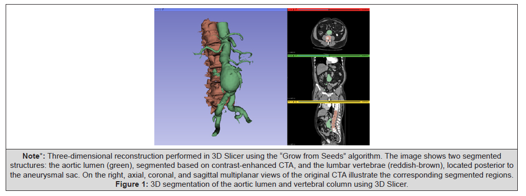
Note*: Three-dimensional reconstruction performed in 3D Slicer using the “Grow from Seeds” algorithm. The image shows two segmented structures: the aortic lumen (green), segmented based on contrast-enhanced CTA, and the lumbar vertebrae (reddish-brown), located posterior to the aneurysmal sac. On the right, axial, coronal, and sagittal multiplanar views of the original CTA illustrate the corresponding segmented regions.
Figure 1:3D segmentation of the aortic lumen and vertebral column using 3D Slicer.
After final review and smoothing, the segmented structures were converted into a 3D surface mesh and exported as an STL (Standard Tessellation Language) file. STL is a widely used file format that represents surface geometry via a triangular mesh, compatible with both 3D visualization and 3D printing software. The inclusion of vertebral landmarks in the model facilitated later anatomical orientation during both AR planning and image fusion.
Augmented Reality Visualization
The STL model was imported into SimLab VR Studio 14 (SimLab Soft, Amman, Jordan), a free cross-platform visualization tool that supports augmented and virtual reality exploration. Visualization was carried out using a Meta Quest 3 headset (Meta Platforms, Inc., Menlo Park, CA, USA), a consumer-grade mixed reality device that allows immersive 3D interaction without tethered workstations.
In SimLab, the STL model was calibrated to true scale, preserving anatomical proportions based on the DICOM voxel dimensions. The AR environment enabled full 360-degree rotation, zoom, and plane slicing for spatial analysis. The SimLab VR Studio environment used for spatial model verification is shown in Figure 2. The surgical team used this immersive platform to explore the patient- specific vascular anatomy in detail, focusing on the trajectory and depth of the accessory renal artery in relation to the aneurysmal sac and bony structures. Figure 3 shows the use of Meta Quest 3 for immersive AR-based planning and team familiarization with the segmented anatomy.
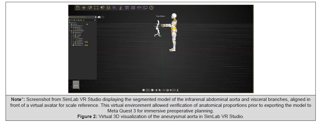
Note*: Screenshot from SimLab VR Studio displaying the segmented model of the infrarenal abdominal aorta and visceral branches, aligned in front of a virtual avatar for scale reference. This virtual environment allowed verification of anatomical proportions prior to exporting the model to Meta Quest 3 for immersive preoperative planning.
Figure 2:Virtual 3D visualization of the aneurysmal aorta in SimLab VR Studio.

Note*: (A) Real-time augmented visualization of the segmented aortic model and visceral branches projected in the clinical environment. (B) Surgical team members using Meta Quest 3 headsets to explore the vascular anatomy in three dimensions prior to the intervention. This setup allowed intuitive spatial understanding of the aneurysm and accessory renal artery.
Figure 3:Use of augmented reality for immersive surgical planning with Meta Quest 3.
This preoperative review enhanced procedural planning by enabling intuitive spatial recognition of critical landmarks. The use of lumbar vertebrae within the 3D model provided consistent anatomical reference points between AR visualization and intraoperative fluoroscopy. Although AR was not integrated in real time during the intervention, the preprocedural cognitive mapping significantly improved the team’s understanding and confidence before catheterization.
The entire process was completed using free or low-cost tools and commercially available devices, demonstrating that advanced AR visualization for vascular planning can be implemented without the need for specialized imaging platforms or proprietary systems.
Intraoperative Image Fusion
Intraoperative navigation was facilitated using the Vessel Navigator system (Philips Healthcare, Best, Netherlands), which enables real-time fusion of preoperative CTA data with live fluoroscopic imaging. The segmented volume from the preoperative CTA was uploaded into the platform prior to the procedure.
Co-registration was performed by aligning anatomical landmarks visible in both the 3D model and the fluoroscopic field. These included the lumbar vertebrae, aortic contours, renal artery origins, and calcified plaques. Rigid transformation was used for alignment, with fine manual adjustment based on fluoroscopic projections and patient positioning to ensure optimal overlap.
Following successful registration, the system generated a dynamic overlay of the vascular anatomy onto live fluoroscopy. This “virtual roadmap” provided continuous visual guidance for catheter navigation and device deployment without the need for repeated contrast-enhanced imaging. The tool proved particularly useful in identifying the ostium of the accessory renal artery, which was catheterized and embolized with precision under fused image guidance. Figure 4 illustrates the fusion workflow and final angiographic results, including successful navigation and embolization of the accessory renal artery

Note*:(A) Segmented aortic lumen reconstruction generated by the Vessel Navigator module (Philips Azurion with Clarity IQ), displaying anatomical landmarks for ostia of major visceral arteries, including bilateral hypogastric arteries, renal arteries, and a left-sided accessory renal artery. (B) Live fusion overlay of the segmented 3D vascular model with fluoroscopy, guiding advancement of a 5 Fr multipurpose catheter toward the accessory renal artery without contrast injection. (C) Selective angiogram confirming the catheter position within the accessory renal artery prior to coil embolization. (D) Final angiographic control showing successful coil embolization of the accessory renal artery and preserved perfusion through both renal arteries and the iliac limbs of the deployed endograft.
Figure 4:Image fusion guidance and final angiographic outcome during EVAR.
The fusion system remained active throughout the intervention, enabling accurate stent graft deployment with minimal radiation exposure and contrast administration. A total of only 34mL of iodinated contrast was used. Final angiography confirmed successful exclusion of the aneurysm and patency of the main renal arteries.
Results and Discussion
The procedure was completed successfully without intraoperative complications. The accessory renal artery was selectively catheterized and embolized using detachable coils under fusion image guidance. A bifurcated stent graft was then deployed with precise alignment and complete exclusion of the aneurysmal sac. The final angiographic control confirmed adequate positioning, exclusion of the aneurysm, patency of both main renal arteries, and absence of endoleaks. Only 34mL of iodinated contrast was required for the entire procedure. The patient remained hemodynamically stable, required no additional support, and was transferred to the general ward on postoperative day one. He was discharged in good condition on postoperative day two.
This case demonstrates the clinical feasibility and potential advantages of combining low-cost 3D modelling, Augmented Reality (AR), and real-time image fusion in the planning and execution of complex EVAR procedures. The immersive preoperative visualization allowed the surgical team to internalize the patient-specific vascular anatomy and plan an effective strategy for embolization and graft deployment. The inclusion of vertebral landmarks in the STL model was particularly valuable, as it enabled seamless orientation between AR and intraoperative fluoroscopy.
The use of image fusion significantly reduced the need for multiple angiographic runs, contrast injection, and radiation exposure. These benefits align with previous studies, such as that of Edsfeldt, et al. [1], who demonstrated that fusion imaging could achieve vascular registration accuracy within 4mm while dramatically reducing radiation dose and contrast use in standard EVAR cases. Similarly, He, et al. [2] and Chen, et al. [3]. have shown that augmented reality-based navigation systems can improve intraoperative precision and planning efficiency.
The synergistic combination of AR and image fusion is especially advantageous in patients with accessory or aberrant renal arteries, where precise localization and catheterization are critical. In our case, the accessory renal artery arose directly from the aneurysmal sac and was successfully embolized without exploratory angiography. This outcome supports the role of fusion imaging not only for graft alignment but also for the selective navigation of visceral branches [4].
Furthermore, the accessibility of open-source software (3D Slicer) and consumer-grade devices (Meta Quest 3) challenges the notion that advanced intraoperative planning requires expensive or proprietary systems. As previously described by Gomez-Rodriguez, et al. [5,6] low-cost digital toolkits can offer reliable
3D planning capabilities in resource-limited settings, enhancing preoperative understanding and procedural safety.
Although the AR component was used exclusively in the preoperative phase and not in real-time intraoperatively, its impact was clear in terms of team preparation, spatial orientation, and procedural confidence. This echoes findings by Wong, et al. [7], who showed that AR visualization improves the learning curve and catheterization times, particularly among less experienced operators.
Limitations include the absence of dynamic AR during the procedure and the need for manual co-registration during image fusion. Nevertheless, ongoing developments in deformable registration algorithms and real-time 3D tracking systems may soon overcome these challenges [8,9]. Emerging tools such as Intraoperative Positioning Systems (IOPS) have also shown promise in complex endovascular navigation, enabling vessel cannulation with minimal use of fluoroscopy or contrast agents [10]. Additionally, 3D-printed aortic models generated using open-source software have shown high accuracy and practical value in the planning of fenestrated EVAR procedures, even in urgent settings [11].
In summary, this case illustrates how a hybrid approach—combining accessible 3D segmentation, immersive visualization, and image fusion—can optimize EVAR procedures in anatomically complex settings, while maintaining low procedural cost, minimal contrast use, and high technical success.
Conclusion
This case highlights the successful integration of low-cost 3D modelling, augmented reality, and real-time image fusion in the endovascular treatment of a complex infrarenal abdominal aortic aneurysm. The approach enabled precise preoperative planning, accurate catheterization of an accessory renal artery, and efficient deployment of the stent graft with minimal contrast use and radiation exposure.
By leveraging open-source segmentation tools and commercially available AR devices, the surgical team was able to achieve a high level of anatomical understanding and procedural precision without the need for expensive proprietary platforms. The synergy between preoperative AR visualization and intraoperative image fusion provided complementary benefits that improved spatial orientation, reduced contrast usage, and enhanced technical confidence.
This experience demonstrates that advanced imaging technologies can be implemented effectively in vascular surgery using cost-effective tools, offering valuable clinical benefits, especially in anatomically challenging cases or in settings with limited resources.
Acknowledgement
The authors would like to thank Dr. Hernan Castro, from the Department of Anaesthesiology at Clínica Medellín, for his valuable support during the procedure. We also extend our gratitude to the entire team of the Hemodynamics Unit at Clínica Medellín for their collaboration and professionalism throughout the intervention. No specific funding was received for this study.
Conflict of Interest
The authors declare no conflict of interest related to this publication.
References
- Edsfeldt A, Sonesson B, Rosén H, Petri MH, Hongku K, et al. (2020) Validation of a New Method for 2D Fusion Imaging Registration in a System Prepared Only for 3D. J Endovasc Ther 27(3): 468-472.
- He F, Ǫi X, Feng Ǫ, Zhang Ǫ, Pan N, et al. (2023) Research on augmented reality navigation of in vitro fenestration of stent-graft based on deep learning and virtual-real registration. Comput Assist Surg (Abingdon) 28(1): 2289339.
- Chen X, Xu L, Wang Y, Wang H, Wang F, et al. (2015) Development of a surgical navigation system based on augmented reality using an optical see-through head-mounted display. J Biomed Inform 55:124-131.
- Patel RJ, Lee AM, Hallsten J, Lane JS, Barleben AR, et al. (2023) Use of surgical augmented intelligence maps can reduce radiation and improve safety in the endovascular treatment of complex aortic aneurysms. J Vasc Surg 77(4): 982-990.e2.
- Gomez Rodriguez JC (2024) Low-Cost Digital Tools for Endovascular Aortic Aneurysm Repair Sizing and Planning. In: Aortic Aneurysms - Screening, Diagnostics and Management. IntechOpen.
- Gómez Rodríguez JC, García LF, Nieto LA, Cáceres D, Contreras M, et al. (2022) Low-cost digital tool kit for planning and sizing with 3D printing of abdominal aortic aneurysms for endovascular aortic repair: A Latinoamerican experience. Cirugia Cardiovascular 29(2): 68-71.
- Wong J, Mesnard T, Vacirca A, George M, Goel V, et al. (2025) Intraoperative position system guided with augmented reality improves the learning curve of endovascular navigation in endovascular-naive operators. J Vasc Surg 81(3): 759-763.
- Chan HHL, Haerle SK, Daly MJ, Zheng J, Philp L, et al. (2021) An integrated augmented reality surgical navigation platform using multi-modality imaging for guidance. PLoS One 16(4): e0250558.
- Czerny M, Grabenwöger M, Berger T, Aboyans V, Corte A Della, et al. (2024) EACTS/STS Guidelines for diagnosing and treating acute and chronic syndromes of the aortic organ. Ann Thorac Surg 118(1): 5-115.
- Muluk SC, Elrakhawy M, Chess B, Rosales C, Goel V (2022) Successful endovascular treatment of severe chronic mesenteric ischemia facilitated by intraoperative positioning system image guidance. J Vasc Surg Cases Innov Tech 8(1): 60-65.
- Barón V, Guevara R (2019) Three-dimensional printing-guided fenestrated endovascular aortic aneurysm repair using open source software and physician-modified devices. J Vasc Surg Cases Innov Tech 5(4): 566-571.




 We use cookies to ensure you get the best experience on our website.