
Research Article 
 Creative Commons, CC-BY
Intraperitoneal Administration of Empagliflozin Reduces Myocardial Infarct Size in Mice
*Corresponding author:Peter S Macdonald, Medical Director, Heart Transplant Unit, St Vincent’s Hospital, Darlinghurst NSW 2010, Australia.
Received:June 12, 2025; Published:June 16, 2025
DOI: 10.34297/AJBSR.2025.27.003561
Abstract
Aims: We have previously shown that Empagliflozin (EMPA) protects the isolated rat heart during prolonged hypothermic ischaemic
storage. We hypothesised that parenterally administered EMPA will acutely protect the heart from normothermic ischaemic
damage. Here, we aimed to investigate the cardioprotective effect of acute EMPA administration in murine models of permanent
ligation and Ischemia Reperfusion Injury (IRI) Myocardial Infarction (MI).
Methods and results: C57BL/6J adult male non-diabetic mice were used. For the permanent ligation model, EMPA (10mg/kg) or
vehicle (2% dimethyl sulfoxide in saline) was administered by Intraperitoneal (IP) injection immediately (0 h) or 2 hours (2h) after
ligation. For the IRI model, the Left Anterior Descending artery (LAD) was transiently ligated for 40minutes, and the same dose of
either EMPA or vehicle was administered IP 10 minutes before reperfusion. In the permanent ligation model, EMPA administration
at 0 h resulted in a 50% decrease in infarct size compared with vehicle (8.4±1.5 vs 16.9±1.0%, P<0.0001). The reduction in infarct
size was attenuated but still significant with EMPA administration 2 h after LAD ligation (12.1±1.4 vs 18.1±1.9 %, P<0.05). A 50%
decrease in infarct size was observed in the IRI model (2.4±1.9 vs 5.1±3.9 %, P<0.05).
Conclusion: A single acute bolus dose of EMPA exerted cardioprotective effects by attenuating MI size in both permanent ligation
and IRI models of acute MI in non-diabetic mice. If these findings are confirmed in a large animal model of acute MI, they would suggest
that early parenteral administration of EMPA during acute MI may provide clinical benefits beyond those already demonstrated
in published clinical trials.
Keywords:SGLT2, Empagliflozin, Myocardial infarction, NHE1, Mice, Permanent ligation, Ischemia Reperfusion Injury (IRI)
Introduction
Cardiovascular Disease (CVD) is the leading cause of death globally [1]. Ischaemic heart disease is the leading risk factor for heart failure, cardiac arrest, and chronic heart disease, with 9.2 million deaths reported in 2021 [2]. Clinically, no drugs currently block the acute injury response to myocardial ischaemia [3]. For patients suffering a heart attack, emergency transfer to a hospital and urgent opening of the blocked coronary artery by percutaneous intervention or surgery is the most effective treatment. However, delays in the recognition of the problem and in transferring patients to hospitals from rural and remote areas, reduce the chance of limiting myocardial damage and/or saving patients’ lives.
Myocardial Ischaemia (MI) leads to anaerobic metabolism resulting in lactic acidosis, which alters the activity of two critical transmembrane ion channels in cardiomyocytes: the sodium hydrogen (Na+/H+) exchanger (NHE) and Acid Sensing Ion Channel 1a (ASIC1a). NHE is activated by intracellular acidosis and ASIC1a by extracellular acidosis. NHE1 is the NHE subtype mainly expressed in cardiomyocytes, and its activity significantly increases under the pathological conditions of diabetes, heart failure, and acute ischaemia-reperfusion injury [4,5]. Activation of NHE1 increases cardiomyocyte intracellular sodium load, resulting in calcium overload during ischaemia-reperfusion and aggravation of reperfusion injury [6,7]. Activation of ASIC1a also results in calcium overload as well as activation of the receptor-interacting serine/threonine kinase (RIP) necroptosis pathway and cardiomyocyte death [8-10].
EMPA, a sodium-glucose co-transporter 2 (SGLT2) inhibitor approved by the FDA for the treatment of type 2 diabetes mellitus (T2DM) in 2014, has been shown to significantly reduce cardiovascular mortality and heart failure hospitalisation in patients with and without T2DM [11,12]. However, the mechanisms underlying these beneficial effects are not clearly understood [11]. Several reports have recently been published demonstrating that SGLT2 inhibitors reduce MI size in murine MI models, however, in these studies, EMPA was delivered chronically by oral gavage commencing before or after MI [13-16]. Most heart attacks happen unexpectedly, so treatment cannot be initiated until the event has taken place. There is an unmet need for drugs that are able to protect the heart from Ischaemia-Reperfusion Injury (IRI) or chronic coronary artery occlusion after onset and to determine optimal timing of administration.
We have previously shown that EMPA protects the isolated rat heart during prolonged hypothermic ischaemia with similar efficacy to the NHE1 inhibitor zoniporide [17] and others have reported that SGLT2 inhibitors also inhibit the NHE1 channel in isolated cardiac myocytes in a manner similar to other NHE1 inhibitors [5,18,19]. Given that NHE1 is activated during and after induction of myocardial ischaemia and reperfusion [6,7], we hypothesised that parenterally administered EMPA will acutely protect the heart from IRI as well as injury resulting from permanent coronary artery occlusion. Thus, here we aimed to investigate the cardioprotective effect of acute Intraperitoneal (IP) EMPA administration in murine models of permanent ligation and IRI MI.
Methods
Animals
All experimental procedures were approved by the Garvan Institute/St. Vincent’s Hospital Animal Experimentation Ethics Committee (AEC22/14) performed in accordance with the National Health and Medical Research Council (NHMRC) of Australia Guidelines on Animal Experimentation. All efforts were made to minimise suffering. C57BL/6J male non-diabetic mice aged 10-12 weeks were maintained under a 12-hour light/dark cycle and given food and water ad-lib.
Experimental Design
We examined the efficacy of EMPA versus vehicle in two murine models of acute MI: permanent ligation of the Left Anterior Descending coronary artery (LAD), and IRI. For the permanent ligation model, mice were divided randomly into vehicle and EMPA groups after LAD ligation and were subjected to early (after closure of the surgical wound – within 5 to 10minutes of LAD ligation, 0h) or delayed (2hours post LAD ligation, 2h) IP administration (Figure 1A). For the IRI model, the LAD was transiently ligated for 40minutes, animals were randomised, and EMPA or vehicle was administered IP 10minutes before reperfusion (Figure 1B).
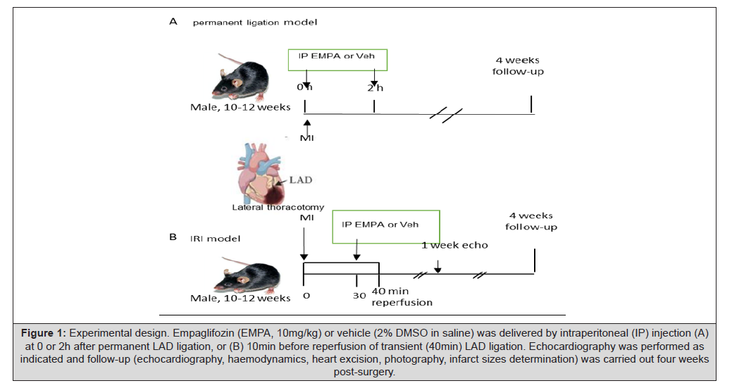
Figure 1:Experimental design. Empaglifozin (EMPA, 10mg/kg) or vehicle (2% DMSO in saline) was delivered by intraperitoneal (IP) injection (A) at 0 or 2h after permanent LAD ligation, or (B) 10min before reperfusion of transient (40min) LAD ligation. Echocardiography was performed as indicated and follow-up (echocardiography, haemodynamics, heart excision, photography, infarct sizes determination) was carried out four weeks post-surgery.
Myocardial infarction surgery
Mice were anaesthetised (ketamine: 100mg/kg; xylazine: 13mg/kg; atropine: 0.5mg/kg) by IP injection, placed on a heating pad for 4-5minutes, then intubated with a 23-gauge catheter into the trachea. Successful intubation was confirmed by connecting the catheter to a positive ventilator and checking that the frequency of chest movement was the same as that of the ventilator. Anaesthesia was induced with 4% isoflurane and maintained at 2% during surgery. Anaesthesia was monitored by the colour of mucous membranes and skin, and the depth of anaesthesia by the pedal withdrawal reflex (from toe pinch). The heart was accessed by an incision into the left wall of the chest at the fourth intercostal space. Blunt dissection of the intercostal tissue was performed to create an opening in the chest wall. The ribs were retracted by a small tissue retractor and the anterior surface of the heart exposed. The pericardial sac was opened, and the LAD was ligated with 8/0 prolene. The chest of the operated mouse was closed with 6/0 prolene suture and the pneumothorax was reduced by blocking the ventilator for two cycles. Mice were monitored continuously during recovery until the righting reflex was regained. They were then housed overnight with the cage half on/half off a heating pad. Post-operative analgesia (buprenorphine, 0.075 mg/kg) was administered subcutaneously twice daily for three days and the mice were monitored daily for another two days. They were then checked twice weekly till 4 weeks after surgery.
Permanent LAD ligation: For studies on the myocardial response to permanent LAD occlusion, the LAD was ligated with 8/0 prolene suture positioned 2mm below the edge of the left auricle. Sustained myocardial pallor and hypokinesia distal to the ligature were assessed visually to confirm cessation of blood flow and ischaemia (Figure 1A).
Ischaemia reperfusion injury surgery: For studies on the effect of IRI, the ligature was tied 1 mm below the edge of the left auricle to ensure discernible infarct sizes. To allow subsequent reestablishment of blood flow, the occlusion was produced by placing a 1-mm length of Polyethylene (PE) tubing (OD=0.61mm; Intramedic PE-10, Clay Adams, Parsippany, NJ) on the artery and fixing it in place with the ligature. The artery was then compressed by tightening the ligature, producing myocardial blanching. After regional ischaemia for 40min, reperfusion of the myocardium was ensured by removing the ligature and confirming resulting hyperaemia by visualisation (Figure 1B). If the myocardium was not reperfused, the animal was euthanised and excluded from the study.
EMPA Administration
EMPA (10mg/kg; Cayman Chemical, No.17375) versus vehicle (2% dimethyl sulfoxide (DMSO) in saline) was delivered by a 30-gauge needle IP into the lower quadrant of the abdomen. The 10mg/kg EMPA dose was chosen based on a pilot study that was performed using a permanent ligation model and increasing doses of EMPA (1mg/kg, 2mg/kg, 5mg/kg, 10mg/kg, 15mg/kg and 20mg/kg) delivered IP at 0 h.
Infarct size determination
Infarct size was determined at 4 weeks after MI surgery. To measure infarct size, the heart was excised immediately following micro- manometry. The atria and right ventricle were trimmed off and weighed, and the Left Ventricle (LV) was cut open and pinned flat to expose the endocardial surface for determination of LV wall scar size by planimetry, expressed as a percentage of the total LV chamber area, as described previously [20]. Infarct sizes were scored in a blinded manner using the ImageJ program by three independent people who were only provided with the eight-digit identification number of the mice to be assessed. Pictures were provided in random order with no physical evidence of which treatment had been administered.
Data Processing and Statistical Analysis
All results are expressed as mean ± SD. GraphPad Prism 10.0 (GraphPad; USA) was used to prepare graphs and for statistical analyses. Differences among groups were tested by unpaired Student’s t-test; P<0.05 was considered statistically significant. The data underlying this article will be shared on reasonable request to the corresponding author.
Results
Surgery Survival
The numbers of mice in each study group and their outcome are shown in Table 1. Of the two vehicle-treated groups in the permanent ligation model, 6 of 32 animals (19%) that survived the initial surgical induction of MI suffered cardiac rupture in the first week post-MI compared with 2 of 28 animals (7%) in the two EMPA- treated groups. There were no cardiac ruptures in either group in the IRI model; 5 mice were euthanised before recovery due to non-reperfusion after ligature removal (Table 1).
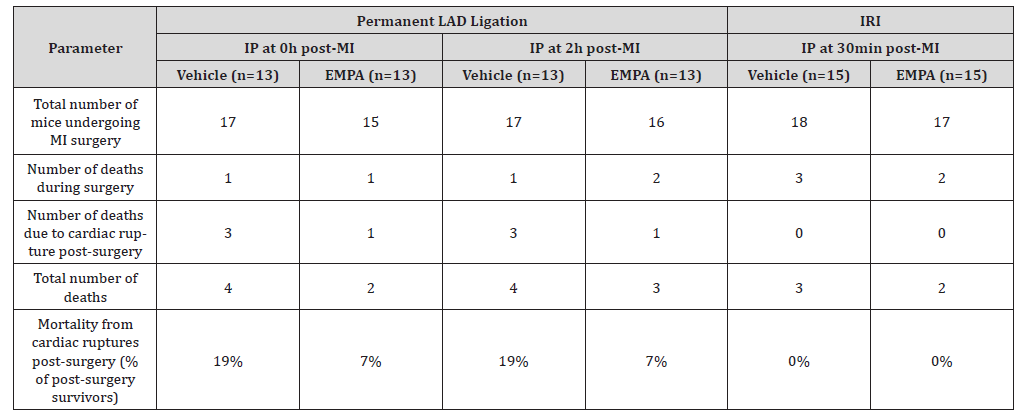
Table 1:Mortality Outcomes after MI surgery.
Note*: Vehicle (2% DMSO in saline) or EMPA (10mg/kg) was administered by Intraperitoneal (IP) injection.
Infarct Size
EMPA delivered by IP injection immediately after LAD occlusion resulted in a significant reduction in infarct size (as a percentage of the left ventricle). In the permanent ligation model, administration of EMPA at 0 h resulted in a 50% decrease in infarct size compared with vehicle (8.4±5.5% vs 16.9±3.8%, P<0.0001, Figure 2). The reduction in infarct size was attenuated although still significant when administration of EMPA was delayed 2 hours after LAD ligation (12.1±5% vs 18.1±6.8%, P<0.05, Figure 2). In the IRI model, pre-reperfusion EMPA treatment also resulted in about a 50% decrease in infarct size measured four weeks after surgery (2.4±1.9% vs 5.1±3.9%, respectively, P<0.05, Figure 2).
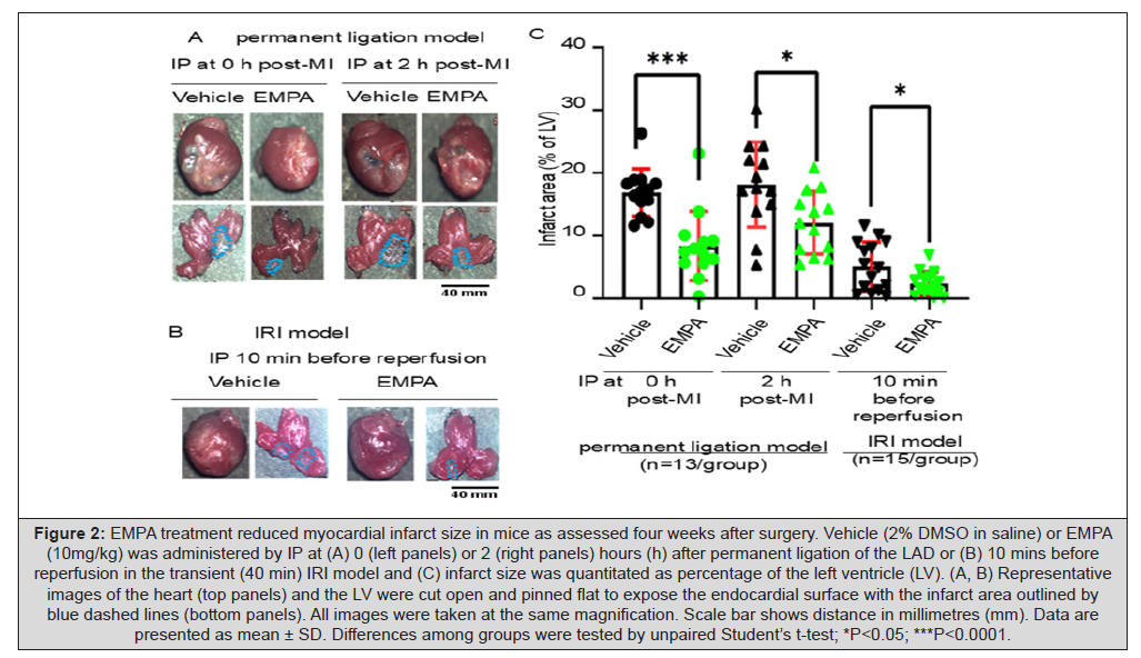
Figure 2:EMPA treatment reduced myocardial infarct size in mice as assessed four weeks after surgery. Vehicle (2% DMSO in saline) or EMPA (10mg/kg) was administered by IP at (A) 0 (left panels) or 2 (right panels) hours (h) after permanent ligation of the LAD or (B) 10 mins before reperfusion in the transient (40 min) IRI model and (C) infarct size was quantitated as percentage of the left ventricle (LV). (A, B) Representative images of the heart (top panels) and the LV were cut open and pinned flat to expose the endocardial surface with the infarct area outlined by blue dashed lines (bottom panels). All images were taken at the same magnification. Scale bar shows distance in millimetres (mm). Data are presented as mean ± SD. Differences among groups were tested by unpaired Student’s t-test; *P<0.05; ***P<0.0001.
Discussion
To our knowledge, this is the first report of the beneficial effects of acute parenteral EMPA administration in reducing infarct size in non-diabetic mice subjected to either IRI or permanent coronary ligation. Although our study was not powered to demonstrate improved survival with EMPA, mortality due to cardiac rupture in the permanent ligation model was almost three times higher in the vehicle- treated animals compared with those receiving EMPA. Cardiac rupture is generally a consequence of a large infarct. Despite the greater loss of animals from cardiac rupture in the vehicle-treated groups, administration of EMPA was still beneficial in reducing infarct size, even when administration was delayed for 2h after LAD ligation in the permanent ligation model.
Previously, Jiang, et al. [14] reported that pre-treatment of both non-diabetic and diabetic mice with high-dose oral EMPA significantly reduced infarct size in a permanent ligation MI model. Our findings extend these observations by demonstrating that acute parenteral bolus administration of EMPA after the onset of ischaemia is also cardioprotective even in the absence of reperfusion. In another murine study, Liu et al. [13] reported that commencement of oral EMPA 24 hours after permanent LAD ligation and continuation for 2 weeks also resulted in a reduction in infarct size and improved myocardial function suggesting a beneficial action of EMPA beyond the period of acute ischaemia and reperfusion.
Our findings in the IRI model are consistent with those of Sayour, et al. [21], who showed that administration of an IV bolus of canagliflozin, also an SGLT2 inhibitor, five minutes after induction of ischaemia significantly reduced infarct size in a rat IRI model. Other investigators have also demonstrated the cardioprotective effect of EMPA in mouse IRI models [15,16], although in those studies, EMPA was administered orally commencing one week pre-MI induction [15], or 6 weeks pre-MI induction [16].
The cardioprotective actions of SGLT2 inhibitors in the setting of acute myocardial infarction have been extensively investigated in pre-clinical models [22,23]. EMPA has been shown to directly inhibit NHE1, which is activated by the intracellular acidosis that occurs during myocardial ischaemia and again during reperfusion [5-7]. Other direct actions of EMPA include activation of AMP kinase and STAT3 and down-regulation of CaMKII activity with downstream effects that include reduced oxidative stress and inflammation [24- 26]. Interestingly, studies of acute administration of SGLT2 inhibitors in ex vivo models of acute myocardial ischaemia have failed to demonstrate a reduction in infarct size [27-29] whereas acute administration in vivo as in our study and that of Sayour et al. [21] showed a significant reduction in infarct size. Based on these findings we speculate that the cardioprotective actions of SGLT2 inhibitors may be mediated by a combination of direct effects of the drug on the heart and systemic effects that are only observed in intact animals.
Clinical trials have demonstrated that EMPA improves survival in patients with Type 2 diabetes mellitus and patients with chronic heart failure regardless of diabetic status [11,12]. EMPA has also been found to reduce incident heart failure in patients with Type 2 diabetes mellitus [11]. More recent clinical trials have examined the safety and efficacy of EMPA administered in the setting of acute MI, but in these trials, oral EMPA was commenced within days of patients presenting with acute MI rather than in the initial hours following infarct onset [22,30]. In the EMMY Trial [30], EMPA produced a greater reduction in NT-pro BNP levels and better LV function as assessed by echocardiography after 6 months, while the larger EMPACT MI Trial [22] demonstrated reduced hospitalisation for heart failure with no impact on overall survival after 18months follow-up. While both trials demonstrated the benefit of EMPA commenced after acute MI, they were not designed to assess any impact of EMPA on infarct size as it was only commenced days after AMI onset.
A question remains regarding the potential impact of EMPA on MI size and subsequent clinical outcome if commenced within the first few hours of the onset of acute MI. Our findings suggest that parenteral administration of EMPA within the first few hours of acute MI, may substantially reduce MI size and have additional benefits beyond those demonstrated in the EMMY and EMPACT MI Trials, regardless of whether the patient is able to undergo emergent revascularisation. This approach challenges existing prescribing guidelines which recommend that SGLT2 inhibitors be withheld when patients present acutely unwell due to the risk of Euglycaemic Keto-Acidosis (EKA) in diabetic patients [31] .We believe that the benefits of acute administration of empagliflozin in the setting of acute MI are likely to far outweigh the risk of EKA, which is rare in non-diabetics and is detectable by monitoring capillary blood ketone levels in diabetic patients and is readily treatable.
Our study has a number of limitations. We did not explore the mechanisms by which empagliflozin reduced MI size. Multiple published studies and reviews have demonstrated the benefit and possible mechanisms whereby SGLT2 inhibitors reduce IRI and MI size in a variety of MI models (13-16,24). The aim of our study was to examine the impact of early (within hours) administration of empagliflozin in both IRI and permanent ligation models. Also, our study was conducted only in male mice. Future studies are needed to validate our findings, to determine the mechanism of benefit and whether the benefits observed with early administration of empagliflozin also occurs in females.
Conclusion
A single acute bolus dose of EMPA by IP injection attenuates cardiac infarct size in both permanent ligation and IRI models of acute MI in non-diabetic mice. Future confirmation in a large animal model of acute MI would suggest that early parenteral administration of EMPA during acute MI may provide clinical benefits beyond those already demonstrated in published clinical trials.
Authors’ Contributions
JW, JV, LG, AD, YJ, SD, CM and PSM conceived the study design. JW performed all animal surgical procedures. SK performed all echocardiographic procedures. JW wrote the first draft of the manuscript. RMG, MPF and SEI provided critical review and revision of the manuscript. All authors approved the final version of the manuscript.
Conflict of interest
Peter Macdonald has received honoraria from Boehringer- Ingelheim for delivering lectures on heart failure. The other authors declare no conflicts of interest.
Funding
This work was supported by a NSW Health Senior Investigator Grant.
References
- Mc Namara K, Alzubaidi, H, Jackson JK (2019) Cardiovascular disease as a leading cause of death: how are pharmacists getting involved? Integr Pharm Res Pract 8: 1-11.
- Martin SS, Aday AW, Almarzooq ZI, Anderson CAM, Arora P, et al. (2024) Heart Disease and Stroke Statistics: A Report of US and Global Data From the American Heart Association. Circulation 149(8): e347-e913.
- Hausenloy DJ, Yellon DM (2013) Myocardial ischemia-reperfusion injury: a neglected therapeutic target. J Clin Invest 123(1): 92-100.
- Packer M (2017) Activation and Inhibition of Sodium-Hydrogen Exchanger Is a Mechanism That Links the Pathophysiology and Treatment of Diabetes Mellitus With That of Heart Failure. Circulation 136(16): 1548-1559.
- Trum M, Riechel J, Lebek S, Pabel S, Sossalla ST, et al. (2020) Empagliflozin inhibits Na(+) /H(+) exchanger activity in human atrial cardiomyocytes. ESC Heart Fail 7(6): 4429-4437.
- Karmazyn M, Gan XT, Humphreys RA, Yoshida H, Kusumoto K, et al. (1999) The myocardial Na(+)-H(+) exchange: structure, regulation, and its role in heart disease. Circ Res 85: 777-7786.
- Allen DG, Xiao XH (2003) Role of the cardiac Na+/H+ exchanger during ischemia and reperfusion. Cardiovasc Res 57(4): 934-941.
- Herrera Y, Katnik C, Rodriguez JD, Hall AA, Willing A, et al. (2008) sigma-1 receptor modulation of acid-sensing ion channel a (ASIC1a) and ASIC1a-induced Ca2+ influx in rat cortical neurons. J Pharmacol Exp Ther 327(2): 491-502.
- Wang YZ, Wang JJ, Huang Y, Liu F, Zeng WZ, et al. (2015) Tissue acidosis induces neuronal necroptosis via ASIC1a channel independent of its ionic conduction. Elife 4: e05682.
- Redd MA, Scheuer SE, Saez NJ, Yoshikawa Y, Chiu HS, et al. (2021) Therapeutic Inhibition of Acid-Sensing Ion Channel 1a Recovers Heart Function After Ischemia-Reperfusion Injury. Circulation 144(12): 947-960.
- Zinman B, Wanner C, Lachin JM, Fitchett D, Bluhmki E, et al. (2015) Empagliflozin, Cardiovascular Outcomes, and Mortality in Type 2 Diabetes. N Engl J Med 373(22): 2117-2128.
- Packer M, Anker SD, Butler J, Filippatos G, Pocock SJ, et al. (2020) Cardiovascular and Renal Outcomes with Empagliflozin in Heart Failure. N Engl J Med 383(15): 1413-1424.
- Liu Y, Wu M, Xu J, Xu B, Kang L, et al. (2021) Empagliflozin prevents from early cardiac injury post myocardial infarction in non-diabetic mice. Eur J Pharm Sci 161:105788.
- Jiang K, Xu Y, Wang D, Chen F, Tu Z, et al. (2022) Cardioprotective mechanism of SGLT2 inhibitor against myocardial infarction is through reduction of autosis. Protein Cell 13(5): 336-359.
- Nikolaou PE, Mylonas N, Makridakis M, Makrecka Kuka M, Iliou A, et al. (2022) Cardioprotection by selective SGLT-2 inhibitors in a non-diabetic mouse model of myocardial ischemia/reperfusion injury: a class or a drug effect? Basic Res Cardiol 117(1): 27.
- Andreadou I, Efentakis P, Balafas E, Togliatto G, Davos CH, et al. (2017) Empagliflozin Limits Myocardial Infarction in Vivo and Cell Death in Vitro: Role of STAT3, Mitochondria, and Redox Aspects. Front Physiol 8: 1077.
- Villanueva JE, Gao L, Doyle A, Scheuer SE, Hicks M, et al. (2020) The cardioprotective potential of the sodium-glucose cotransporter 2 inhibitor empagliflozin in donor heart preservation. J Heart Lung Transplant 39(10): 1151-1153.
- Baartscheer A, Schumacher CA, Wust RC, Fiolet JW, Stienen GJ, et al. (2017) Empagliflozin decreases myocardial cytoplasmic Na(+) through inhibition of the cardiac Na(+)/H(+) exchanger in rats and rabbits. Diabetologia 60(3): 568-573.
- Uthman L, Baartscheer A, Bleijlevens B, Schumacher CA, Fiolet JWT, et al. (2018) Class effects of SGLT2 inhibitors in mouse cardiomyocytes and hearts: inhibition of Na(+)/H(+) exchanger, lowering of cytosolic Na(+) and vasodilation. Diabetologia 61(3): 722-726.
- Iismaa SE, Li M, Kesteven S, Wu J, Chan AY, et al. (2018) Cardiac hypertrophy limits infarct expansion after myocardial infarction in mice. Sci Rep 8(1): 6114.
- Sayour AA, Korkmaz Icöz S, Loganathan S, Ruppert M, Sayour VN, et al. (2019) Acute canagliflozin treatment protects against in vivo myocardial ischemia-reperfusion injury in non-diabetic male rats and enhances endothelium-dependent vasorelaxation. J Transl Med 17(1):127.
- Butler J, Jones WS, Udell JA, Anker SD, Petrie MC, et al. (2024) Empagliflozin after Acute Myocardial Infarction. N Engl J Med 390(16): 1455-1466.
- Martin TP, MacDonald EA, Elbassioni AAM, O Toole D, Zaeri AAI, et al. (2022) Preclinical models of myocardial infarction: from mechanism to translation. Br J Pharmacol 179(5): 770-791.
- Andreadou I, Bell, RM, Bøtker, HE and Zuurbier, CJ (2020) SGLT2 inhibitors reduce infarct size in reperfused ischemic heart and improve cardiac function during ischemic episodes in preclinical models. Biochim Biophys Acta Mol Basis Dis 1866(7): 165770.
- Kleinbongard P (2023) Perspective: mitochondrial STAT3 in cardioprotection. Basic Res Cardiol 118(1): 32.
- Ye Y, Jia X, Bajaj M, Birnbaum Y (2018) Dapagliflozin Attenuates Na(+)/H(+) Exchanger-1 in Cardiofibroblasts via AMPK Activation. Cardiovasc Drugs Ther 32(6): 553-558.
- Uthman L, Nederlof R, Eerbeek O, Baartscheer A, Schumacher C, et al. (2019) Delayed ischaemic contracture onset by empagliflozin associates with NHE1 inhibition and is dependent on insulin in isolated mouse hearts. Cardiovasc Res 115(10): 1533-1545.
- Yoshii A, Nagoshi T, Kashiwagi Y, Kimura H, Tanaka Y, et al. (2019) Cardiac ischemia-reperfusion injury under insulin-resistant conditions: SGLT1 but not SGLT2 plays a compensatory protective role in diet-induced obesity. Cardiovasc Diabetol 18(1): 85.
- Paelestik KB, Jespersen NR, Jensen RV, Johnsen J, Botker HE, et al. (2017) Effects of hypoglycemia on myocardial susceptibility to ischemia-reperfusion injury and preconditioning in hearts from rats with and without type 2 diabetes. Cardiovasc Diabetol 16(1): 148.
- von Lewinski D, Kolesnik E, Tripolt NJ, Pferschy PN, Benedikt M, et al. (2022) Empagliflozin in acute myocardial infarction: the EMMY trial. Eur Heart J 43(41): 4421-4432.
- Le Neveu F, Hywel B, Harvey JN (2013) Euglycaemic ketoacidosis in patients with and without diabetes. Practical Diabetes 30: 167-171.




 We use cookies to ensure you get the best experience on our website.