
Case Report 
 Creative Commons, CC-BY
Laparoscopic Combined Resection of Sigmoid Colon Cancer Complicated with Renal Oncocytoma: A Case Report and Literature Review
*Corresponding author: Hu Bin, The First Clinical Medical College of Nanjing University of Chinese Medicine, Jiangsu Nanjing 210029, China.
Received:May 20, 2025; Published:May 26, 2025
DOI: 10.34297/AJBSR.2025.27.003534
Abstract
Objective: Sigmoid colon cancer is one of the common digestive tract tumors. However, sigmoid colon cancer complicated with
renal oncocytoma is relatively rare, and the preoperative diagnosis is often unclear. This paper aims to report a case of laparoscopic
combined resection of sigmoid colon cancer and renal oncocytoma, providing a reference for the diagnosis, treatment, and prognosis
of this type of disease.
Methods: A retrospective analysis was conducted on the diagnosis and treatment of a 54 - year - old male patient with sigmoid colon
cancer complicated with renal oncocytoma. The clinical data, such as the patient’s imaging and pathological results, were combined
with relevant literature to analyze this case.
Results: The patient’s main complaint was hematochezia that had lasted for more than half a month. Colonoscopy in an external
hospital suggested sigmoid colon cancer; the pathological result showed adenocarcinoma with necrosis in the sigmoid colon. Abdominal
CT showed local thickening of the sigmoid colon wall and enlargement of some lymph nodes, considering metastasis, and
a space - occupying lesion in the upper pole of the right kidney. After admission, the preliminary diagnosis was “sigmoid colon
cancer”, and laparoscopic combined resection of sigmoid colon cancer and renal oncocytoma was performed on March 26, 2024.
The postoperative pathological results showed ulcerative moderately - poorly differentiated adenocarcinoma of the colon and renal
oncocytoma. No signs of tumor recurrence or metastasis were observed during the one - year follow - up, and the patient recovered
well after the operation.
Conclusion: Combined with the existing literature reports, sigmoid colon cancer complicated with renal oncocytoma is rare, with
few clinical reports, and the preoperative diagnosis is not yet clear. Synchronous resection is the best treatment method. Adopting
the Multidisciplinary Team (MDT) model and laparoscopic minimally invasive treatment for sigmoid colon cancer complicated with
renal oncocytoma is safe and feasible. Under the management of Enhanced Recovery After Surgery (ERAS) during the peri - operative
period, the patient can be discharged smoothly after recovery, which can benefit the patient.
Keywords:Sigmoid colon cancer, Renal oncocytoma, Neoadjuvant chemotherapy, Multidisciplinary team, Enhanced recovery after surgery
Introduction
Sigmoid colon cancer is one of the common digestive tract tumors. Renal Oncocytoma (RO) is a rare benign renal tumor. There are few reports of colorectal cancer complicated with renal tumors. Two cases of synchronous colorectal adenocarcinoma and renal oncocytoma have been reported [1,2]. The best treatment for colorectal cancer complicated with renal tumors is synchronous resection. Currently, the laparoscopic approach has been proven to be feasible and safe. Due to the advantages of minimally invasive surgery, it has become the gold standard for synchronous resection. We report a case of a 54 - year - old male patient with simultaneous sigmoid colon cancer and renal oncocytoma, who underwent laparoscopic and robotic partial nephrectomy, radical resection of sigmoid colon cancer, and colostomy. To our knowledge, this is the first case in the literature of simultaneous laparoscopic surgery for colorectal cancer and renal tumors.
Case Presentation
This 54 - year - old male patient was admitted to the hospital in January 2024, with the chief complaint of “hematochezia for more than half a month”. Colonoscopy in an external hospital showed sigmoid colon cancer; the pathological result showed adenocarcinoma with necrosis in the sigmoid colon. Abdominal CT showed local thickening of the sigmoid colon wall and enlargement of some lymph nodes, considering metastasis, and a space - occupying lesion in the upper pole of the right kidney (Figure 1A). After admission, necessary examinations were carried out for the patient. Contrast - enhanced CT scan of the whole abdomen showed: 1. Combined with the medical history, it was consistent with sigmoid colon cancer with surrounding invasion and lymph node metastasis; 2. A nodule in the upper pole of the right kidney, renal cancer could not be excluded. Magnetic Resonance Imaging (MRI) of the middle abdomen (both kidneys) with contrast enhancement showed: a space occupying lesion in the upper pole of the right kidney, small renal cancer was considered possible, and adenoma was to be excluded. The preliminary diagnosis was sigmoid colon cancer complicated with renal cancer.
After completing relevant preoperative examinations, contrast - enhanced CT scan of the whole abdomen showed the same results as above. MRI of the middle abdomen (both kidneys) with contrast enhancement also showed the same findings. Considering the medical history, symptoms, and physical signs, renal oncocytoma was highly suspected. After consultation with the urology department, it was considered that the patient had surgical indications, and surgical treatment was recommended.
Preoperative preparations were actively completed. The blood pressure was controlled at 130/80 mmHg, and the heart rate was maintained at 70 - 80 beats per minute to prevent hypertensive crisis after tumor resection during the operation. Following the principles of Enhanced Recovery After Surgery (ERAS) during the peri - operative period, health education was provided before the operation to relieve the patient’s tension and fear. At the same time, fasting and water deprivation were not routinely carried out to improve the patient’s comfort.
On March 26, 2024, laparoscopic combined resection of sigmoid colon cancer and renal oncocytoma was performed. During the operation, the patient was first placed in the left lateral position with the waist elevated, in a low - head and low - foot jackknife position, tilted backward by 15°, and the position was fixed. The resection of renal oncocytoma was performed referring to the Expert Consensus on Laparoscopic and Robotic Partial Nephrectomy for Renal Tumors in China (Figure 1). A tumor about 2.0×1.5 cm in size was found in the upper pole of the right kidney during the operation, with clear boundaries.

Figure 1: Laparoscopic partial nephrectomy
After the operation on the kidney, the patient was changed to the supine position with the head 15° lower than the feet. Trocar was inserted, and it was found that there was a small amount of ascites in the abdominal cavity. The tumor was located at the peritoneal reflection, about 5×4 cm in size, invading the extra - colonic fat, especially obvious at the mesenteric margin of the colon. The lymph nodes around the rectum and sigmoid colon mesentery were enlarged, and the lymph nodes at the root of the inferior mesenteric artery and vein were enlarged and partially fused. No obvious lesions were found in the liver, gallbladder, pancreas, spleen, small intestine, and the rest of the colon. No obvious metastatic lesions were found in the bladder and pelvic cavity. “Laparoscopic radical resection of sigmoid colon cancer, lymph node dissection, and colostomy” were performed referring to the Guidelines for Laparoscopic Radical Resection of Colorectal Cancer (2023 Edition) (Figure 2).
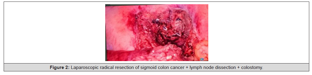
Figure 2: Laparoscopic radical resection of sigmoid colon cancer + lymph node dissection + colostomy
Results
During the operation, the anesthesia was good, and the operation was smooth. The operation time was 470 minutes. The blood pressure fluctuated between 160 - 140/60 - 80 mmHg during the operation. The intraoperative blood loss was about 100 ml, the total fluid intake was 2500 ml, and the total fluid output was 1280 ml. Considering that the patient underwent combined resection of multiple organs and the operation time was long, and the blood pressure fluctuated greatly during the operation, the patient was transferred to the Intensive Care Unit (ICU) after the operation to stabilize vital signs and reduce the risk of postoperative infection.
The patient was transferred back to the general surgery ward on the second day after the operation. Following the ERAS management measures, the urinary catheter was removed, the electrocardiogram monitoring and pulse oximetry monitoring were stopped, the patient was advised to drink a small amount of water, and parenteral nutrition support was provided. The patient was also encouraged to get out of bed early. The colostomy regained ventilation on the third day after the operation, the patient started to eat liquid food on the fourth day, the drainage tube was removed on the seventh day, and the patient was discharged smoothly on the 11th day. The patient did not complain of any special discomfort, could move freely, the physical examination was normal, and the laboratory and imaging examinations showed no obvious abnormalities. There was no recurrence during the 12 - month follow - up.
Pathological Diagnosis
Colon tumor resection specimen (Figure 3): Ulcerative moderately - poorly differentiated adenocarcinoma of the colon, with a tumor size of 4 cm×2.5 cm×1.5 cm. The cancer tissue invaded the serosa layer, and vascular invasion was observed, but no definite nerve invasion was found. Immunohistochemical results: The cancer tissue retained the expression of MMR proteins MLH1, MSH2, MSH6, and PMS2. Her - 2 was weakly to moderately positive in about 70% of the cells. CD31 and D2 - 40 showed blood vessels (Figure 4).

Figure 3: The resected sigmoid colon tumor specimen.

Figure 4: Post - operative Pathology of Sigmoid Colon Cancer.
Right renal tumor resection specimen (Figure 5): Combined with routine and immunohistochemical results, it was consistent with renal oncocytoma, with a size of 1 cm×1 cm×0.8 cm. No tumor residue was found at the surgical margin. The ypTN stage was T4a N1b. Immunohistochemical results : In the No. 13 paraffin block, the tumor cells expressed CA9 (-), CD10 (weakly positive locally), CD117 (strongly positive), CK7 (-), Ki67 (about 3% positive), P504s (-), TFE3 (-), E - Cad (+), CK20 (-), TFEB (weakly to moderately positive in some areas), SDHB (retained expression), and FH (retained expression) (Figure 6).

Figure 5: The resected renal tumor specimen.

Figure 6: Post - operative Pathology of Renal Tumor.
Discussion
Sigmoid colon cancer is one of the common digestive tract tumors, but sigmoid colon cancer complicated with renal oncocytoma is relatively rare, with few clinical reports. Colorectal cancer is a malignant tumor caused by abnormal gene expression in the cells of the colorectal region. Neoadjuvant Chemoradiotherapy (NACRT) or chemotherapy (NACT) + radical resection + adjuvant therapy is considered the best treatment mode for Locally Advanced Colorectal Cancer (LACRC). Compared with postoperative treatment, neoadjuvant chemotherapy for colorectal cancer patients before surgery can shrink the tumor, reduce the tumor stage, and increase the R0 resection rate. At the same time, it can reduce tumor invasion, lower the risk of tumor metastasis, reduce the scope of radical resection required, thereby reducing the risk of complications and the potential impact on survival, improving the patient’s prognosis, prolonging the survival period, and enhancing the quality of life [3- 5]. In this case, considering the advanced preoperative stage, the patient received neoadjuvant chemotherapy before surgery. The specific regimen was “oxaliplatin 200 mg on day 1 + capecitabine 1.5 g from day 1 to day 14”. After two cycles of chemotherapy, the tumor markers decreased effectively, which provided conditions for the smooth progress of the operation.
Renal oncocytoma (RO) is a rare benign renal tumor [6], accounting for about 3% - 7% of all renal tumors. It was first reported by Zippel in 1941. RO is generally sporadic and has an insidious onset. Most patients have no obvious clinical symptoms, and a few present with abdominal distension, low back pain, hematuria, hypertension, etc. [7]. Surgical resection is usually required for treatment, and the prognosis is good. However, due to the lack of specificity in blood biochemical and imaging examinations, it is difficult to distinguish RO from renal malignant tumors before surgery. On non - contrast CT, most ROs appear as isodense or slightly hyperdense lesions, and a few are slightly hypodense. The mass has clear boundaries and a capsule. On contrast - enhanced CT, it shows “rapid - in and slow - out” or “progressive” enhancement [6]. Some scholars have also pointed out that MRI has unique value in the diagnosis of renal oncocytoma.
The MRI of RO usually shows: on T1WI, it is mainly medium - low signal, and the signal of some lesions is slightly inhomogeneous, with small patches of lower signal visible; on T2WI, it shows heterogeneous signal, mainly high signal, with local or marginal low signal visible, and obvious enhancement is seen after contrast enhancement [8]. The high - incidence age of renal cancer is mostly between 50 - 70 years old. Renal cancer grows rapidly, has rich blood supply, and is prone to bleeding, necrosis, and cystic change. Therefore, its density is mostly inhomogeneous, and on contrast - enhanced CT, it shows a “rapid - in and rapid - out” imaging manifestation [9]. In this case, the non - contrast and contrast - enhanced CT of the abdomen showed a slightly hypodense nodule about 14 mm in diameter in the upper pole of the right kidney, and internal progressive enhancement was seen after contrast enhancement. The MRI of both kidneys with contrast enhancement showed a small nodule protrusion in the upper pole of the right kidney, with slightly low signal on T1WI and high signal on T2WI fat - suppressed sequence. Therefore, the diagnosis was basically clear through enhanced CT and MRI, and metastatic tumors and malignant tumors were excluded.
The Multidisciplinary Team (MDT) model [10] is a patient - centered diagnosis and treatment model. By organizing experts from relevant disciplines to comprehensively discuss and analyze the patient’s condition, the most suitable treatment strategy is jointly formulated to strive for the best prognosis. Resecting the intestinal and adrenal tumors in one operation can not only reduce the total surgical cost, relieve the patient’s pain, and reduce the patient’s surgical trauma and economic burden but also improve the surgical efficiency, reduce the risk of complications, shorten the hospital stay, and improve the patient’s prognosis. However, for this, the attending doctor needs to make a clear diagnosis and accurate assessment before the operation and cooperate with the multidisciplinary team to formulate the surgical plan and prognosis plan.
First, before the operation, the attending doctor needs to communicate fully with the anesthesiology department to predict the possible risks during the operation and formulate emergency plans. Second, cooperation with the urology team is required to pre - determine the operation time, operation method, and position selection to ensure the complete and smooth progress of the operation. Finally, after the operation, the attending doctor needs to cooperate with the intensive care medicine department to help the patient safely wean from the ventilator early after the operation, stabilize the condition and vital signs, reduce the incidence of pulmonary infection and complications, and improve the quality of the patient’s postoperative recovery.
In this case, after consultation with the anesthesiology department, it was planned to perform the operation under general anesthesia, which would take a long time. The patient was fully communicated with before anesthesia to exclude absolute surgical contraindications. After discussion with the urology department, considering factors such as blood pressure and anastomosis, laparoscopic renal tumor resection was performed first, followed by laparoscopic colorectal resection. During the operation, the patient was first placed in the left lateral position and then adjusted to the supine position. The umbilical and right mid - clavicular line trocars were shared during the operation, which reduced the trauma and intraoperative risks. Since the patient underwent combined resection of multiple organs and the operation time was long, the ICU was communicated in advance to prepare a bed, and the patient was transferred to the ICU for continued treatment after the operation to stabilize vital signs.
The intervention of Enhanced Recovery After Surgery (ERAS) further reduces surgical stress, decreases postoperative complications, and accelerates the patient’s postoperative recovery. Professor Jiang Zhiwei was the first to introduce the concept of ERAS in China. ERAS is different from traditional peri - operative management. Multiple clinical studies have confirmed that ERAS can effectively reduce the incidence of Postoperative Ileus (POI) [11]. Mechanistic studies have shown that the application of minimally invasive techniques, multimodal analgesia, shortening of fasting and water - deprivation time, and reduction of stress sources in the ERAS strategy can promote the patient’s postoperative recovery through the optimization of peri - operative stage plans and reduce the occurrence of related gastrointestinal complications.Strengthening preoperative health education helps patients improve their understanding of their own diseases, relieve their negative emotions, and assist patients in actively cooperating with various treatment measures during the peri - operative period, thereby reducing the rate of postoperative readmission [12].
ERAS recommends laparoscopic surgery and robotic surgery, emphasizing the implementation of minimally invasive surgery with function priority. Compared with traditional open colorectal cancer surgery, laparoscopic surgery has significant advantages such as small surgical trauma, less intraoperative blood loss, and milder postoperative pain. It can reduce the stimulation of the vagus nerve and effectively shorten the hospital stay [13]. Shortening the preoperative fasting and water - deprivation time helps reduce the adverse reactions of hunger, thirst, and restlessness in patients before surgery, improves patient comfort, reduces postoperative insulin resistance, alleviates catabolism, and maintains the body’s homeostasis [14]. Professor Jiang Zhiwei proposed a multimodal analgesia strategy combining “surgical incision injection of ropivacaine + oral administration of oxycodone for visceral pain control + intravenous injection of non - steroidal anti - inflammatory drugs (NSAIDs) + methylprednisolone for anti - inflammation”, which can reduce the use of opioid drugs and their related adverse reactions such as respiratory and gastrointestinal function inhibition, effectively prevent and reduce the incidence of postoperative delirium, inhibit the early postoperative inflammatory response, and promote postoperative recovery [15-17].
Postoperative guidance for patients to drink water and eat early, allowing them to drink a small amount of water 6 hours after anesthesia recovery without any special circumstances and then gradually transitioning to a liquid diet, can accelerate gastrointestinal function recovery, improve wound healing, and reduce the hospital stay [18]. Encouraging early ambulation after surgery is an important part of promoting postoperative recovery. It can effectively reduce the incidence of postoperative intestinal paralysis, promote gastrointestinal function recovery, advance the time of exhaust and defecation, relieve the patient’s weakness after surgery, and improve the patient’s clinical prognosis [19].
At the same time, early extubation is advocated. Early removal of nasogastric tubes, urinary catheters, and abdominal drainage tubes can reduce the risk of abdominal effusion, infection, and postoperative fistula formation. It also facilitates the patient’s early ambulation and accelerates gastrointestinal function recovery [20]. Therefore, peri - operative nursing plays a crucial role in the patient’s recovery management during the entire peri - operative period [21]. By ensuring surgical safety, minimizing complications, providing necessary postoperative care, and promoting patient education and publicity, it can significantly affect the patient’s prognosis. Therefore, the implementation of peri - operative ERAS management requires close cooperation with the nursing team to accelerate the postoperative recovery process.
Cases of patients with combined gastrointestinal and urinary tract tumors are relatively rare. There has been no previous literature report on laparoscopic combined resection of sigmoid colon cancer complicated with renal oncocytoma. In this case, through ERAS peri - operative management combined with the multidisciplinary team model, the surgical resection of abdominal organ tumors was successfully carried out, demonstrating the safety and effectiveness of this method. This model can not only reduce the total cost but also is beneficial to the patient’s postoperative recovery, which may indicate the future development direction of surgical treatment and has popularization value.
Of course, there are also some deficiencies in this case. Since the patient was transferred to the ICU after the operation, it affected the ERAS management process. Cooperation with the intensive care medicine department should be strengthened to coordinate the postoperative management measures. In actual clinical work, the treatment plan needs to be adjusted in a timely manner according to the specific situation, and inter - departmental communication and cooperation should be strengthened to provide patients with a better diagnosis and treatment experience and effect.
Acknowledgements
None..
Conflict of Interest
None.
References
- Dalli J, Camilleri Brennan J (2011) Synchronous colonic adenocarcinoma and renal oncocytoma: a case report and literature review. Scott Med J 56(3): 181.
- Cullinane CA, Jarrahy R, Wilson TG, Ellenhorn JD (2001) Asymptomatic renal neoplasms in the rectal cancer patient. Am Surg 67(12): 1162-1164.
- Zi Tong Zhang, Wei Wei Xiao, Li Ren Li, Xiao Jun Wu, Qiao Xuan Wang, et al. (2024) Neoadjuvant chemoradiotherapy versus neoadjuvant chemotherapy for initially unresectable locally advanced colon cancer: short - term outcomes of an open - label, single - centre, randomised, controlled, phase 3 trial. EClinicalMedicine 76: 102836.
- Morton D, Seymour M, Magill L (2023) Preoperative Chemotherapy for Operable Colon Cancer: Mature Results of an International Randomized Controlled Trial. J Clin Oncol 41(8): 1541-1552.
- Hindson J (2023) Neoadjuvant chemotherapy for operable colon cancer. Nat Rev Gastroenterol Hepatol 20(3): 131.
- Wang Yu (2021) Analysis of imaging and pathological characteristics of different pathological types of renal tumors [D]. China Medical University.
- Qiu Xiang, Guo Liang, Ding Xiaobo (2020) Imaging diagnosis and differential diagnosis of solid renal cell carcinoma. Chinese Journal of Radiology 54(9): 917-920.
- Ma Liya, Li Zhen, Shen Yaqi (2018) Analysis of CT and MRI imaging features of renal oncocytoma and metanephric adenoma. Journal of Contemporary Urologic and Reproductive Oncology 10(04): 216-221.
- Zhao Hongfei, Shao Xiaodong, Zhang Xiaohui (2024) Analysis of CT signs of renal oncocytoma. Chinese Journal of CT and MRI 22(02): 118-120.
- Jin Qunlong, Wang Guixian, Sun Zhenqiang (2020) Influence of the multidisciplinary team model on the diagnosis, treatment, and prognosis of locally advanced rectal cancer. Journal of Zhengzhou University (Medical Sciences) 55(1): 13-17.
- Chen HT, Hung KC, Huang YT (2024) Efficacy of electroacupuncture in improving postoperative ileus in patients receiving colorectal surgery: a systematic review and meta - analysis. Int J Surg 110(2): 1113-1125.
- Lü Xiaomiao (2019) Application of comprehensive preoperative holistic education in patients undergoing colorectal cancer surgery. China Continuing Medical Education 11(20): 174-177.
- Shen Danli, Cheng Hui, Wang Gang, Jiang Zhiwei (2021) Enhanced recovery after surgery and the protection of vagus nerve function. Journal of Medical Postgraduates 34(02): 200-204.
- Ljungqvist O, Nygren J, Thorell A (2002) Modulation of post - operative insulin resistance by pre - operative carbohydrate loading. Proc Nutr Soc 61(3): 329-336.
- Jiang Zhiwei, Zhou Jiahui, Cheng Hui (2019) The role of multimodal analgesia in enhanced recovery after surgery. Journal of Shandong University (Health Sciences) 57(09): 1-4
- Li Li, Zhang Lina (2019) Optimizing analgesia and sedation to accelerate the recovery of critically ill patients. Journal of Medical Postgraduates 32(11): 1130-1134.
- Cao L, Zhang L, Chen B, Yan L, Shi X, et al. (2024) Application of multimodal standardized analgesia under the concept of enhanced recovery after surgery in laparoscopic radical colorectal cancer surgery. Front Oncol 14: 1381809.
- Herbert G, Perry R, Andersen HK (2019) Early enteral nutrition within 24 hours of lower gastrointestinal surgery versus later commencement for length of hospital stay and postoperative complications. Cochrane Database Syst Rev 7(7): CD004080.
- Liu Lin, Xu Qin, Chen Li (2013) Analysis of the first out - of - bed activity status and its influencing factors in patients after gastrointestinal surgery. Journal of Nursing Science 28(04): 17-19.
- K C Conlon, D Labow, D Leung, A Smith, W Jarnagin, et al. (2001) Prospective randomized clinical trial of the value of intraperitoneal drainage after pancreatic resection. Ann Surg 234(4): 487-494.
- van der Vlies E, Smits AB, Los M (2020) Implementation of a preoperative multidisciplinary team approach for frail colorectal cancer patients: Influence on patient selection, prehabilitation and outcome. J Geriatr Oncol 11(8): 1237-1243.




 We use cookies to ensure you get the best experience on our website.