
Research Article 
 Creative Commons, CC-BY
Neurosteroid 17β-estradiol reduces motor dysfunctions in a 6-OHDA-induced rat model of Parkinson’s disease
*Corresponding author: Cabrera RJ, Valdéz SR, INBIOMED, University of Mendoza, Huarpes 630, 5500 Mendoza-Argentina
Received: May 15, 2025; Published: May 21, 2025
DOI: 10.34297/AJBSR.2025.27.003522
Abstract
Several experimental models have been developed to fulfill the clinical, morphological, and neurochemical criteria, mimic the neuropathological lesion, and allow defining therapeutic strategies for Parkinson’s Disease. The unilateral injection of 6-OHDA in the left corpus striatum produces a progressive degeneration of the nigrostriatal system on the injured side, allowing internal comparison with the intact contralateral side. This condition is called hemiparkinsonism. Motor impairments generated in the unilateral administration model can be assessed by tests that examine, among other signs, akinesia, body axis deviation, drug-induced asymmetric rotational behavior, and spontaneous motor behaviors. Some evidence has suggested that female sex hormones may influence the onset and severity of the symptoms, suggesting a possible protective influence of estradiol on disease predisposition. The present work’s objective is to evaluate the potential effects of 17β-estradiol on motor dysfunction in a 6-OHDA-induced rat model of Parkinson’s disease. Treatment with 17β-estradiol produced some interesting effects on activity and functionality in motor performance. If degeneration is not severe, this treatment could be a neuroprotective agent. In conclusion, the neurosteroid 17β-estradiol reduces motor dysfunctions in a 6-OHDA-induced rat model of Parkinson’s disease. These investigations contribute to the recognition of the potential impact of steroid sex hormones on nervous system injuries.
Keywords: Parkinson’s disease, 17β-estradiol, 6-OHDA, Motor activity, behaviour
Introduction
Several experimental models have been developed to fulfill the clinical, morphological, and neurochemical criteria, mimic the neuropathological lesion, and allow defining therapeutic strategies for Parkinson’s Disease [28]. Among these models, neurotoxins’ most frequently employed intracerebral application is [41,30,7]. The neurotoxic 6-OHDA is widely used as a fundamental tool to model the pathophysiology of dopaminergic denervation [35]. When injected into the nigrostriatal pathway of rodents, it selectively destroys catecholaminergic neurons and results in the loss of nigral dopaminergic neurons and reduced dopamine levels in the striatal terminals [45,46]. These lesions lead to spontaneous motor and sensory-motor function changes in rodents, similar to those occurring in PD in humans. Particularly, unilateral injection of 6-OHDA produces a progressive degeneration of the nigrostriatal system on the injured side, allowing internal comparison with the intact contralateral side. This condition is called hemiparkinsonism, which involves degeneration of the nigrostriatal pathway in only one cerebral hemisphere [16,14,39].
Motor impairments generated in the unilateral administration model can be assessed by tests that examine, among other signs, akinesia, body axis deviation, drug-induced asymmetric rotational behavior, and spontaneous motor behaviors [34,16,26,49]. Dopaminergic decrease leads to akinesia, bradykinesia, and body asymmetries [15, 36]. It is also possible to see and quantify ambulatory behavior in the open field test [12,24]. The rotatory response to drug administration is an indicator of the degree of severity of the nigrostriatal lesion. On the one hand, the administration of substances that increase DA release (such as amphetamine) produces ipsilateral rotation towards the injured side. On the other hand, administering a non-selective agonist of dopaminergic D1 and D2 receptors, such as apomorphine (Apo), induces contralateral behavior as a compensatory response. In the last decades, several studies have been carried out to elucidate the neuroprotective effects of estrogens in diseases and nervous system injuries. These investigations led to the recognition of the potential impact of steroid sex hormones on such disturbances [21]. Intracellular steroid receptors, involved in specific gene transcription, have been identified in most structures that regulate neuroendocrine functions. These receptors have a unique pattern of distribution in the complex anatomy of the brain and exert their functions through steroid-induced feedback mechanisms that correlate with behavioral expression [8,9,29,22,23]. Investigations have proved that the brain is an important target organ for steroid hormones and an organ where steroid metabolism is considerable [32,20,2,3].
Particularly, as neuroactive steroids are lipophilic hormones with low molecular weight, they easily cross the blood-brain barrier and are available for their functions in the brain, including regulation of the reproductive neuroendocrine system, mood, and cognition, as well as neuroprotective effects [4,40]. In particular, 17β-estradiol (E) can be synthesized directly in the brain and exert its functions there or come from the periphery. It directly promotes cell survival and synaptic plasticity, prevents axonal and dendritic loss, and helps prevent neuronal malfunction by altering neurotransmitters, receptors, and second messengers [17,18]. Concerning Parkinson’s disease, basic science, epidemiology, and clinical evidence have suggested that female sex hormones may influence the onset and severity of the symptoms [48,27]. Furthermore, the prevalence is higher in men than in women, at a ratio of approximately 3:2 Mayeux et al., (1992) [31], suggesting a possible protective influence of estradiol on disease predisposition [17,18]. One clinical study conducted on PD in estrogen replacement in the menopausal years suggests that symptoms may be exacerbated after menopause and may be delayed or alleviated by hormone replacement therapy (POETRY).
The neurosteroid estradiol is neuroprotective because it modulates the function indices of mesencephalic dopaminergic neurons and prevents neurotoxin-induced dopaminergic neuronal depletion in animal models of PD. Modulation can occur at synthesis, release, DA reuptake, and binding to specific receptors.
As neuroprotective results are treatment-dependent on the period of administration and dose and the gonadal status of the animals studied, test evaluation on different stages allows individual behavior characterization according to the stage of the disease [16] as well as the response to the proposed treatment. Suppose the specific mechanisms by which estrogen exerts its neuroprotective effects in PD are established. In that case, new combinations of targeted therapies with structural modifications can be developed to maximize neuroprotection and minimize unwanted systemic effects.
Materials and Methods
Animals
We used adult male Sprague Dawley rats from our breeding colony. They were 60 days old at the beginning of the study and weighed 280-340 g. Experimental subjects were housed under controlled temperature (22 ± 3 ºC) and lighting (12-hour cycle beginning at 7:00am), with food and water made available ad libitum.
Animals were kept and handled according to the Guide for the Care and Use of Laboratory Animals of the National Research Council (National Academies, USA, 8th edition, 2011) and Comité Institucional para el Cuidado y Uso de Animales de Laboratorio (CICUAL), Facultad de Ciencias Médicas de la Universidad Nacional de Cuyo, Argentina, 86/2016.
Surgical Procedures
At the beginning of the experiment, the rats were anesthetized with an intraperitoneal injection of ketamine (100mg/kg) and xylazine (20mg/kg) and placed into a stereotaxic frame (David Kopf, USA). The neurotoxin, 6-OHDA (Sigma-Aldrich, St. Louis, MO, USA), was dissolved at a 2μg/μl saline concentration in 0.1% ascorbic acid. A group of rats received 6-OHDA injections to achieve unilateral lesions, and another group received vehicle (V) into the left caudate-putamen (CPu). The lesion was performed by injecting the neurotoxic vehicle with a Hamilton syringe at the following coordinates: AP: +1.2mm; ML: +2.5mm; DV: -5.0mm; TB at 0mm. The injection was conducted at a rate of 0.5μl/min, and the needle was left in place for another 5min before it was slowly drawn back. After surgery, when the animals were fully recovered, they were taken to a room where they rested for seven days (Figure 1).
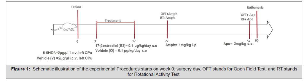
Figure 1: Schematic illustration of the experimental Procedures starts on week 0: surgery day. OFT stands for Open Field Test, and RT stands for Rotational Activity Test.
Treatment
From day 7 to 17, animals received chronic asymmetry treatment with 17β-estradiol (E=0.1μg/kg/day s.c) or corn oil as vehicle (O). Groups were formed as HP (6-OHDA lesion + O), HP+E (6-OHDA lesion+ E treatment), E (V lesion+ E treatment), and C (V lesion+ O). From 6 to 12, animals were finally assigned to each experimental group.
Motor Behavioural activity
Behavioral assessments were performed to evaluate the presence of alterations in motor function involved in the 6-OHDA lesion. In week 2 (day 27), after amphetamine injection (Amph), an Open Field Test (OFT) and Rotational Activity Test (RT) were performed and evaluated. The same tests were performed on week 4 post-lesion (day 57) after apomorphine injection (Apo). All behavioral tests were performed from 10am to 5pm during the daytime. In week 5 (day 60), animals were euthanized, and samples were prepared for further assays.
Open Field Test
The experiment involves exposing the animal to a novel situation for 5 minutes, during which time the animal’s exploration, locomotion, and non-ambulatory activity are assessed [24]. In every performance, each animal was exposed to an open field without additional stimuli, allowing it to explore the area freely. The open field is a wooden box 60cm wide x 80cm long x 35cm high. The box floor is black and divided with white lines into 48 squares of 9.5cm sides each. The box must be in a purpose-built room [24,12]. The animals were brought to the behavioral evaluation room one hour before the test. At the evaluation, they were placed in the centre of the box and filmed for 5 minutes (300 seconds). Each video was processed in real-time with Anymaze v4.99 software Figure 3,2,1,2, where the following variables were assessed:
Ambulatory activity: movements detected as displacement.
a. Line crossing [nº]: frequency of crossing with the four limbs in the grid.
b. Time in each zone [s]: center, the corners, and the edges.
c. Average speed [m/s]: average space-time relationship of the exploratory activity.
d. Total Distance traveled [m]: space traveled during the test.
e. Trajectories (track plots)
Non-ambulatory activity: Number of times the animal stops and does not move.
a) Number of episodes [nº] and time [s] of immobility; taking by convention as a reference for the start of this activity, 2000ms.
b) Freezing: the Number of times [nº] the animal remains in the same place in a stationary state, with or without piloerection, and the total time [s] during which this is carried out. By convention, it is counted when the time exceeds 250ms.
Rotational Activity Test
The drug-induced rotational test is the most widely used and the best-characterized animal model system of PD because of its simplicity and sensitivity. The circular walking pattern in the 6-OHDA rats can be easily evoked by dopaminergic agonists. Behavioural records were all performed by an observer blinded to the experimental condition of the group. Two weeks after surgery, all groups were tested for amphetamine-induced ipsilateral rotation. Rats received 1mg/kg amphetamine i.p.. They were placed in an individual plastic bowl (20cm diameter) and attached via a specially adapted harness to an automated rotameter (Rotamex, Columbus Instruments, Columbus, OH). They were allowed to habituate to their dimly lit environment for 10min before contralateral and ipsilateral turns, regarding the side of the lesion, were recorded over 60min. Four weeks after surgery, all groups were tested for apomorphine-induced contralateral rotation using the same experimental setup as for amphetamine-induced rotation. Apomorphine was injected s.c. At a 2mg/kg dose, rotation was monitored for 60 min.
The following variables were determined:
i. Rotations to the right [nº].
ii. Rotations to the Left [nº].
iii. Total rotations [nº].
When this parameter is induced with amphetamine, it is calculated as the difference between the Number of rotations to the left side minus the Number of rotations to the right side. For apomorphine- induced rotations, it is calculated as the difference between the Number of rotations to the right side minus the Number of rotations to the left side.
Data analysis
Data from behavioral trials were Analyzed by One-Way Analysis of Variance (ANOVA). When statistical significance was found between groups, Tukey’s post hoc multiple comparisons. Tests were performed to determine points of significant difference. Open Field results were expressed as the mean and standard error of the mean difference (Mdiff ±SEdiff). In Rotational tests, data were expressed as Means± S.E.M.; the total Number of turns/ 60 min was calculated as right minus left turns. With Welch’s correction, Left and proper parameters were compared with the Unpaired t-test. For all tests, p<0.05 was considered statistically significant.
Results
Open Field Test
Amphetamine Induction: As shown in Table 1, amphetamine administration (Amph) induced differences among experimental groups and increased ambulatory activity. 6-OHDA-injured animals that did not receive neurosteroid treatment (HP) traveled shorter distances than the other experimental groups (p<0.05), and they decreased the average speed compared to the C and HP+E groups. HP animals increased the total freezing episodes, statistically higher (p<0.05) than C, E, and HP+E. Moreover, HP+E animals did not show significant differences in freezing episodes compared to E or C groups (Table 1).

Table 1: Mobility parameters measured with the OFT (Amph). Results are expressed as Mean and standard error of the mean difference (Mdiff±SEdiff). Group C (n=6), E (n=6), HP (n=8), HP+E (n=8). Statistics: 1-way ANOVA, Tukey post hoc test; *p<0.05, **p<0.01, ***p<0.001, ns = not significant.
Table 2 shows that the injured animals that did not receive 17β-estradiol treatment (HP) spent more time freezing compared to the other experimental groups (p<0.05), moved less, and spent more time in the corner zone of the open field. In contrast, 17β-estradiol treatment significantly improved times of movement performance for the HP+E group, p<0.05 (Table 2).

Table 2: Times measured with the OFT (Amph). Results are expressed as Mean and standard error of the mean difference (Mdiff±SEdiff). Group C (n=6), E (n=6), HP (n=8), HP+E (n=8). Statistics: 1-way ANOVA, Tukey post hoc test; *p<0.05, ***p<0.001, ****p<0.0001, ns = not significant.
As shown in Figure 2, with Amph, the trajectories of the HP animals were more sparse and peripheral. The group of HP+E animals showed increased locomotion due to greater entries to the central zone and edges and decreased episodes in the corner zones. Track plot density and movement patterns in HP+E were similar to those in E (Figure 2).
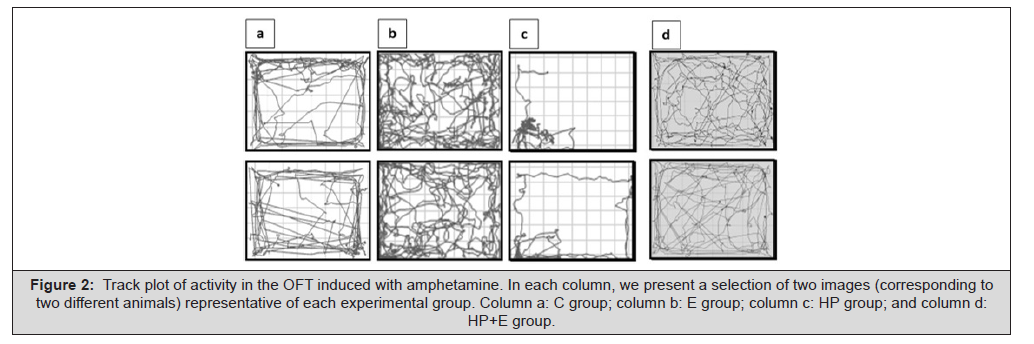
Figure 2: Track plot of activity in the OFT induced with amphetamine. In each column, we present a selection of two images (corresponding to two different animals) representative of each experimental group. Column a: C group; column b: E group; column c: HP group; and column d: HP+E group.
Apomorphine Induction: The ambulatory activity of HP animals decreased significantly compared to C, E, and HP+E (Table 3). Long-term treatment with 17β-estradiol (57 days post-injury) caused HP+E animals to behave significantly differently from the HP group and similar to the vehicle-injured animal groups (C and E). No significant results were seen in this test for the analysis of non-ambulatory activity.

Table 3: Times measured with the OFT (Amph). Results are expressed as Mean and standard error of the mean difference (Mdiff±SEdiff). Group C (n=6), E (n=6), HP (n=8), HP+E (n=8). Statistics: 1-way ANOVA, Tukey post hoc test; *p<0.05, ***p<0.001, ****p<0.0001, ns = not significant.
Table 4 shows that C, E, and HP+E groups spent more time moving than HP (p<0.05). In the corners, groups E and HP+E stayed less than HP, p<0.05. Furthermore, the comparison of freezing times between HP and E showed that E had a significantly longer freezing time (Table 4).

Table 4: Times measured with the OFT (Apo). Results are expressed as the mean and standard error of the mean difference (Mdiff± SEdiff). Group C (n=10), E (n=8), HP (n=10), HP+E (n=11). Statistics: 1-way ANOVA, Tukey post hoc test; *p<0.05, **p<0.01, ns= insignificant.
Activity track plots after apomorphine induction differed for HP animals compared to the other groups. They exhibit a poor performance with minimum movements and longer corner stops. In contrast, animals in the other experimental groups exhibited random movements throughout the open field. Interestingly, even at this late stage of neurodegeneration, treatment with 17β-estradiol showed positive effects on the motor activity of the HP+E animals (Figure 3).
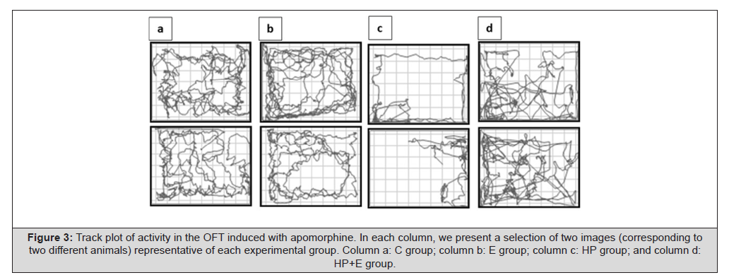
Figure 3: Track plot of activity in the OFT induced with apomorphine. In each column, we present a selection of two images (corresponding to two different animals) representative of each experimental group. Column a: C group; column b: E group; column c: HP group; and column d: HP+E group.
Rotational Activity Test
Amphetamine Induction: After Amph. induction, we observed that the HP animals decreased contralateral turns (Figure 4a), and they showed a marked “laterality,” increasing movement towards the side of the lesion (Figure 4b). As shown in Figure 4c, HP animals increased the Number of turns performed towards the ipsilateral side. The total Number of rotations for this group was statistically significant compared to the other experimental groups (p<0.05). These results support the ones obtained with the open field. Specifically, the compensatory response to denervation of the HP+E group reached values similar to those of vehicle-injured animals (C and E) (Figure 4).
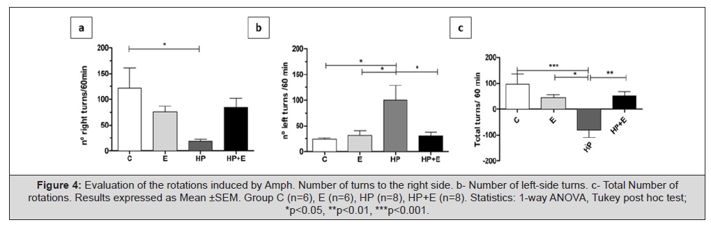
Figure 4: Evaluation of the rotations induced by Amph. Number of turns to the right side. b- Number of left-side turns. c- Total Number of rotations. Results expressed as Mean ±SEM. Group C (n=6), E (n=6), HP (n=8), HP+E (n=8). Statistics: 1-way ANOVA, Tukey post hoc test; *p<0.05, **p<0.01, ***p<0.001.
Figure 5 shows the differences between the left and right sides for all parameters evaluated in each experimental group. Ipsilateral locomotor imbalance induced by Amph in HP animals favored the uninjured (left) side, and 17β-estradiol administration prevented this marked asymmetry for the HP+E group (Figure 5).
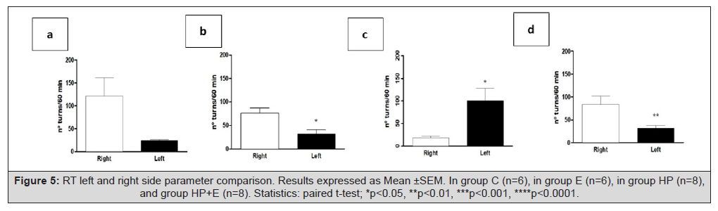
Figure 5: RT left and right side parameter comparison. Results expressed as Mean ±SEM. In group C (n=6), in group E (n=6), in group HP (n=8), and group HP+E (n=8). Statistics: paired t-test; *p<0.05, **p<0.01, ***p<0.001, ****p<0.0001.
Apomorphine Induction
Apomorphine is a direct agonist that increased contralateral rotations in HP animals (p<0.01) (Figure 6a) and decreased the Number of Left turns (p<0.05) (Figure 6b). Results were statistically significant compared to HP+E. Figure 6c shows that the total Number of rotations HP animals performed was higher than all experimental groups (p<0.01) (Figure 6).
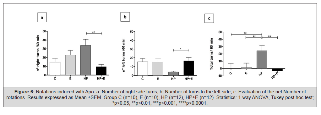
Figure 6: Rotations induced with Apo. a. Number of right side turns; b. Number of turns to the left side; c. Evaluation of the net Number of rotations. Results expressed as Mean ±SEM. Group C (n=10), E (n=10), HP (n=12), HP+E (n=12). Statistics: 1-way ANOVA, Tukey post hoc test; *p<0.05, **p<0.01, ***p<0.001, ****p<0.0001.
The comparisons between the left and right sides are presented in Figure 7. A sign of neurodegeneration in HP animals is the imbalance generated by apomorphine administration; this situation was delayed by 17β-estradiol treatment, as shown in the HP+E group performance (Figure 7).
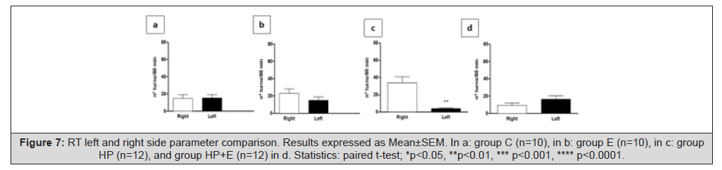
Figure 7: RT left and right side parameter comparison. Results expressed as Mean±SEM. In a: group C (n=10), in b: group E (n=10), in c: group HP (n=12), and group HP+E (n=12) in d. Statistics: paired t-test; *p<0.05, **p<0.01, *** p<0.001, **** p<0.0001.
Conclusion
The tests outline the asymmetry in the hemiparkinsonism model generated by the loss of dopamine after a neurotoxic injury with 6-OHDA in the left CPu. This unpaired activity is interpreted as a motor sign of degeneration of the nigrostriatal dopaminergic pathway of the disease in rodents. Treatment with 17β-estradiol produced some interesting effects on activity and functionality in motor performance. If degeneration is not severe, this treatment could be a neuroprotective agent.
Discussion
In our experiments, the 17β-estradiol dose of 0.1μg/kg/day administered for 10 days is within the physiological range of a regular estrous cycle compatible with rising levels in early proestrus in female rats [10,42]. Selecting an appropriate dose is important when examining the safety and tolerability of estrogens as potential pharmacological tools to ameliorate neurodegenerative motor symptoms [38]. Regarding administration protocol, Smith, et al., conducted a study in which animals were treated with estrogen compounds at different times, including before, concurrently with, and after toxin treatment. Their results demonstrated that treatment must precede toxin administration to observe a neuroprotective effect [44]. While this research contrasts our results, we agree that treatment after neurotoxic damage cannot reverse the injury, so its effect is neuroprotective rather than neuroreparative. To confirm that administration of 6-OHDA in the striatum triggers the motor symptoms characteristic of the disease, we developed a battery of both spontaneous and pharmacologically induced behavioral tests performed at different stages. The results revealed that the administration of 17β-estradiol may influence the motor signs of neurodegeneration characteristic of PD. Behavioral assessment is a powerful tool for in vivo animal models. It allows inclusion/ exclusion criteria to demonstrate the efficacy of treatments and the identification of sedative, stimulant, or toxic influences of different substances, among others. However, the effects of experimenters are responsible for a significant proportion of behavioural research. The operator requires patience, experience, and animal handling and restraint skills. Although it is a potentially stressful performance for the animal, standardizing test conditions as much as possible in the environment, handling, training, and the test itself is how to acquire consistent results [6]. The open field study demonstrated that, with the administration of dopamine agonists (amphetamine and apomorphine), there are differences in rodents’ responses in different groups when encountering a novel situation. We assessed the animals’ exploration, locomotion, and non-ambulatory activity in two periods. We observed that unilateral striatal administration of 6-OHDA produces deficits in motor activities in injured animals that did not receive 17β-estradiol treatment. That group (HP) made shorter movement episodes and moved less than the other experimental groups. In a model of hemiparkinsonism previously evaluated in our laboratory [12] Casas et al., (2013), no significant differences were found between the study groups (6-OHDA-injured, controls, and progesterone- treated groups). We estimate that the main difference with these results lies in the program they used to measure behavior. Casas, et al., worked with Etholog [37] Ottoni, et al., (2000), and the parameters measured were limited (in quantity and precision) compared to our assessment method. Moreover, the results obtained with OFT coincided with those we observed in rotational behaviour with the rotational test. It is known that the magnitude of rotation is proportional to dopaminergic system degeneration, so it is used as an indicator of the degree of the lesion.
In the literature, there is contradictory evidence regarding the Number of turns corresponding to higher striatal dopaminergic deficits. Some authors indicate that a number greater than or equal to 200 turns per hour, in rodents, corresponds to a greater than 90% depletion of striatal DA [25,1] Hudson et al., (1993), Barnéoud et al., (1995); while other authors report a loss of close to 80% given by approximately 100 turns per hour. Da Cunha, et al,. observed that animals with partial midbrain dopaminergic loss turned ipsilateral to the side of the lesion, and the ones that lost almost all midbrain dopaminergic neurons exhibited contralateral turning behavior. As in our results with dopaminergic agonists, amphetamine and apomorphine, it is possible to demonstrate that the animals manifest marked laterality in turning. This magnitude is differential for HP compared to the other study groups. Two main factors influence the Number of turns performed: the degree of injury and the experimental group condition of the animals. Additionally, in our experiments, we observed how those conditions affect the turning time concerning the maximum and total duration of the turns. We consider that these parameters must be compared to the Number of turns and turning time to talk about laterality.
Regarding estrogens as a neuroprotective treatment, they are well-characterised agents, but the mechanisms by which they exert their actions are still unknown. They have abundant cellular effects including nuclear activation of receptors, increased expression of anti-apoptotic proteins, the interaction of second messenger cascades, alterations in glutamatergic activation, maintenance of intracellular calcium homeostasis, and antioxidant activity [32,17,20,19]. Although several of these effects are involved in neuronal survival, the exact role of each of these pathways in neuroprotection remains unclear [21]. According to Morale et. al., in Parkinson’s Disease, there are two pathways; while one is detrimental, the other is beneficial [33]. They suggest that this occurs because 17β-estradiol has the primary role of a “glial switch”. Harmful cytotoxic effects in situations of inflammatory stress can lead to the disappearance of nigral dopamine and neuronal death. On the other hand, beneficial effects protect nigral neurons and occur mainly through the action of astrocytes. These cells are responsible for removing glutamate from the extracellular space, producing antioxidant enzyme-inducing factors, and expressing crucial neurotrophic molecules, regulating neurons’ growth, differentiation, and survival as part of bidirectional, neuro-glial interactions.
Animal studies have shown pre- and postsynaptic modulation by 17β-estradiol in the nigral dopaminergic pathway, as well as the ability to protect dopaminergic neurons from the effects of neurotoxins such as 6-OHDA [43,44]. Neuroprotection after neurotoxin exposure is either related to 17β-estradiol prevention of striatal dopamine and its related metabolites reduction, or neuronal integrity of the dopamine synthesis pathway [44]. Depending on the viability of cells in response to the action of 17β-estradiol, if neurons are healthy at the time of treatment, their response is beneficial for both neurological function and survival. In contrast, if the neuronal function is impaired, exposure to treatment over time exacerbates neurological injury [7]. At an early stage of degeneration, when the neurons of the dopaminergic pathway are injured but not killed, a therapeutic window opens for neuroprotection with steroids. Indeed, in humans, it is in the early stages of PD, before initiating levodopa therapy, that 17β-estradiol therapy is reported to be beneficial.
Declaration of Conflicting Interests
The authors declared no potential conflict of interest concerning this article’s research, authorship, and publication.
Acknowledgments:
None.
References
- Dai C, David D, and Wu D (2025) A layman’s strategy of investing in ordinary mutual funds. American Journal of Biomedical Sciences & Research 26(5).
- Morningstar Ratings 101: What You Need to Know. https://www.morningstar.com/company/morningstar-ratings-faq.
- Yahoo Finance. https://finance.yahoo.com/.
- R software. https://www.r-project.org/.




 We use cookies to ensure you get the best experience on our website.