
Research Article 
 Creative Commons, CC-BY
The Phenomenon of Fast Empting of an Isolated Vacuole Via Holes Forming in Its Membrane, and Perspectives of Application of the Respective Knowledge in Organelle-Level Treatment of Heavy Diseases
*Corresponding author:Michael Yu. Chernyshov, Siberian Institute of Plant Physiology and Biochemistry, Russia and Irkutsk Scientific Center, Russian Academy of Sciences, Russia
Received:June 03, 2025; Published:June 11, 2025
DOI: 10.34297/AJBSR.2025.27.003555
Abstract
The present publication describes a discovery. This discovery is bound up with the phenomenon of fast empting of isolated vacuoles. It is important to note that the effect of empting of the vacuolar content has never before been investigated. This empting has the form of fast (during decades of seconds) efflux of the vacuolar soluble and non-soluble content. In hundreds of observations (conducted with aid of laser scanning confocal microscopy), it has been revealed that isolated red beetroot vacuoles can eject their internal content in course of a sequence of events bound up with opening holes of micron diameter in the vacuolar membrane. The phenomenon discovered presumes a sequence of intermittent processes, which include: (i) Formation of a hole (diameter: 0.5- 1.9μm) in the vacuolar membrane; (ii) Efflux of the vacuolar content into the outer space; (iii) Sealing of the hole; and, (iv) If the vacuole has not been completely devastated, formation of another hole at the same or at a different location in the membrane of the same vacuole; (v) Its sealing; etc. Such (or similar) phenomenon has never been earlier discussed in the literature bound up with intracellular trans-membrane transport.
Keywords:Isolated vacuoles, Natural decay, Efflux of nutrients, Hole in the membrane, Membrane hole resealing, Biophysical characteristics of vacuoles, Laser scanning confocal microscopy
Abbreviations:ABC Transporters: ATP Binding Cassette Type Transporters; ANS: 8-Anylino-1-Naphtalensulphonic Acid; CPP: Cell Penetrating Peptide; Cryo-EM: Cryo-Electron Microscopy; DOPC: Dioleoylphosphatidylcholine; DPH: Diphenilhexatrien; FLSCM: Fluorescent Laser Scanning Confocal Microscopy; GABAARs: γ-aminobutyric acid A receptors; GPCR: G Protein-Coupled Receptor; GUV: Giant Unilamellar Vesicle; KB-ideology: Knowledge-Based Ideology; LUV: Large Unilamellar Vesicle; NA transporters: Nucleobase- Ascorbate Transporters; NMR: Nuclear Magnetic Resonance; PCV: Plant Cell Vacuole.
Introduction
The present paper discusses the phenomenon of fast empting of an isolated Plant Cell Vacuole (PCV) expressed in the form of fast stepwise efflux of substances (soluble and non-soluble content) through the PCV membrane. This phenomenon was for the first time noticed by the author’s team of researchers in 2015, in the confocal microscopy observations bound up with analysis of peculiar properties of some components in the PCV morphological structure [1]. The phenomenon was specially observed and registered in 2017 [2] and later confirmed in numerous experiments from 2018 till 2024. The phenomenon discovered is new. In this connection, there appears the need to consider biophysical characteristics of PCVs, characteristics of PCV membranes, which are bound up with this phenomenon (e.g. mechanical membrane stability [3] and other important properties [4], and analyse the opportunities of applying the discovered phenomenon in the aspect of transport of substances from PCVs, which could be useful from the viewpoint of contemporary goals of medicine.
A PCV and its membrane are capable of dynamical morphological reconstructions [4-7]. It is known that principal functions of the central vacuole living inside a plant cell are determined by its participation in processes of redox ionic homeostasis of the cytosol [8], storage of primary and secondary metabolites, osmotic regulation, detoxification of xenobiotics, formation of protective responses of the cell under the conditions of biotic and abiotic stress, as well as participation in the metabolism of poisons and in the processes of programmed cell death [9]. Furthermore, already in the 2000s, it was shown that vacuoles were bound up with cytoskeletal elements [10]. The role of vacuoles in the stomatal movement was emphasized [11]. As far as vacuolar membranes are concerned, there have appeared declarations about insights into the architecture of membranes of PCVs [12], and into the diversity of systems providing for transport of water and substances via vacuolar membranes.
Vacuolar membranes form a protective layer against (i) External mechanical, osmotic and toxic forms of stress, while providing for mechanical stability; (ii) Bacterial infecting and even (iii) Viral infecting. Furthermore, vacuolar membranes are involved in ongoing signaling, trafficking, and morphogenesis, while providing for high degree of plasticity. Biochemical diversity of membranes, which are comprised by hundreds of lipid species, contributes to membrane stability and plasticity. Many properties of membranes are to be understood better. Furthermore, vacuolar membranes participate in provision of intra-cellular redox homeostasis [3,8,13].
In diverse investigations conducted by researchers from many countries, the authors dared to analyze several known concepts describing transport of water, substances and compounds (a) Inside the cytoplasm, (b) Via cellular and organelle transport channels, (c) Via organelle membrane pores into the organelles, etc.
A number of molecular-level trans-membrane transport mechanisms have been identified and described. It has been postulat ed that transport of soluble substances and compounds from the cellular cytosol into the vacuole may be provided by such transport systems as: tonoplast-bound ABC transporters [14-28], nucleobase- ascorbate transporters, tonoplast-bound ABC transporters [14-17], nucleobase-ascorbate transporters [18-21], tonoplast transporters using the proteomic mechanism [22-24], vacuolar H+-ATPase driven potassium transport channels [25-28], solute/H+ antiporters [26,27]. C. Roos, et al., (2012) discussed nanodisc complexes with small multidrug transporters [29] etc. Noteworthy, the proteomic approach studied for the transport process in the vacuoles isolated from cauliflower buds was represented as providing for a novel (in its time) form of transport [23].
The list of above transport systems includes peptide-based transporters. These transporters are represented by amphipathic Cell Penetrating Peptides (CPPs) [30,31]. Activity of this type of transport channels is known to be mediated also by pore forming peptides and pore forming proteins [32,33]. CPPs, which are known to be capable of inducing dynamical phase separation of phospholipid bilayers, formation and growth of membrane’s negative curvatures resulting (say, according to [34,35]) in undulations, and even membrane thinning or thickening, provide for the peptide-based transport. Transport of acid-glutathione conjugate into the vacuole was discussed by N. Ohkama-Ohtsu, et al., (2011) [36].
The list of above transport systems may also be complemented with SWEET and SemiSWEET transporters [37-39], which provide for or facilitate the respective diffusion mechanism. Special attention of well-known researchers was concentrated also on transport processes mediated by protein-bases transporters, which were discussed as mediators providing for transport of nutrients inside and outside a vacuole [26,40,41]. The issues bound up with invaginations in the membrane, which were related to the transport processes, were not forgotten in discussions. Some of abovementioned transport systems bound up with vacuoles may play important roles in the processes of cell growth and in cellular developmental processes. These issues are planned to be discussed in our forthcoming publications. It was stated also that some of abovementioned transport systems were needed, for example, in formation of (i) Fe/S clusters, (ii) So called stomatal movement [11], and probably also (iii) Ion fluxes. Furthermore, we have to emphasize that according to the accepted conception, transport of soluble substances from the cytosol into the vacuole may also be mediated by ion channels. The widely discussed type of the channel for transport of soluble substances is represented by pores forming in membranes owing to various influences (factors and processes).
In the literature, one will not find any plausible discussions bound up with observations of dynamics of pores in biological membranes of natural cells. Meanwhile, discussed are various forms of experiments, which presume hypothetical formation of pores in such artificial objects as Giant Unilamellar Vesicles (GUVs). Known experiments bound up with formation of such pores include:
(i) Tension-induced formation of transient pores [42-46]
(ii) Pore formation under external stress factors [47];
(iii) Ion-induced transient potential fluctuations provoking
pore formation [48];
(iv) Pore formation owing to membrane electroporation and
electropermeabilization [49,50];
(v) Pore formation owing to mechanical factors (accompanied
with lateral sorting of membrane lipids) [51];
(vi) Pore formation owing to membrane thinning (as the process
in course of which peptide-induced pore formation can
take place) [52];
(vii) Application of bacterial pore-forming toxins (for example,
one of the models good for plasma membrane damage
assessment implies that the cells are exposed to the bacterial
pore-forming toxin Listeriolysin O (LLO), which forms rather
large (30-50nm in diameter) protein pores in cholesterol-containing
membranes).
(viii) Furthermore, in [53], analysis of γ-Aminobutyric Acid A
Receptors (GABAARs) was conducted. The respective pentameric
ion channels were revealed with the aid of cryo-electron
microscopy (cryo-EM).
Unfortunately, the corresponding conclusions of the researchers were bound up mainly with hypothetically assumed possibility of appearance and activity of membrane pores. The conclusion about the hypothetical character of practically all the known conclusions bound up with membrane pores is explained very simply: (1) Practically all the attempts to discuss behaviour of membrane pores have been conducted on GUVs (not on membranes of natural organelles); (2) Membrane pores are very small (1-3nm) and, so, their dynamics (opening, sealing) is absolutely unobservable. Such traditional methods as cryo-electron microscopy, X-ray crystallography, atomic force microscopy, Nuclear Magnetic Resonance (NMR) spectroscopy may allow one to observe something, which may be qualified as a pore. Meanwhile these methods do not allow researchers consider nano-dimensional pores in dynamics. Observations of dynamic processes are presently available only for the objects of 103 larger. Such observations may be conducted only with the aid of Fluorescent Laser Scanning Confocal Microscopy (FLSFM). But, unfortunately, the resolution of FLSCM is very low. FLSCM does not allow one to obtain useful results of observations of any objects smaller than 0.5μm. Furthermore, structures of biological membranes necessitate application of the tools more powerful than FLSCM [54].
Noteworthy, practically all the trans-membrane transport mechanisms mentioned above were discussed mainly in the aspect of transport of water and solutes. Unfortunately, in 99%, the researchers discussed the trans-membrane transport almost exclusively in one way only. As obvious from titles of many articles, this was the way “to the vacuole” (note, not into the vacuole). A really plausible mechanism responsible for trans-membrane transport of non-soluble substances into the vacuole and in the reverse direction was discussed, for example, in one of the author’s articles as transports via micro-tubes [2]. This is an absolutely realistic transport mechanism observed in hundreds of observations. This mechanism is responsible for transport of not only solutes but also non-soluble substances and even nutrient protein globules through vacuolar membranes. And this mechanism is responsible for transport (i) Into vacuoles. (ii) From vacuoles outside, and (iii) Inside vacuoles. No wonder that this mechanism was ignored by the community of biologists. This consequence is absolutely understandable. Consideration of biological publications bound up with vacuoles published during the recent 25 years gave evidence that the problem was as follows.
The dominating majority of biologists were (and are) involved in (i) Investigations bound up with the trans-membrane transport of water and solutes (this transport being mediated by transporters), and (ii) Experiments bound up with accidental membrane transformations (formation of invaginations, curvatures, pearling in GUVs, formation of membrane tubes on the basis of curvatures, etc.). Noteworthy, the related experiments were conducted mainly on GUVs. Doubtful results of such experiments may not be applied to membranes of natural organelles. Meanwhile, the author’s new transport mechanism [2] studied on isolated vacuoles came in contradiction with several hypothetical conceptions of trans-membrane transport. It is necessary to emphasize that widely discussed forms of trans-membrane transport have never been practically observed. These could be only hypothetically simulated, and the results could be represented in the form of computations and diagrams. Nevertheless, all abovementioned transport mechanisms somehow bound up with invaginations, curvatures, pearling, formation of membrane tubes have been accepted by the biomedical community as the only possible forms of transport. Furthermore, there is an impression that the systems responsible for transport of non-soluble substances in the direction from the organelle were considered by many researchers as either problematic or hardly ever representing any substantial interest.
It is important to emphasize also the fact traditionally ignored by practically all the researchers involved in the investigations bound up not with GUVs, but with natural organelles, in particular, with isolated vacuoles. These talented researchers all conduct (and earlier conducted) their investigations on isolated vacuoles undergoing the process of natural decay. And this is not the same as to work with central vacuoles of living cells. Nevertheless, majority of the researchers constructed their investigations as if the vacuoles were not isolated. They ignore (and earlier ignored) to remark about the issue of “isolatedness”. Sometimes, the researchers briefly remark the fact that they work with “isolated” vacuoles (vacuolar “isolatedness”) and, nevertheless, do not (and earlier did not) take into account the facts of:
(a) Existence of the vacuole observed and studied in the ‘saving
liquid’ (surely, not ‘conservation liquid’, because isolated vacuoles
do not undergo any conservation); furthermore, according to some
strange tradition, such liquid is mechanistically qualified as either
some ‘solution’ or some ‘buffer’;
(b) Existence of the vacuole observed and studied in ‘some
mass’ (which according to another strange tradition has been illogically
(and without any shade of doubt) qualified formally, as ‘some
suspension’) represented by other isolated vacuoles (meanwhile,
as we have already ascertained, this really is some mass of specifically
interacting isolated vacuoles) (the author is planning to consider
this issue in one of his forthcoming papers);
(c) Vacuole undergoing the process of natural decay.
Anyway, the researchers inevitably encounter (and earlier encountered): (i) Specific behavior of the decaying isolated vacuoles (and this is a variant of collective behavior, which considering some circumstances that may not be easily understood (as somebody would like to), has been completely ignored by biologists of the world); (ii) Specific processes bound up with a form of collective behavior of decaying isolated vacuoles (the issues of collective behavior and the related processes have been observed by the author, and will be described in the author’s forthcoming papers); (iii) Specific biophysical characteristics of isolated vacuoles (which characterize both the collective behavior and the processes bound up with these vacuoles); (iv) Specific principles and laws of behavior typical of membranes of isolated vacuoles, which dominate over primitive osmotic laws (noteworthy, almost all the researchers concentrate their attention on osmotic regularities, while completely ignoring any other factors, which surely influence the behavior of vacuoles and, furthermore, having forgotten that these vacuoles are isolated ones); (v) Specific behavior of membranes of the vacuoles (undergoing the process of natural decay), which according to our experience does not look like a form of accidental behavior that implies accidental processes bound up with, e.g., liquid character of the lipid layer, which is qualified as its fluidity. The researchers are sure that the property of fluidity forms the basis of the membrane’s matrix function, but this is a false statement. Noteworthy, almost all the researchers involved in the discussion of these problems do not (and did not) pay any serious attention to all the issues mentioned above. Moreover, some of the researchers dare to openly state that “investigation of vacuoles as isolated objects is unimportant”.
In their chapter to the monograph “Membrane Biophysics: New Insights and Methods”, outstanding researchers J. Gao and H. Wang, et al., (2018) have logically stated: “We must realize that the membrane structure is far from being completely understood. Biological details are generally more complicated than the resolving power of a simple model, which describes generalized, uniform behavior of molecules in the membrane… Even the cell membrane structure needs to be refined and modified continually…” [54].
Furthermore, J. Gao and H. Wang are sure that many issues bound up with membrane proteins are still unknown. These issues include “localization of proteins in the membrane” (even in 2018-2025, in 99% of the respective publications, strange and ugly inclusions of proteins in membranes were still drawn on a shit of paper with the aid a pencil). Unknown are “relationships between membrane proteins”, “relationships between membrane proteins and membrane lipids”, etc. Unknown is even “the mechanism that underlies the formation of the protein pattern” [54].
Development of biological science during the recent 70 years has shown that even experiments conducted by A. Hodgkin and A. Huxley in the 1950s and bound up with ‘functional reconstruction of ion channels’ necessitate reconsideration. Today it is possible to state only that such reconstruction has proved that the membrane transport may aid to maintaining the ionic balance on membranes of nerve cells (and, so, forms the conditions for distribution of the nerve impulse) [55]. Understanding of the membrane ion channels is as uncertain as understanding of invisible membrane pores, which presumes elements of fantasy.
Moreover, J. Gao and H. Wang have stated that “Being limited to (current) methods and techniques available, there are still many unanswered questions about cell membranes.” “It is not clear how the cell membranes efficiently and precisely accomplish the intricate functions” [54]. And the author has to agree with these respected researchers regarding the issues noted. Indeed, on the whole, the level of the research conducted in the direction discussed may not be considered as high. The author has to emphasize again and again that the majority of researchers prefer to conduct their observations on rather specific objects (such as the giant axon of Loligo [56,57] or giant unilamellar vesicles). And, nevertheless, the researchers easily apply the conclusions obtained in observations and experiments on these specific objects to the issues of behavior and functions of cell organelles. Some of the known attempts of numerous researchers to construct formal approaches to investigations of GUVs and construct formal (presently quasi-mathematical) functional models (the results for these models being easily distributed onto organelles), sometimes cause surprise. For example, when constructing mathematical models of processes in the membranes, practically all the researchers compute so called ‘free energy’, and this is very good. Meanwhile, knowledge of this function does not give anything definite to any interested researcher.
Furthermore, even well-known and respected researchers manage to complement their investigations with the mathematical models, wherein the energy, which is interpreted as “a negative quantity” [58], is discussed without any shade of smile. Such conclusions necessitate long discussions. Obviously, there are many issues bound up with the membrane structural details and membrane functions, which are waiting for their detailed investigations and deeper understanding. In our investigations, the issues related to isolation of vacuoles, which undergo the process of natural decay, were from the very beginning taken into account as very important issues. The present publication discusses the issue of fast empting of isolated vacuoles, to be exact, the issue of very fast efflux of soluble and non-soluble vacuolar content from isolated vacuoles.
Problem Statement
In over than 500 observations, it has been revealed that isolated red beetroot vacuoles can eject their internal content in course of a sequence of events bound up with opening holes (having an average diameter of 0.5 to 1.9μm) in the vacuolar membrane. It is necessary to study the process of fast empting of isolated vacuoles at the expense of these holes in the process of natural vacuolar decay and understand the corresponding regularities.
Materials and Methods
The Materials Used
Our investigation was conducted on isolated vacuoles of Beta vulgaris L. dormant storage red beetroots, variety Bordeaux. The seeds were obtained from Sibirskii Sad, Inc., Novosibirsk, Russia. These were planted on an experimental field of Siberian Institute of Plant Physiology and Biochemistry, Siberian Branch of RAS. The beetroots were stored at +4 to 5°C.
The Technique Used for Isolation of Vacuoles
The technique of isolation of vacuoles by cutting the beetroot tissues in various saving liquids containing KCl (as the main component) was described in [59]. The mass (suspension) of isolated vacuoles was placed into the saving liquid (some researchers inadequately call it ‘conservation liquid’, meanwhile, no conservation is implied) containing 300mM KCl, 10mM EDTA, 25mM NaH- 2 PO4+KOH up to pH 8.0, β-alanine (650mOsm∙kg-1 H2 O)). The result of isolation was purified each time. This technique of isolation is preferable (without doubt) in comparison with application of some “buffered sucrose solution”. The latter statement may be explained
KCl is generally used as the main component of the saving liquid for maintaining both the electro-chemical potential (it maintains the charges on both sides of the membrane) and the osmotic potential (KCl represents a good aid to structure water). Meanwhile, sucrose solutions used in the capacity of the saving liquid are capable of maintaining only the osmotic potential. The experience has shown that the life of isolated vacuoles in the sucrose solution is obviously shorter; potassium goes out of the vacuole according to the law of the concentration gradient. As a result, (i) The electro-chemical potential is violated, (ii) Depolarization of the membrane takes place, and (iii) The membrane’s collapse takes place.
In this connection, there have appeared an idea to add alanine. This addition presumes the objective to apply a lower size molecule of the osmotically active substance (the size of the molecule defines the pressure inside the space filled with these molecules). Note, a molecule of sucrose is a large one. A molecule of КСl is an optimally small molecule. A molecule of alanine has the size intermediate between the sizes of the two abovementioned molecules (sucrose and КСl). Vacuoles placed into the saving liquid containing alanine live longer indeed.
The results of isolation of the medium composition, pH and the temperature of the yield, stability of isolated vacuoles and other biophysical characteristics were analyzed. When the micro-method was used, the vacuolar yield was 800-1200 vacuoles per 1sq. cm of the cutting area, and in case of application of the macro-method, it was 4-6×10⁸ vacuoles per 1kg of beetroot tissue, what corresponded to 0.8-1.2% of the pigment released from the tissue cut. In all our experiments, the isolated vacuoles were kept in the saving liquid during the time period shorter than 1hour before the beginning of observations. Targeted observations of the mass of isolated vacuoles in the process of their natural decay have given the following non-typical results (Figures 1-5). As obvious from Figure 1, inside the mass of fresh isolated vacuoles (first five minutes after isolation), it was possible to observe the vacuoles filled with the vacuolar content (nutrients, possibly, protein globules).
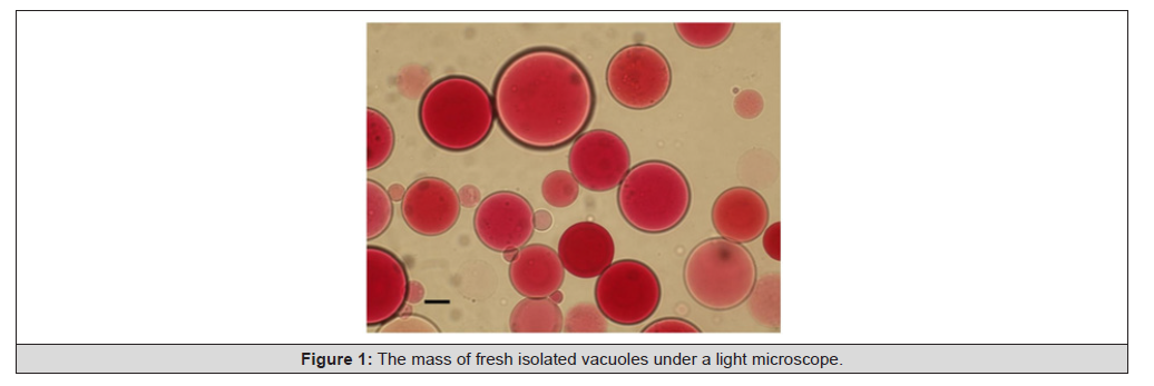
Figure 1:The mass of fresh isolated vacuoles under a light microscope.
The Equipment and Techniques Applied for Observation of the Processes Bound up with Transport of Substances Through the Vacuolar Membrane
The dynamic localization of the scrutinized objects was routinely followed with the aid of FLSCM and the algorithms oriented to single-object tracking. The locations and trajectories of the isolated vacuoles observed could be traced, mapped. The locations and trajectories were bound up with the respective biophysical characteristics. On this basis, the corresponding physiological processes were observed, assessed, analyzed and understood (Figure 1).
In routine observations we applied a laser scanning confocal microscope LSM710 (Carl Zeiss, Germany) (laser 405nm, objective plan-apochromat 63×/1.40 Oil DIC M27, pinhole 43μm, Ch1: 420- 460nm, Ch2: 470-530nm). In various observations the author and his outstanding team used such fluorescence molecular probes as filipin (a well-known sterol-binding antibiotic having an expressed affinity to sterols); laurdan (or 2-(dimethylamino)-6-dodecanoilnaphtaline), a lipophilic probe actively fluorescing in contact with hydrophobic domains; Diphenilhexatrien (DPH) (all-Sigma-Aldrich, USA). ANS (8-anylino-1-naphtalensulphonic acid) and bis-ANS known to have affinity mainly to proteins (but surely retaining the affinity to lipids) were used in the cases, when it was necessary to confirm the presence of proteins in the objects observed.
The probes mentioned above were added for marking the objects (with the dye) and, so, allowing the researcher identify the vacuoles (0.2% solution in DMSO, the final concentration being 5μM). The size of each confocal microscope’s snapshot was 500×500 pixels (1pixel corresponding to 0.1μm). In each case of observations, the author and his team tried to choose the fluorescent molecular probe providing for (i) Better fluorescence intensity and (ii) More explicit snapshot. To the end of binding the probe’s molecules and the vacuolar membrane molecules, the probe (diluted in methanol down to the final concentration of 10μM) was added to the mass of isolated vacuoles. The mass of vacuoles was incubated at 20°C±2°C during 10 min and, next, observed via the FLSCM used.
The equipment and the techniques used for observations allowed the author observe the activity of the scrutinized isolated vacuoles, the processes in the membranes, the process parameters, and assess some of biophysical characteristics of the vacuoles. Observed and assessed were (i) External structures of isolated vacuoles, (ii) Structures and behavior of vacuolar membranes, (iii) Internal contents of isolated vacuoles, (iv) Relations between isolated vacuoles, (v) Structures and functions of the objects occurring in the isolated vacuoles and in the inter-vacuolar space.
Results
Fluorescence microscopy was applied for capturing and tracing of scrutinized objects on the snapshots at 20±2°C. The author and his team obtained video films, some sequences of snapshots from which were chosen for demonstration of the observed processes in the present article. The observation equipment described above and the fluorescence molecular probes used have given the author an opportunity to observe the process of empting of isolated vacuoles. Targeted observations of individual vacuoles in the mass of isolated vacuoles have given the following results. The phenomenon of fast empting of isolated red beetroot (Beta vulgaris L.) vacuoles (i.e. fast efflux of the vacuolar content during decades of seconds) in the process of their natural decay was first registered by the author’s team of researchers in January, 2017. This phenomenon, which was later periodically observed from 2018 to 2024, necessitated deep understanding and plausible explanation. Consider the snapshots given in (Figure 2).
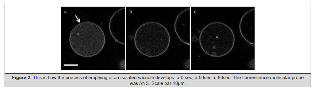
Figure 2:This is how the process of emptying of an isolated vacuole develops. a-0 sec; b-50sec; c-60sec. The fluorescence molecular probe was ANS. Scale bar-10μm.
As obvious from Figure 2 (snapshots a-c), the internal space of the scrutinized vacuole is gradually blanching. This means that the vacuole is gradually and quickly losing its internal content. But no channel, through which the vacuolar content is going out, is visible. Meanwhile, when considering the fast character of the process of blanching (blanching has taken place in a minute, and, so, we may speak that a very substantial part of the vacuolar content (nutrients) has been lost in a minute), one (on account of the corresponding unobvious indirect indicators) may speak about the fact of existence of some hole or holes (of course, not pores, never!) in the vacuolar membrane. It is completely dishonest even to start to speak about any fantastic nano-dimensional pores in the membrane, which would have never provided for vacuolar empting in one minute.
As obvious from Figure 3 (snapshots a-d), the internal space of the scrutinized isolated vacuole is gradually blanching. This means that the vacuole is gradually losing its internal content. In snapshot d, the vacuole is practically empty. Likewise, above (in Figure 2), no channel, through which the vacuolar content is going out, is visible. Meanwhile, when considering the fast process of blanching (the vacuolar content has been lost in less than one third of a minute), any researcher (on account of the corresponding indirect indicators) may speak about the fact of existence of some hole (or holes) in the membrane. Noteworthy, the majority of other vacuoles observed in Figure 3 remain filled with the vacuolar content (Figure 3).
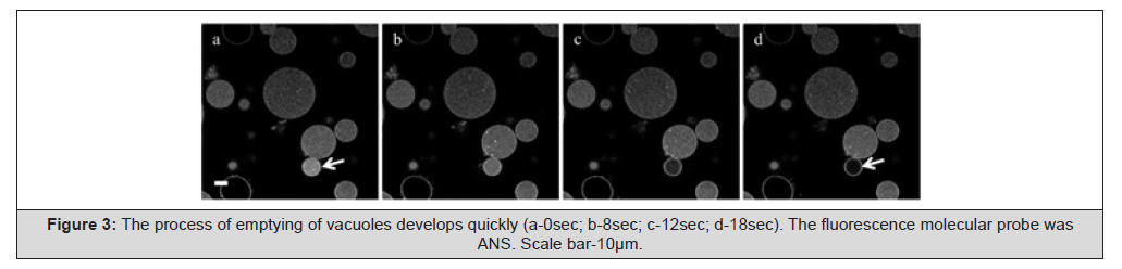
Figure 3:The process of emptying of vacuoles develops quickly (a-0sec; b-8sec; c-12sec; d-18sec). The fluorescence molecular probe was ANS. Scale bar-10μm.
Now, consider the snapshots given in Figure 4. As obvious from Figure 4 (snapshots a-f), the internal space of the scrutinized isolated vacuole is gradually blanching. This means that the vacuole is gradually losing its content. But the channel, through which the vacuolar content is going out, is not visible. Meanwhile, when considering the fast character of the process of blanching (some substantial part of the vacuolar content has been lost in less than a minute), one (on account of the corresponding indirect indicators) may speak about the fact of existence of some invisible hole in the membrane (Figure 4).
The indirect indicators are as follows: (i) The flow of the vacuolar content from the isolated vacuole is not only fast, it is rather intensive (probably, what one observes on snapshot b (see the object indicated by the asterisk) is a hole); (ii) The flow is even turbulent, and (iii) The turbulent flow from the hole, which is practically invisible, exerts pressure upon an “empty” (and, so, flexible) isolated vacuole, which is located below, on the way of this turbulent flow. This turbulent flow has distorted the form of this small empty vacuole (see Figure 4, snapshots b-e), and the latter starts to slowly acquire the spherical form again only after sealing (healing) of the hole in the active isolated vacuole, which has lost much of its internal content (Figure 4, snapshot f). So, it is possible to state that the author has observed the process of fast efflux of the vacuolar content from the vacuole. One can make sure how quick and intensive the process of efflux really is. In course of numerous subsequent observations, the author has made sure that the process of disappearance of the vacuolar content from isolated vacuoles is conditioned by observed (NB!) opening of holes (more than obviously, not large pores) in vacuolar membranes. This process presumes fast efflux of the vacuolar content.
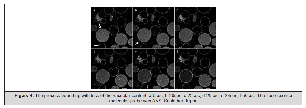
Figure 4:The process bound up with loss of the vacuolar content: a-0sec; b-20sec; c-22sec; d-25sec; e-34sec; f-50sec. The fluorescence molecular probe was ANS. Scale bar-10μm.
For our subsequent observation and analysis, we have intentionally chosen a fragment of the video-film (see Figure 5), which represents continuation of the events shown in the fragment given above in Figure 4. In this case, empting of another isolated vacuole takes place (Figure 5).
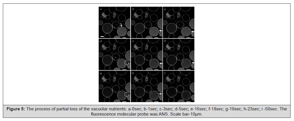
Figure 5:The process of partial loss of the vacuolar nutrients: a-0sec; b-1sec; c-3sec; d-5sec; e-16sec; f-18sec; g-19sec; h-23sec; i -50sec. The fluorescence molecular probe was ANS. Scale bar-10μm.
This time, the equipment described above has given our team an opportunity to observe the processes of intermittent formation of clearly visible channels in vacuolar membranes (visible under a fluorescent laser scanning confocal microscope) (see Figure 5 b, c; e, f). These channels have the form of observable holes. An average diameter of the observed holes in approximately five hundred of observations conducted by my team of researchers was assesses as 0.5-1.9μm.
The sequence of snapshots in Figure 5 on the whole demonstrates a really fast process of emptying of the scrutinized isolated vacuole via holes intermittently forming in the vacuolar membrane. This process is intermittent: opening of a hole in the membrane alternates with its sealing (the places of sealing are indicated with small rounds (Figure 5 d, g)).
On stage 1, after opening of a hole in the membrane (Figure 5 b-c) one can observe the process of its sealing (Figure 5 d). On stage 2, one can observe an explicit (rather large) hole in the membrane (Figure 5 e-f) and the result of its subsequent sealing (Figure 5 g). On stage 3, one observes opening of another large hole in the membrane (Figure 5 h) and its sealing (Figure 5 i). So, the process of empting of the vacuole turns out to be gradual and intermittent. It is obvious from (Figure 5 i) that the process of empting of the scrutinized vacuole is incomplete after 50 sec. In principle, the process of empting is going on until the isolated vacuole turns out to be optimally (probably, from the viewpoint of the process of vacuolar decay) emptied. Meanwhile, presently, on account of the present level of knowledge, it is not possible to rigorously judge about completeness of the observed vacuolar empting.
It is worth noting that processes of the observed type have never been discussed in the world biological literature earlier. The biophysics of the observed efflux (and, so, loss) of the vacuolar content via opening holes is also quite specific. The issues and the specificity of this biophysics are planned to be discussed in the author’s forthcoming articles.
The characteristics of efflux of the vacuolar content from an isolated vacuole are now relatively obvious: (i) The flow of the content from the vacuole is not only fast, it is rather intensive and even turbulent; (ii) The turbulent flow from the hole surely exerts pressure upon the vacuoles, which are located on the way of this turbulent flow. But in the case considered in Figure 5, the shapes of the vacuoles located on the way of this turbulent flow are not distorted because these vacuoles are not empty (filled with the vacuolar content) (see Figure 5, e-f). When resuming the results of this investigation, it is possible to state that the author observed the process of efflux of the vacuolar content from the vacuole. When considering the time scale, one can make sure how intensive the process of efflux really is and how quickly the process of empting proceeds.
So, it is possible to state that the present manuscript describes a discovery bound up with the phenomenon of fast empting of isolated vacuoles via holes forming in their membranes. The mechanism responsible for opening holes in the vacuolar membrane is planned to be discussed in the forthcoming publications. It is very probable that this process takes place owing to the regularities bound up with the process of natural decay of the mass of isolated vacuoles. So, explications of the processes have incurred, (a) Mathematical models of these processes, and (b) Explanations of the phenomenon discovered have already been planned to be discussed in the author’s forthcoming publications.
Discussion
Many researchers consider the PCV as a temporary storage compartment. Probably, in this connection, they limit their investigations with the mechanisms of transport of solutes into the vacuole. And they represent this mechanism within the frames of the understanding, which is guaranteed by the related investigations conducted in the world. Formation of pores in organelle membranes has for long time been considered as the main candidate mechanism for trans-membrane transport of substances. This mechanism was interpreted as mediated by (i) Artificial membrane poration, (ii) Internalizing of amphipathic peptides, (iii) Other mediators.
It is known that experiments conducted on model membranes have shown that antennapedia peptide-based transport in Large Unilamellar Vesicles (LUVs) is less efficient than that in GUVs [60]). This difference may be bound up with the facts that membrane curvatures formed is larger (and membrane tension is higher) in LUV than in GUV.
Discussion of Importance of the Discovered Phenomenon of Fast Empting of the Vacuole from the Viewpoint of Biology. Resume.
Since the early 1970s, several teams of talented researchers tried to make steps forward, i.e. to undertake attempts to understand the processes bound up with trans-membrane transport in cells and in organelles. Many researchers published results, which seemed rather interesting. One can understand their wish to go deeper into the details. But not all the results obtained were either grounded or explained sufficiently well. The researchers concentrated on the complex issues considered by them as the most important. It is important to note that investigations of the issues of trans-membrane transport presumed almost exclusively transport of solutes due to osmotic conditions and owing to hypothetical formation (opening) and sealing of membrane pores.
Everything bound up with membrane pores seemed absolutely clear to the researchers themselves. At the background of this understanding, any processes in the vacuole (and we consider processes bound up with the vacuole) were considered by many wellknown researchers as natural and not necessitating explanations. And, as far as the author of the present publication understands, the result expressed in empting of vacuoles (loss of the vacuolar content) seemed to them absolutely natural. Furthermore, this process seemed as something secondary. It did not necessitate any explanations. In this connection, the process of vacuolar empting produced an impression that it did not represent any special interest. So, numerous researchers working in the field concentrated their efforts on the issues of mathematical descriptions and mathematical modeling of trans-membrane transport of solutes in their understanding, with the use of various transporters.
Only two or three publications bound up with transport processes across the vacuolar membrane, which may be found in publications of a number of outstanding researchers in 2002-2025 [5,6,11,12,23,26,27,43,44,51,54,61], contained descriptions of trans-membrane transport out of the vacuole. It is natural that in investigations of 2010s and later, many researchers concentrated their attention and efforts bound up with understanding of the mechanisms of transport of soluble substances through organelle membranes on the mechanisms of “Direct Transport” (DT). Unfortunately, understanding of such DT was bound up with transport via membrane pores. And everything would have been well if opening of pores, their sealing and resealing could have been observed with the aid of FLSCM. Obviously, investigation of membrane pores did not lead the researchers to any obvious progress in understanding of processes of trans-membrane transport.
No wonder that in the conclusion to an article written in 2012, such outstanding researchers as E. Etxeberria and his co-authors honestly stated:
“The vacuole is still considered as the main reservoir of metabolites; the majority of whose functions remain unidentified” [6]. “Despite the enormous strides accomplished over the past decades, our understanding of an integrated functioning vacuole is still rudimentary. Based on our present knowledge, it is difficult to draw firm conclusions regarding solute transport to and from the cytosol, apoplast and vacuole of storage cells” [6]. This team of talented authors laid their hopes on the “combined transporter/vesicle-mediated systems for solute transport to distinct cell compartments” [6].
The issues of intracellular delivery of substances at the expense of membrane disruption have been discussed by M. Stewart and his co-authors (2018) [62]. So, in all the cases the problem is obvious: (i) The time for true understanding of the processes bound up with vacuolar trans-membrane transport of substances has not yet come; (ii) The corresponding knowledge has not yet formed into a system.
In this connection, there were obvious reasons for the author (a) To take a PCV in the capacity of the object under scrutiny; (b) To study in greater detail one of the forms of trans-membrane transport, i.e. the process of transport of the vacuolar content through the membrane out of the vacuole. The necessity of such an investigation was conditioned by the following important circumstances.
Many investigations bound up with trans-membrane transport of substances, which may be found in the literature, are reduced to solutes, and the results may not be applied to real biological membranes of cellular or organelle levels (the point is that approximately 90% of such investigations have been conducted on GUVs). The issues of transport of solutes in 99% of investigations have been considered for only one way: through membrane pores into the organelle (most frequently, into GUV).
It is necessary to emphasize the following circumstances, which define biological importance of the present investigation. The discovered phenomenon of fast efflux of the vacuolar content via halls is exclusively important because for the first time in the history of contemporary biological investigations it shows that potentially there exists a real mechanism of obviously efficient efflux of substances and compounds (both solutes and solid ones) from the vacuole into the cytosol, and, consequently, potentially there exists a real opportunity of efficient delivery of these substances to some target place in the cell.
The discovered phenomenon of fast empting of isolated vacuoles via holes forming in their membranes has been described. According to our preliminary conclusion, this empting is bound up with the process of the natural decay, which these vacuoles undergo. So, the discovery made (bound up with empting of vacuoles) suggests an approach to understanding of the phenomenon of natural decay of vacuoles.
Discussion of Importance of the Discovered Phenomenon of Fast Empting of the Vacuole from the Viewpoint of Perspectives of Application of the Respective Knowledge in Medicine
When speaking about perspectives of medicine in principle, it is necessary to emphasize that in the perspective implied is elaboration and refinement of methods oriented to treatment of heavy diseases on the cellular and subcellular levels. The author has oriented his teams of researchers and medical practitioners to treatment of oncogenic diseases, sugar diabetes, cardiovascular diseases, to treatment of retinitis pigmentosa and restoration of retina of the eye.
The problem of restoration of retina of the eye represents a very important problem of investigations, whose solution may be obtained only in investigations conducted on the subcellular (organelle) level [63]. Perspective approaches to understanding and deeper knowledge of the problems bound up with degradation of retina of the eye and with oncogenic diseases may, in our opinion, be obtained via understanding what dysfunctions and dysregulations, which happen at the subcellular (membrane) level, contribute in comparison with the normal state [64,65]. One of the most perspective directions is bound up with delivery of medicinal drugs to the desired sites in the cell. Nevertheless, from the very beginning, it is important to emphasize that almost all known statements about medical applications of mechanisms of drug delivery to some objects (targets) of the cellular or subcellular levels carry experimental character.
Already in the 2000s, researchers started to apply ultrasound- mediated techniques in their attempts (a) To deliver drugs or (b) Deliver of genes for the purposes of treatment of some forms of cancer [66]. Later, the attempts followed to treat some forms of cardiovascular diseases [67]. In the early 2010s, one could already find in the literature data about ultrasound-mediated delivery of doxorubicin into tumor cells [68]. Simultaneously, ultrasound techniques were used for ultrasound-targeted microbubble destruction. Such a technique was proposed, for example, to deliver siRNA cancer therapy aids [69]. It was ascertained in 2015 that ultrasound targeted microbubble destruction-mediated delivery of a transcription factor decoy inhibits STAT3 signaling and cancer tumor growth [70]. In the early 2020s, the MR-guided focused ultrasound technique, and the issue of delivery of trastuzumab to Her2-positive brain metastases were discussed [71].
Meanwhile, it is not possible to state that the already discussed and other respective treatment tools are presently really and widely available in practice of clinics. It is hardly ever possible to state that any really persuasive results of successful practical application of the advertised tools (aids of drug delivery, membrane transporters, membrane poration, etc.) in real treatment of oncogenic (or some other heavy) diseases of people have been obtained. Presently there are no techniques, bound up with trans-membrane transport, which may really be applied in any branches of practical human-oriented medicine. Meanwhile, being driven by the wish to represent their often-modest results in their publications in the form, which has received the qualification “in-depth”, some researchers, having forgotten about the reality, state, for example, that their “attempts have already been successful”, and their “descriptions and models may help in treatment of real diseases” (even in treatment of oncogenic diseases). Furthermore, known researchers state that they have already successfully applied their interpretations of known approaches (say, known forms trans-membrane transport, known techniques of electroporation, well-known drugs, etc.) in real treatment of heavy diseases (oncogenic and some other diseases).
In the literature one can find articles stating that various vacuoles participate in the cell functions directed against infecting of this cell. Moreover, some authors state that transport processes bound up with PCVs prevent (i) Bacterial infecting and (ii) Viral infecting of the cell. Nevertheless, even when discussed are processes oriented to protection of only a given cell or a given PCV against infecting, this does not mean that all the issues of such protection have been studied and are known. In the literature, one can also find articles, in which the processes bound up with transport via membrane pores (while including also transport via pores in membranes of GUVs), are directly bound up with the vacuolar ATPase driven potassium transport in highly metastatic breast cancer cells [25]. And it appears obvious that there is a real opportunity to orient medical specialist to the “vacuolar ATPase driven potassium transport” in treatment of women in cases of highly metastatic breast cancer cells. The respected researchers speak about vacuolar ATPase driven potassium transport, but… what kind vacuole (PCV or a digestive vacuoles of animals) is implied? As it has been honestly stated by outstanding researchers J. Gao and H. Wang, et al., (2018) [54], such statements are doubtful already because transport processes via pores in membranes have never been observed. Pores are nano-dimensional. Presently no technical aids good for their observations in dynamics really exist. In the literature, one can find information about drugs modulating multidrug resistance of cancer cells [72]. The author of the present publication may only hope that this information is at least somehow plausible. In the literature, one can find articles stating that membrane electroporation represents “a very perspective direction”. This statement necessitates due analysis.
Already in 2003, attempts of constructing of a theory of electroporation and assessment of perspectives of electroporation in drug delivery and even in gene therapy were undertaken [73]. It is not so easy to apply electroporation in gene therapy. Meanwhile, investigations of the mechanisms and attempts of constructing models of irreversible membrane electroporation and electro permeabilization were conducted during the following 20 years, and these investigations had an experimental character [74]. Nevertheless, electroporation techniques have been declared and even recommended as “the best techniques in treatment of real diseases of people”. Presently, there are several targeted drug delivery strategies already developed on the basis of electroporation and electric permeabilization [75].
For example, the technique of electroporation realized with the use of endoscopic needle-electrode in the case of porcine pancreas [76] is obviously experimental. Nevertheless, according to some publications found in the literature, the approach bound up with irreversible electroporation has been verified in the attempts to work with destruction of prostate cancer [77], fighting against brain tumors [78], and some other problems of clinical oncology [79]. In this connection, does this mean that poration has already become not an experimental technique, but a practical method of treatment?
In turn, S. He and his co-authors (2022) state that localized and, note, reversible plasma membrane disruption can become a promising technique for targeted delivery (and possibly deposition) of (exogenous) therapeutic compounds needed for treatment of some diseases [80]. These talented authors discuss possible methods of physics, which may be used to generate plasma membrane perforations (electroporation, photoporation, sonoporation). They discuss possible electroporation results bound up with formation of pores having radii generally < 1nm. And they have computed that the theoretical density of such pores obtained with the use of electroporation might be up to 109 pores per cm2 (ibid., p. 2). While accepting the traditional understanding, in 2025, L. Starke and his co-authors state that, in the norm, complexity of the membrane’s lipid composition and lateral sorting of lipids ensure mechanical stability of the membrane, and “this prevents from formation of pores, while allowing for ongoing membrane remodeling” [81]. Meanwhile, these talented authors emphasize that violation of cytoplasmic membrane’s integrity achieved via induced formation of pores in the membrane “may trigger either uncontrolled depolarization or influx of toxic compounds, causing a fatal threat to the living cell” [81]. On account of these conditions, can one speak about any useful treatment techniques bound up with such electroporation?
Meanwhile, very serious problems existing with electroporation and formation of cell membrane pores remain the same (and this fact has numerously been emphasized by biologists): (a) When electroporation is applied to a cell membrane, and the desired pore forms, the cell dies, and no further treatment may be realized; (b) It is not possible to ascertain how many pores appear in the membrane as a result of one act of electric discharge in the process of electroporation (the computed density of 109 pores per cm2 is hardly ever realistic because this is practically a hole in the membrane); (c) The process of electroporation has never been confirmed by practical observations of the facts of formation of pores.
Indeed, poration is a dynamical process. And as any other dynamical process with the objects, which are nano-dimensional, it cannot be observed with the aid of any presently available tools. Nevertheless, publications, which advertise poration in various respects, regularly appear in the literature. And these publications avoid any explanations of the issues (i) Non-observability of both the process and the result of electroporation (if poration really takes place), and (ii) Irreversible character of electroporation. But if electroporation is irreversible, it may not be applied in treatment of people.
It is known that anthocyanin accumulates in PCVs. When coming out of the cellular vacuole, anthocyanin is capable of producing some healing effect (if not to say treatment effect) upon the cell. Trans-membrane transport of anthocyanin in connection with the vacuole was discussed in the biomedical literature already in the early 2010s. But is so happened, in connection with some strange circumstances, discussed was the transport in the reverse direction: “delivery of anthocyanin from cytosol to tonoplasts” [41]. This statement (in the title of the article [41]) and this publication seemed strange to the author.
The problem is that anthocyanin can approach to a vacuole (as stated in the title of article [41]). But anthocyanin cannot simply get into the vacuole. It is important to emphasize that anthocyanin molecules are rather large, and, therefore, their transport even through rather large (and still nano-dimensional) lipid pores is completely impossible. Furthermore, formation of pores, if formation really takes place, anyway, represents an accidental process. In reality, anthocyanin is generated and, in the final result, is concentrated inside the vacuole. The process of such concentration takes substantial time.
Meanwhile, really important (from the viewpoint of healing/ treatment issues) is the process of fast (not slow) efflux of anthocyanin from the vacuole. Anthocyanin is thrown out through the vacuolar membrane into the cytosol to be (with time) delivered to the target site in the cell. Anthocyanin participates in treatment of a diseased part (an organelle) of the cell. So, the phenomenon (and the respective mechanism) of fast efflux of substances from the vacuole, which has been discovered by our team, may really represent a form of organelle-level auto-treatment of the cell.
Furthermore, whereas concentration of anthocyanin inside the vacuole may represent a long-time process, efflux of anthocyanin in the direction of the target (an object in the cell, which needs immediate treatment) must be fast. The effect described by the author is really fast (decades of seconds). Noteworthy, the opportunity of the fast process of efflux of the vacuolar content (which surely contains anthocyanin and surely some other substances) through the holes opening in the vacuolar membrane has been discovered only now (and it is described in the present publication for the first time).
In this connection, it is probably possible to state that (i) Anthocyanin possesses antimicrobial, anti-inflammatory and antioxidant properties and (ii) Anthocyanin molecules act as a form of bio-protection against ROS forming in the photosynthesis process [82], but these statements necessitate due verification of the facts declared, when one speaks about actual, practical activity of anthocyanin in the cell.
The case considered above for the trans-membrane transport of anthocyanin represents only one of a large number of cases, which necessitate detailed consideration. The issues of delivery of substances (e.g., some drugs for the purposes of protection of the cell against heavy viruses) or delivery of some genetic materials to the desired points in the cell [83,80] are important in the aspect of perspectives of cellular-level and organelle-level medical biology.
According to opinions of a number of researchers, processes of changes and possibly elastic remodeling in organelle membranes may be bound up with conformational transitions of proteins [84,85]. Biochemical details of these processes may be found in [86]), where it is stated that: (i) These elastic and conformational changes influence the membrane’s stability; are capable of inducing changes of the organelle’s shape (e.g., folding) (ion channels and transporters of soluble substances participate in such changes). Furthermore, M. Brown and his co-authors state that processes, which take place within the membrane’s stress field during such changes, include budding of such viruses as influenza, dengue, HIV [85,86]. Unfortunately, this statement does not coincide with the author’s knowledge obtained in practice and regarding dengue and HIV [87,88].
There is a hope that understanding of the processes bound up with membrane proteins will with time lead to understanding of not only the logic of structures, but also to understanding of the functions and the corresponding deep biological mechanisms. Unfortunately, today, in many other cases, specialists in medicine may only dream of applying cell-level, organelle-level and advanced molecular- level methods of treatment of various diseases to people. Let us hope that with time such methods will become traditional in the medical practice.
Conclusion
The present publication has described a little discovery. Still, this is a discovery, and this discovery is bound up with the phenomenon of fast empting of isolated vacuoles. Meanwhile, when studying the corresponding biomedical literature, the author has come to the conclusion that many researchers (despite the fact that they often work with isolated vacuoles as with the material of their investigations) consider special (separate) investigations of isolated vacuoles as either uninteresting or even backward, and, so, unnecessary. Anyway, no special observations of isolated vacuoles oriented to at least superficial understanding of the mechanisms of their natural decay have been conducted by numerous representatives of the biological community. The author knows for sure that there were no corresponding investigations (experiments) conducted for understanding of the behavior and biophysical characteristics of isolated vacuoles. Such investigations (complemented with mathematical modeling approaches) have been conducted by the author. The results of these investigations will appear in author’s forthcoming publications.
According to the phenomenon discovered by the author and his team, the process of empting of an isolated vacuole is expressed in the form of efflux of the content from the vacuole via the vacuolar membrane. When speaking more exactly, such empting has the form of fast (during decades of seconds) efflux of the vacuolar content (and it is important to emphasize that this may be both some soluble content and some non-soluble content) through a hole (or a number of holes) in the vacuolar membrane. Such a phenomenon has not been earlier described or discussed in the literature by any other researchers, which do not belong to the author’s team.
The result in the form of a discovery has been obtained on the basis of application of the knowledge-based ideology (KB-ideology) elaborated for conducting scientific investigations, analyses and medical treatment. This ideology has been developed by the author and described in his articles [64,65,87,88]. The author asks the readers to trust him. The KB-ideology is not a sort of specific general-character chatter about trivial issues. This is a way to more knowledge-saturated approaches (not simply methods) both in scientific investigations and in medical treatment problems.
The effect of fast efflux, which is the core of the phenomenon discovered, has been revealed in 2017. An outstanding scientist Dr. Vadim Nurminsky was the first person to register the fact of opening of a hole in the vacuolar membrane is course of high-resolution FLSCM observations of PCVs. It is important to emphasize that later, in hundreds of observations (conducted with aid of high-resolution FLSCM during 2018-2024), the non-accidental character of this phenomenon has been numerously confirmed. It has been practically and concretely ascertained that isolated red beetroot vacuoles can eject their internal content into the outer (extra-vacuolar) space in course of a sequence of events bound up with opening holes of micron diameter in the vacuolar membrane. These events produce an impression of non-accidental ones. In our experiments with observations, opening of holes in membranes of isolated vacuoles was bound up with the process of natural decay, which these vacuoles underwent.
The phenomenon discovered presumes a sequence of intermittent processes, which includes: (i) Formation of a hole (diameter of 0.5-1.9μm) in the vacuolar membrane; (ii) Efflux of the vacuolar content the outer (extra-vacuolar) space; (iii) Sealing of the hole; and, if the vacuole has not been completely devastated, (iv) Formation of another hole at the same or at a different location in the membrane of the same vacuole; (v) Efflux of the residual vacuolar content (or some part of this content) into the extravacuolar space; (vi) Resealing of the hole; etc. The sequence of these processes produces an impression of a non-accidental sequence.
Noteworthy, the discovered channel of fast efflux of the vacuolar internal (soluble and non-soluble) content from the vacuole (i.e. real observable holes) is more efficient than any other well-known transport channels studied by the biological community as responsible for delivery of substances through the vacuolar membrane.
There is no doubt that knowledge and application of new methods of biophysics, biochemistry, cell and organelle biology, microbiology complemented with contemporary microscopy techniques may bring the researchers closer to discoveries. E. Etxeberria and his co-authors (2012) [6] placed their hopes upon methods and techniques in connection with development of definite patterns of intracellular transport systems, and, particularly, systems bound up with transport of substances across organelle membranes. It is expedient to repeat that, in the present case, the author has relied not upon new methods themselves, but upon the KB-ideology. The author has also relied upon the factor of the necessity of due completeness of the programs of investigations (observations and experiments), which cannot be reduced only to some well-known or “universally accepted” (by the biomedical community) programs (and also to the conceptions and theories bound up with these programs). Only an extended research platform can bring the researcher to a desired result useful for the humankind.
Furthermore, the corresponding biophysical processes (bound up with efflux of nutrients from the hole in the vacuolar membrane and the influence of the turbulent flow from the hole upon other vacuoles) have been explained by the author. Within the frames of the present article, it is not possible to describe the respective biophysical mechanisms responsible for (i) The formation of the vacuolar membrane holes, which provides for efflux of nutrients from the vacuole, and (ii) Sealing the holes formed. The issues bound up with biophysics of the processes discussed will be considered in the author’s forthcoming articles. The author would like to repeat that the plausibility of the phenomenon described above has been confirmed in more then 500 of observations conducted on isolated red beetroot vacuoles, and also in hundreds of observations conducted on other organelles all with the aid of laser scanning confocal microscopy (during 2018-2025).
The author has shown the present manuscript in its anonymous form (without identifying the author) to one of the wellknown experts in the field. And the reaction of this expert has been as follows: “The author and his research team are outstanding. The result they’ve obtained, when considered in the aspect of its possible further medical applications, and possible development of this result, is exclusively innovative. This result has an obvious “driving potential” from the viewpoint of stimulating the developments possible in the given field science. The overall logic, the structure (design) of this investigation, the standards of (i) Constructing of the text and (ii) Choosing the illustrations are excellent”. So, the author may hope that the phenomenon of fast empting of an isolated vacuole expressed in the form of fast efflux of soluble and non-soluble content from the vacuole (which represents the discovery described in this article) will in future occupy its rightful place in the list of phenomena providing for useful approaches to cell-level and organelle-level treatment of hazardous diseases.
Acknowledgements
The author is grateful to the administration of the Department of the Institute of Laser Physics (Irkutsk Scientific Center, Siberian Branch of Russian Academy of Sciences) for friendly co-operation.
The author would like to express his cordial gratitude to his colleagues and friends, outstanding scientists Dr. Vadim Nurminsky and Dr. Elena Pradedova, who have decided not to take part in the present publication addressed to AJBSR. So, the author expresses his gratitude for their contribution into the final result of our joint investigations and, to some extent, into this publication. Dr. Vadim Nurminsky was the first one to notice the holes formed in the membrane in 2015. Some hints of the outstanding researcher Dr. Elena Pradedova bound up with various membrane transporters, transport channels, and with application of membrane transport mechanisms in medical aspects were very useful. The author hopes that Dr. Vadim Nurminsky and Dr. Elena Pradedova will participate in following publications of our team.
Funding
The author declares that no funds, grants, or other support were received by him during fulfilment of the research work and preparation of this manuscript.
Declarations
The author declares that he has no conflicts of the interests, no competing interests to declare, which might be related to the idea or to the content of this manuscript. The author has neither financial nor non-financial competing interests to disclose. There are no personal relationships that could negatively influence the investigation reported in the manuscript.
Data availability
Data sharing is not available.
References
- Nurminsky VN, Rakevich AL, Martynovich EF, Ozolina NV, Nesterkina IS, et al. (2015) Peculiar properties of some components in a plant cell vacuole morphological structure revealed by confocal microscopy. Cell and Tissue Biology 9(5): 406-414.
- Chernyshov MY, Nurminsky VN, Ozolina NV (2017) Lipid-protein microinclusions in the morphological structures of organelle membranes and studied by fluorescence confocal microscopy. Advances in Biological Chemistry 7(1): 42-59.
- Nurminsky VN, Ozolina NV, Nesterkina IS, Kolesnikova EV, Korzun AM, et al. (2011) Stability of plant vacuolar membranes under osmotic stress and in the presence of redox agents. Supplement Series A: Membrane and Cell Biology (Pleiades Publishing, Ltd.) 5(2): 185-190.
- Ozolina NV, Nesterkina I S, Kolesnikova EV, Salyaev R K, Nurminsky VN, et al. (2013) Tonoplast of Beta vulgaris L. contains detergent-resistant membrane microdomains. Planta 237(3): 859-871.
- Oda Y, Higaki T, Hasezawa S Kutsuna N (2009) New insights into plant vacuolar structure and dynamics. Int Rev Cell Mol Biol 277: 103-135.
- Etxeberria E, Pozueta Romero J, Gonzalez P (2012) In and out of the plant storage vacuole. Plant Sci 190: 52-61.
- Etxeberria E, Gonzalez P, Pozueta Romero J (2013) Architectural remodeling of the tonoplast during fluid-phase endocytosis. Plant Signal Behav 8(7): e24793.
- Andreyev IM (2012) The role of vacuole in redox homeostasis of plant cells. Phiziologiya rastenyi 59(5): 660-667.
- Flores Romero H, Ros U, Garcia Saez AJ (2020) Pore formation in regulated cell death. EMBO J 39(23): e105753.
- Sheahan MB, Rose RJ, McCurdy DW (2007) Actin-filament-dependent remodeling of the vacuole in cultured mesophyll protoplasts. Protoplasma 230(3-4): 141-152.
- Gao XQ, Wang X L, Ren F, Chen J, Wang XC, et al. (2009) Dynamics of vacuoles and actin filaments in guard cells and their roles in stomatal movement. Plant Cell Environ 32(8): 1108-1116.
- Reisen D, Marty F, Leborgne Castel N (2005) New insights into the tonoplast architecture of plant vacuoles and vacuolar dynamics during osmotic stress. BMC Plant Biology 5(13).
- Pradedova EV, Chernyshov MY (2024) Redox systems of the plant cell vacuole. Russian Journal of Plant Physiology 71: 186.
- Martinoia E, Klein M, Geisler M, Bovet L, Forestier C, et al. (2002) Multifunctionality of plant ABC transporters - more than just detoxifiers. Planta 214(3): 345-355.
- Kang J, Park J, Choi H, Burla B, Kretzschmar T, et al. (2011) Plant ABC transporters. Arabidopsis Book 9: e0153.
- Kretzschmar T, Burla B, Lee Y, Nagy R, Martinoia E, et al. (2011) Functions of ABC transporters in plants. Essays in Biochemistry 50(1): 145-160.
- Theodoulou FL, Carrier DJ, Schaedler TA, Baldwin SA, Baker A, et al. (2016) How to move an amphipathic molecule across a lipid bilayer: different mechanisms for different ABC transporters? Biochem Soc Trans, 44(3): 774-782.
- Rautenkranz AAF, Li L, Machler F, Martinoia E, Oertli JJ, et al. (1994) Transport of ascorbic and dehydroascorbic acid across protoplasts and vacuole membranes isolated from barley (Hordeum vulgare L. cv.Gerbel) leaves. Plant Physiol 106(): 187-193.
- Marina VG, Grubu E, Zielinski J, Schild A, Fischer K, et al. (2006) Identification and expression analysis of twelve members of the nucleobase-ascorbate transporter (NAT) gene family in Arabidopsis thaliana. Plant Cell Physiol 47(10):1381-1393.
- Minton JA, Rapp M, Stoffer AJ, Schultes NP, Mourad GS, et al. (2015) Herologous complementation studies reveal the solute transport profiles of a two-member nucleobase cation symporter 1 (NCS1) family in Physcomitrella patens. Plant Physiol Biochem 100: 12-17.
- Hoang MTT, Almeida D, Chay S, Alcon C, Corratge Faillie C, et al. (2021) AtDTX25, a member of the multidrug and toxic compound extrusion family, is a vacuolar ascorbate transporter that controls intracellular iron cycling in Arabidopsis. New Phytol 231(5): 1956-1967.
- Carter C, Pan S, Zouhar J, Avila EL, Girke T, et al. (2004) The vegetative vacuole proteome of Arabidopsis thaliana reveals predicted and unpredicted proteins. Plant Cell 16(12): 3285-3303.
- Schmidt UG, Endler A, Schelbert S, Brunner A, Schnell M, et al. (2007) Novel tonoplast transporters identified using a proteomic approach with vacuoles isolated from cauliflower buds. Plant Physiol 145(1): 216-229.
- Jaquinod M, Villiers F, Kieffer Jaquinod S, Hugouvieux V, Bruley C, et al. (2007) A proteomics dissection of Arabidopsis thaliana vacuoles isolated from cell culture. Mol Cell Proteomics 6(3): 394-412.
- Salyer SA, Olberding JR, Distler AA, Lederer ED, Clark BJ, et al. (2013) Vacuolar ATPase driven potassium transport in highly metastatic breast cancer cells. Biochim Biophys Acta 1832(10): 1734-1743.
- Neuhaus HE, Trentmann O (2014) Regulation of transport processes across the tonoplast. Front Plant Sci 5(460).
- Martinoia E (2018) Vacuolar Transporters-Companions on a Longtime Journey. Plant Physiology 176(2): 1384-1407.
- Mansour MMF (2023) Role of vacuolar membrane transport systems in plant salinity tolerance. Journal of Plant Growth Regulation 42:1364-1401.
- Roos C, Zocher M, Muller DJ, Munch D, Schneider T, et al. (2012) Characterization of co-translationally formed nanodisc complexes with small multidrug transporters, proteorhodopsin and with the E. coli MraY translocase. Biochemica et Biophysica Acta 1818(2): 3098-3106.
- Chen FY, MT Lee, Huang HW (2003) Evidence for membrane thinning effect as the mechanism for peptide-induced pore formation. Biophys J 84 (6): 3751-3758.
- Bippes CL, Ge M, Meury D, Harder Z, Ucurum H, et al. (2013) Peptide transporter DtpA populates two alternate conformations one of which is promoted by inhibitor binding. Proc Natl Acad Sci USA 110(42): E3978-3986.
- Fuertes G, Giménez D, Esteban Martin S, Garcia Sáez A, Sánchez O, et al. (2010a) Role of membrane lipids for the activity of pore forming peptides and proteins. Adv Exp Med Biol 677:31-55.
- Fuertes G, Giménez D, Esteban Martín S, García Sáez A, Sánchez O, et al. (2010b) Role of membrane lipids for the activity of pore forming peptides and proteins. Adv Exp Med Biol 677: 31-55.
- Lamazière A, Chassaing G, Trugnan G, Ayala Sanmartin J (2006) Transduction peptides: structural-functional analyses in model membranes. J Soc Biol 200(3): 229-233.
- Lamazière A, Maniti O, Wolf C, Lambert O, Chassaing G, et al. (2010) Lipid domain separation, bilayer thickening and pearling induced by the cell penetrating peptide penetratin. Biochimica et Biophysica Acta 1798 (12): 2223-2230.
- Ohkama Ohtsu N, Sasaki Sekimoto Y, Oikawa A, Jikumaru Y, Shinoda S, et al. (2011) 12-oxo-phytodienoic acid-glutathione conjugate is transported into the vacuole in Arabidopsis. Plant Cell Physiol 52(1): 205-209.
- Wang J, Yan C, Li Y, Hirata K, Yamamoto M, et al. (2014) Crystal structure of a bacterial homologue of SWEET transporters. Cell Res 24(12): 1486-1489.
- Lee Y, Nishizawa T, Yamashita K, Ishitani R, Nureki O, et al. (2015) Structural basis for the facilitative diffusion mechanism by SemiSWEET transporter. Nat Commun 6:6112.
- Feng L, Frommer WB (2015) Structure and function of SemiSWEET and SWEET sugar transporters. Trends Biochem Sci 40(8): 480-486.
- Jolliffe N A, Craddock CP, Frigerio L (2005) Pathways for protein transport to seed storage vacuoles. Biochem Soc Trans 33(Pt 5): 1016-1018.
- Sun Y, Li H, Huang JR (2012) Arabidopsis TT19 functions as a carrier to transport anthocyanin from the cytosol to tonoplasts. Mol Plant 5(2): 387-400.
- Karatekin E, Sandre O, Guitouni H, Borghi N, Puech PH, et al. (2003) Cascades of transient pores in giant vesicles: Line tension and transport. Biophys J 84(3): 1734-1749.
- Levadny V, Tsuboi T, Belaya M, Yamazaki M (2013) Rate constant of tension-induced pore formation in lipid membranes. Langmuir 29(12): 3848-3852.
- Karal MAS, Levadndy V, Tsuboi T, Belaya M, Yamazaki M (2015a) Electrostatic interaction effects on tension-induced pore formation in lipid membranes. Phys Rev E Stat Nonlin Soft Matter Phys 92(1): 012708.
- Karal MAS, Yamazaki M (2015b) Communication: activation energy of tension-induced pore formation in lipid membranes. J Chem Phys 143(8): 081103.
- Shibly SUA, Ghatak С, Karal MAS, Moniruzzaman Md, Yamazaki M, et al. (2016) Experimental estimation of membrane tension induced by osmotic pressure. Biophysical Journal 111(10): 2190-2201.
- Akimov SA, Volynsky PE, Galimzyanov TR, Kuzmin PI, Pavlov KV, Batishchev OV (2017) Pore formation in lipid membrane II: Energy landscape under external stress. Sci Rep 7(1): 12509.
- Roesel D, Eremchev M, Poojari CS, Hub JS, Roke S (2022) Ion-induced transient potential fluctuations facilitate pore formation and cation transport through lipid membranes. J Am Chem Soc 144(51): 23352-23357.
- Kotnik T, Rems L, Tarek M, Miklavc̆ic̆ D (2019) Membrane electroporation and electropermeabilization: mechanisms and models. Annu Rev Biophys 48: 63-91.
- Kasparyan G, Hub JS (2024) Molecular simulations reveal the free energy landscape and transition state of membrane electroporation. Phys Rev Lett 132(14): 148401.
- Starke LJ, Allolio C, Hub JC (2025) How pore formation in complex biological membranes is governed by lipid composition, mechanics, and lateral sorting. PNAS Nexus 4(3): pgaf033.
- Chen FY, Chen MT, Lee H, Huang HW (2003) Evidence for membrane thinning effect as the mechanism for peptide-induced pore formation. Biophys J 84(6): 3751-3758.
- García-Nafría J, Tate CG (2020) Cryo-electron microscopy: Moving beyond X-Ray crystal structures for drug receptors and drug development. Annual Review of Pharmacology and Toxicology 60: 51-71.
- Gao J, Wang H (2018) History and Traditional Techniques of Studying the Structure of Cell Membranes. Chapter 2. In: Hongda Wang & Guohui Li (Editors), Membrane Biophysics: New Insights and Methods. Springer Nature Singapore Pte Ltd 421: 21-43.
- Hodgkin AL, Huxley AF (1952a) A quantitative description of membrane current and its application to construction and excitation in nerve. J Physiol 117(4): 500-544.
- Hodgkin AL, Huxley AF (1952b) Currents carried by sodium and potassium ions through the membrane of the giant axon of Loligo. J Physiol 116(4): 449-472.
- Hodgkin AL, Huxley AF (1952c) The components of membrane conductance in the giant axon of Loligo. J Physiol 116(4): 497-506.
- Nguyen TT, Gopal A, Lee KYC, Witten TA (2005) Surface charge relaxation and the pearling instability of charged surfactant tubes. Physical Review E Stat Nonlin Soft Matter Phys 72(5-1): 051930.
- Kuzevanov V Y, Katkov BB, Salyaev RK (1985) General principles of isolation of vacuoles and vacuolar membranes. In: The Structure and Functions of Biological Membranes of Plants. Novosibirsk: Nauka Publ: 93-107.
- Thoren PE, Persson D, Karlsson M, Norden B (2000) The antennapedia peptide penetratin translocates across lipid bilayers - the first direct observation, FEBS Lett 482(3): 265-268.
- Uemura T, Yoshimura SH, Takeyasu K, Sato MH (2002) Vacuolar membrane dynamics revealed by GFP-AtVam3 fusion protein. Genes to Cells 7(7): 743-753.
- Stewart MP, Langer R, Jensen KF (2018) Intracellular delivery by membrane disruption: Mechanisms, strategies, and concepts. Chem Rev 118(16): 7409-7531.
- Brown MF, Chawla U, Perera Suchithranga MDC (2017) Membrane lipid-protein interactions. Chapter 3. In: Richard M. Epand & Jean Marie Ruysschaert (Eds.) The Biophysics of Cell Membranes: Biological Consequences. Springer Series in Biophysics Springer Nature 219: 61-84.
- Chernyshov MY (2023) A systemic knowledge-based approach to understanding and treatment of cancer diseases. Am J Biomed Sci & Res 20(5): 554-63.
- Chernyshov MY (2025) Key elements of the knowledge-based ideology as a basis for defining the objectives bound up with prevention and treatment of oncogenic diseases. Am J Biomed Sci & Res 26(1): 20-27.
- Hynynen K (2008) Ultrasound for drug and gene delivery to the brain. Adv Drug Deliv Rev 60(10): 1209-1217.
- Chen HH, Matkar PN, Afrasiabi K, Kuliszewski MA, Leong Poi H, et al. (2016) Prospect of ultrasound-mediated gene delivery in cardiovascular applications. Expert Opin Biol Ther 16(6): 815-826.
- Escoffre JM, Piron J, Novell A, Bouakaz A (2011) Doxorubicin delivery into tumor cells with ultrasound and microbubbles. Mol Pharm 8(3): 799-806.
- Carson AR, Mc Tiernan CF, Lavery L, Grata M, Leng X, et al. (2012) Ultrasound targeted microbubble destruction to deliver siRNA cancer therapy. Cancer Res 72(23): 6191-99.
- Kopechek JA, Carson AR, Mc Tiernan CF, Chen X, Hasjim B, et al. (2015) Ultrasound targeted microbubble destruction-mediated delivery of a transcription factor decoy inhibits STAT3 signaling and tumor growth. Theranostics 5(12): 1378-1387.
- Meng Y, Reilly RM, Pezo RC, Trudeau M, Sahgal A, et al. (2021) MR-guided focused ultrasound enhances delivery of trastuzumab to Her2-positive brain metastases. Sci Transl Med 13(615): eabj4011.
- Hendrich A, Michalak K (2003) Lipids as a target for drugs modulating multidrug resistance of cancer cells. Curr Drug Targets 4(1): 23-30.
- Gehl J (2003) Electroporation: Theory and methods, perspectives for drug delivery, gene therapy and research. Acta Physiol Scand 177(4): 437-447.
- Kotnik T, Rems L, Tarek M, Miklavcic D (2019) Membrane electroporation and electro permeabilization: mechanisms and models. Annu Rev Biophys 48: 63-91.
- Manzari MT, Shamay Y, Kiguchi H, Rosen N, Scaltriti M, et al. (2021) Targeted drug delivery strategies for precision medicines. Nat Rev Mater 6: 351-370.
- Lee JM, Choi HS, Chun HJ, Kim ES, Keum B, et al. (2019) EUS-guided irreversible electroporation using endoscopic needle-electrode in porcine pancreas. Surg Endosc 33(2): 658-662.
- Rubinsky J, Onik G, Mikus, Rubinsky B (2008) Optimal Parameters for the Destruction of Prostate Cancer Using Irreversible Electroporation. J Urol 180(6): 2668-2674.
- Fang Z, Chen L, Moser MAJ, Zhang W, Qin Z, et al. (2021) Electroporation-based therapy for brain tumors: A Review. J Biomech Eng 143(10):100802.
- Aycock KN, Davalos RV (2019) Irreversible electroporation: Background, theory, and review of recent developments in clinical oncology. Bioelectricity 1(4): 214-234.
- He S, Singh D, Helfield B (2022) An overview of cell membrane perforation and resealing mechanisms for localized drug delivery. Pharmaceutics 14(4): 886.
- Starke LJ, Allolio C, Hub JC (2025) How pore formation in complex biological membranes is governed by lipid composition, mechanics, and lateral sorting. PNAS Nexus 4(3): 1-13.
- Mackon E, Ma Y, Jeazet Dongho Epse Mackon GC, Li Q, Zhou Q, et al. (2021) Subcellular localization and vesicular structures of anthocyanin pigmentation by fluorescence imaging of black rice (Oryza sativa L.) Stigma Protoplast. Plants (Basel)10(4): 685.
- Ziegler A (2008) Thermodynamic studies and binding mechanisms of cell-penetrating peptides with lipids and glycosaminoglycans. Adv Drug Deliv Rev 60(4-5): 580-597.
- Brown MF (1994) Chem Phys Lipids 73: 159-180.
- Brown MF (1997) In: Epand RM (ed) Current topics in membranes. Academic Press San Diego 44: 285-356.
- Brown MF (2012) Biochemistry 51: 9782-9795.
- Chernyshov MY (2023) Carcinogenic impact of HPVs upon people, its epidemiological character, and a systemic knowledge-based approach to understanding and treatment of the corresponding cancer diseases. J Clin Case Rep Med Images Health Sci 6(3): 1-11.
- Chernyshov MY (2025) A knowledge-based ideology as a basis for constructing a complement to the grounds of contemporary world health care and health surveillance platforms. Am J Biomed Sci & Res 26(2): 231-243.




 We use cookies to ensure you get the best experience on our website.