
Case Report 
 Creative Commons, CC-BY
A Case of Abdominal Wall Reconstruction After Gunshot Wound in the Setting of Abdominal Compartment Syndrome and Loss of Domain
*Corresponding author:Atta Nawabi MD, MBA, MPH, FACS, FICS, Professor of Surgery Division of HPB, and Transplant, Department of Surgery, 3901 Rainbow Boulevard Kansas City, KS 66160, USA.
Received:September 15, 2025; Published:September 22, 2025
DOI: 10.34297/AJBSR.2025.28.003709
Abstract
A critical issue in trauma and reconstructive surgery is managing Abdominal Compartment Syndrome (ACS) and loss of domain following major abdominal trauma. ACS is a life-threatening complication that often necessitates damage control surgery, including an open abdomen. However, this approach increases the risk of further complications, such as significant loss of domain, which complicates subsequent abdominal closure. Our case highlights the successful use of a porcine dermal matrix as a biologic mesh in a patient with significant loss of domain following a traumatic injury. This innovative approach facilitated complete mesh granulation prior to autografting. By providing temporary support for the patient’s loss of domain, our technique allowed for stabilization and resolution of operative edema, creating the opportunity for a more definitive myofascial approximation once the patient has achieved full optimization in recovery and rehabilitation.
Introduction
After operative repair of major abdominal trauma patients are at an increased risk of Abdominal Compartment Syndrome (ACS). ACS is a well described phenomenon defined by elevated intraabdominal pressures above 20mmHg causing organ dysfunction. The abdomen is a defined anatomical compartment, and elevated pressures within it compress surrounding organs causing ischemia and ultimately organ dysfunction. Patients will present with hemodynamic instability secondary to decreased cardiac output and respiratory distress [1]. Therefore, it can be necessary in trauma patients requiring emergent laparotomy to not close the fascia, leaving the abdomen open.
Open abdomen is the current standard of care for management of intraabdominal catastrophes in critically ill patients with the goal of fascial closure within 10-14 days. The European Hernia Society (EHS) developed clinical practice guidelines strongly recommending use of dynamic techniques, Negative Pressure Wound Therapy (NPWT) and fascial closure, rather than static techniques, fascial closure alone [2]. However, methods to achieve final closure of the abdomen vary including primary fascial repair, component separation, mesh repair, or flap reconstruction [3,4].
Mesh repair may be necessitated by significant loss of domain. Mesh material can be synthetic, biosynthetic, or biologic. Synthetic mesh is avoided in traumatic patients due to high rate of contamination and subsequent surgical site infection [5]. Biologic meshes, composed of a collagen-based acellular dermal matrix, were created to reduce risk of infection, though evidence is limited [6-8]. Antibiotic-coated acellular dermal matrixes have been produced including the XenMatrix AB Surgical Graft for which industry-funded studies show in vitro and in vivo evidence of reduced mesh infection [9-12].
Case Presentation
A 37-year-old male presented to an outside hospital as a trauma activation for gunshot (GSW) wound to the abdomen causing injuries to his right colon and rectosigmoid colon. At the outside hospital an exploratory laparotomy was performed with right hemicolectomy, rectosigmoid resection and creation of a sigmoid colostomy. At that time primary closure of fascia was achieved. Post-operatively, however, the patient developed features concerning for abdominal compartment syndrome and septic shock including hemodynamic instability, elevated bladder pressure, abdominal distension, and respiratory distress. Therefore, the patient was transferred to our institution for a higher level of care. Upon emergent laparotomy for re-exploration of his abdomen, the patient was found to have significant necrosis along the bullet tract, including skin, soft tissue, fascia, bowel, and rectus abdominus with concern for infection of abdominal wall. The patient required several operative debridements and washouts, resection of the ileocolic anastomosis and creation of an ileostomy. The abdomen was left open with a vacuum-assisted wound device, and he was determined to have a Ventral Hernia Working Group Grade IV. Notably, the patient had a large, full-thickness defect in his anterior abdominal wall including loss of rectus abdominus.
Once the patient was stabilized, we determined his abdominal defect to be 32 x 44 cm in size with complete loss of abdominal domain. Primary closure was therefore not possible, and an antibacterial porcine dermal matrix provided by XenMatrix was selected to close the hernia [12]. Following debridement of wound edges, a 20 x 40cm xenograft was secured to the anterior rectus fascia in a running fashion beginning superiorly with 2-0 PDS suture. The mesh was folded and sutured to itself with PDS sutures to appropriately cover the wound. The bowel was entirely covered by and in direct contact with mesh, and negative wound pressure therapy was applied in the interim.
Subsequently, nutrition and thorough wound care were prioritized to promote wound healing and mesh granulation. The patient was weaned from total parenteral nutrition, and ileostomy output and electrolyte derangements were managed during the transition period to optimal oral intake. During this interim the wound was dressed in clean, Vaseline-soaked gauze covered by saline-soaked Kerlix and changed twice daily.
After approximately six weeks, the mesh achieved near complete granulation. The patient was returned to the operative theater for debridement of granulation tissue and placement of the MAXXEUS cadaver allograft meshed 2:1 to optimize the wound prior to skin grafting. One week post-operatively, wound closure was achieved with a Split-Thickness Skin Graft (STSG) and an epidermal graft using a donor site from the right thigh, and the allograft was removed. The STSG was meshed in a 3:1 fashion and the epidermal graft was processed using Recell. The STSG was applied using ARTISS fibrin sealant and Dermabond, and the epidermal autograft was sprayed onto the wound and donor site to promote healing. Two weeks post-operatively the patient was stabilized, and the autograft was healing adequately, so the patient was discharged with outpatient follow up.
Discussion
We present a case of abdominal wall reconstruction using an antibiotic-coated porcine dermal matrix in a patient with significant loss of domain after traumatic injury to the abdomen with failure of primary fascial closure complicated by ACS. Our case is unique in that abdominal closure was achieved with biologic mesh and delayed wound closure allowed for complete mesh granulation prior to autografting.
Our patient sustained a high-energy trauma caused by a GSW to the abdomen. This trauma gave rise to a traumatic abdominal wall hernia, abdominal compartment syndrome and associated necrosis of small bowel, necessitating small bowel resection and creation of an ileostomy. The additional abdominal wall infection resulted in significant loss of domain and necessitated abdominal wall reconstruction using mesh.
Various techniques have been described to achieve closure of ventral hernias with loss of abdominal domain. Failure to reduce abdominal contents with sufficient area in the abdominal cavity can result in ACS [13]. Skin and soft tissue deficits may be addressed with tissue expansion and locoregional flap coverage. Fascial closure, however, requires components separation or mesh. The components separation is a technique involving separation of myofascial layers of the abdominal wall for medial advancement of fascia and rectus abdominis up to 10cm at the umbilicus [14]. Numerous modifications to this technique have been described including posterior components separation, developed in 2008 by Carbonell et al., wherein lateral dissection is performed between the internal oblique and transversus abdominus [15]. However, local flap advancement is insufficient to close larger defects particularly with significant loss of abdominal domain. Mesh can be used to directly cover bowel and achieve tension-free closure. Biological mesh has become the current standard for contaminated fields in high-risk patients due to its superior antimicrobial properties when compared to synthetic mesh [6-8] (Figure 1).
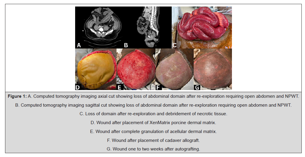
Figure 1:A. Computed tomography imaging axial cut showing loss of abdominal domain after re-exploration requiring open abdomen and NPWT.
B. Computed tomography imaging sagittal cut showing loss of abdominal domain after re-exploration requiring open abdomen and NPWT.
C. Loss of domain after re-exploration and debridement of necrotic tissue.
D. Wound after placement of XenMatrix porcine dermal matrix.
E. Wound after complete granulation of acellular dermal matrix.
F. Wound after placement of cadaver allograft.
G. Wound one to two weeks after autografting.
Conclusion
This case report presents an interesting challenge in surgical planning and reconstruction due to the complex course after traumatic injury involving abdominal compartment syndrome, abdominal wall infection and loss of domain. A defect of this size typically requires fascial repair with mesh [16]. Due to the inherent field contamination a biologic acellular dermal matrix was selected for fascial closure [16,17]. Patient specific factors required the creation of an ileostomy and medical stabilization from critical condition necessitating a delay in hernia repair. At six weeks after abdominal wall reconstruction the mesh was fully granulated and prepared for split thickness skin grafting for wound closure. Our technique described above allowed temporization of the loss of domain in this patient such that once the patient stabilizes and all operative edema subsides attempts at a more definitive myofascial approximation may be obtained.
Acknowledgements
None.
Conflict of Interest
None.
References
- Saggi BH, Sugerman HJ, Ivatury RR, Bloomfield GL (1998) Abdominal Compartment Syndrome. J Trauma 45(3): 597-609.
- López Cano M, García Alamino JM, Antoniou SA, D Bennet, U A Dietz, et al. (2018) EHS clinical guidelines on the management of the abdominal wall in the context of the open or burst abdomen. Hernia 22(6): 921-939.
- Shestak KC, Edington HJ, Johnson RR (2000) The Separation of Anatomic Components Technique for the Reconstruction of Massive Midline Abdominal Wall Defects: Anatomy, Surgical Technique, Applications, and Limitations Revisited. Plast Reconstr Surg 105(2): 731-738.
- Sharrock AE, Barker T, Yuen HM, Rickard R, Tai N (2016) Management and closure of the open abdomen after damage control laparotomy for trauma. A systematic review and meta-analysis. Injury 47(2): 296-306.
- Sagar A, Tapuria N (2022) An Evaluation of the Evidence Guiding Adult Midline Ventral Hernia Repair. Surg J (N Y) 8(3): e145-e156.
- Bondre IL, Holihan JL, Askenasy EP, Jacob A Greenberg, Jerrod N Keith, et al (2016) Suture, synthetic, or biologic in contaminated ventral hernia repair. J Surg Res 200(2): 488-494.
- Lee L, Mata J, Landry T, Kosar A Khwaja, Melina C Vassiliou, et al. (2014) A systematic review of synthetic and biologic materials for abdominal wall reinforcement in contaminated fields. Surg Endosc 28(9): 2531-2546.
- Hsu PW, Salgado CJ, Kent K, Matthew Finnegan, Mark Pello, et al. (2009) Evaluation of porcine dermal collagen (Permacol) used in abdominal wall reconstruction. J Plast Reconstr Aesthet Surg 62(11): 1484-1489.
- Majumder A, Scott JR, Novitsky YW (2016) Evaluation of the Antimicrobial Efficacy of a Novel Rifampin/Minocycline-Coated, Noncrosslinked Porcine Acellular Dermal Matrix Compared With Uncoated Scaffolds for Soft Tissue Repair. Surg Innov 23(5): 442-455.
- Baker EH, Lepere D, Lundgren MP, Patrick J Greaney, David A Ehrlich, et al. (2016) Early Clinical Outcomes of a Novel Antibiotic-Coated, Non-Crosslinked Porcine Acellular Dermal Graft after Complex Abdominal Wall Reconstruction. J Am Coll Surg 223(4): 581-586.
- Cohen LE, Imahiyerobo TA, Scott JR, Spector JA (2016) Comparison of Antibiotic-Coated versus Uncoated Porcine Dermal Matrix. Plast Reconstr Surg 138(5): 844e-855e.
- Becton, Dickenson and Company (2024) XenMatrix AB Surgical Graft. https://www.bd.com/en-us/products-and-solutions/products/product-families/xenmatrix-ab-surgical-graft.
- Parker SG, Halligan S, Blackburn S, A A O Plumb, L Archer, et al (2019) What Exactly is Meant by "Loss of Domain" for Ventral Hernia? Systematic Review of Definitions. World J Surg 43(2): 396-404.
- Ko JH, Wang EC, Salvay DM, Paul BC, Dumanian GA (2009) Abdominal wall reconstruction: lessons learned from 200 "components separation" procedures. Arch Surg 144(11): 1047-1055.
- Carbonell AM, Cobb WS, Chen SM (2008) Posterior components separation during retromuscular hernia repair. Hernia 12(4): 359-362.
- Patel NG, Ratanshi I, Buchel EW (2018) The Best of Abdominal Wall Reconstruction. Plast Reconstr Surg 141(1): 113e-136e.
- Gentile P, Colicchia GM, Nicoli F, Giulio Cervelli, Cristiano Beniamino Curcio, et al (2013) Complex abdominal wall repair using a porcine dermal matrix. Surg Innov 20(6): NP12-NP15.




 We use cookies to ensure you get the best experience on our website.