
Case Report 
 Creative Commons, CC-BY
A Suggestive Case of Complicated Acute Appendicitis: CT Findings and Review of the Literature
*Corresponding author:Francesco Messina, Unit of Radiology, Riuniti Hospital, Azienda Ospedaliera Grande Ospedale Metropolitano, Bianchi– Melacrino–Morelli, Via Giuseppe Melacrino n.21, 89124 Reggio Calabria, Italy.
Received:October 28, 2025; Published:October 31, 2025
DOI: 10.34297/AJBSR.2025.28.003747
Abstract
Acute appendix is one of the most important cause of “acute abdomen”, representing a potential surgical emergency; so its correct diagnosis is important for the clinical and methodological management of patients. We describe the case of a 43-year-old male patient presented with vomiting, fever and abdominal pain. Computed Tomography (CT) scan showed a complicated acute appendicitis and, consequently, an emergency appendectomy was performed.
Keywords:Appendicitis, Peritoneum, Abdomen, Computed Tomography
Introduction
Acute appendicitis is one of the most prevalent general surgical emergencies globally, with an incidence rate ranging from 100 to 233 cases per 100,000 individuals per year. Acute appendicitis refers to the inflammation of the appendix that has not yet ruptured. There are two types of acute appendicitis: “uncomplicated” and “complicated”. A complicated appendicitis is characterized by a peri-appendiceal phlegmon with or without perforation, gangrene, or a perityphlitis abscess, and fluid. Acute appendicitis may impact anyone at any age; however, the age-range between 5 and 45 is the most common for cases to occur. Nonetheless, appendicitis can affect both sexes, and the differences in occurrence between males and females are not significant [1-4]. CT is better for anatomically viewing of the appendix with a higher sensitivity and specificity for acute, and the use of CT imaging in the diagnosis and follow-up is very important for monitoring the abdominal clinical conditions of patients.
Case Presentation
A 43-year-old male patient presented recently to the Emergency Department of our Hospital complaining of abdominal pain of acute onset, located in the right iliac fossa toward the hypogastrium. The pain lasted a few hours and was accompanied by nausea, vomiting and fever. No history of previous abdominal surgery or any health problems was reported, and he wasn’t taking any drug. The patient referred habit on smoking but denied alcohol consumption. Laboratory tests reported high levels of C-reactive protein (125.5 mg/L) and neutrophilic leukocytosis (WBC 15.600 103/μL). The patient, presenting an “Alvarado score” of 7 (probable appendicitis), and so after an inconclusive Echography (US) because of too much intestinal bloating, an abdominal contrast-enhanced Computed Tomography (CT) was urgently made and the images so obtained were analyzed with a slice-thickness of 1.2mm and MPR reconstructions (axial, sagittal, and coronal). CT revealed (Figure 1a-e) the presence of a pathological distension and wall thickening of the cecal appendix (15mm), which also showed contrast-enhancement wall impregnation. The course of the appendix is suggestive and atypical, posteromedial retrocecal, with its most distal portion coming into contact with a distal ileal loop (which appears slightly thickened). Appendicular coprolite is also identifiable. Concomitant diffuse fat infiltration, with thickenings of the adjacent peritoneal fascial planes in the right iliac fossa region. Presence of numerous enlarged, inflammatory, adjacent lymphnodes (max diameter 10mm). Minimal fluid was also identifiable in the pelvic cavity.
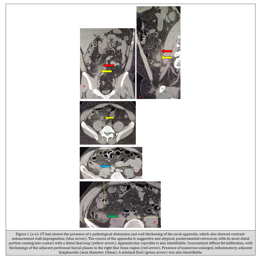
Figure 1:(a-e): CT had shown the presence of a pathological distension and wall thickening of the cecal appendix, which also showed contrastenhancement wall impregnation (blue arrow). The course of the appendix is suggestive and atypical, posteromedial retrocecal, with its most distal portion coming into contact with a distal ileal loop (yellow arrow). Appendicular coprolite is also identifiable. Concomitant diffuse fat infiltration, with thickenings of the adjacent peritoneal fascial planes in the right iliac fossa region (red arrow). Presence of numerous enlarged, inflammatory, adjacent lymphnodes (max diameter 10mm). A minimal fluid (green arrow) was also identifiable.
The patient, after understanding the severity of his medical situation, was urgently operating for appendectomy and was discharged on the 6th post-operative day in a stable condition.
Discussion
The appendix is described as a tubular structure attached to the large intestine. Acute appendicitis is the most common cause of sudden, severe abdominal pain. There are two types of acute appendicitis: “uncomplicated” and “complicated”. An “uncomplicated” case of appendicitis is characterized by inflammation without gangrene, free purulent effusion, phlegmon, or abscess. A “complicated” appendicitis is characterized by a peri-appendiceal phlegmon with or without perforation, and gangrene. Acute appendicitis has been linked to luminal obstruction, infectious agents, abscess, sex, genetic, and environmental variables, though the specific etiological agent has not been found [5,6]. Acute appendicitis may impact anyone at any age; however, the age range between 5 to 45 years old is the most common for cases to occur. The increased incidence of appendicitis in childhood, adolescence, and the early stage of adulthood is reflected in this age distribution. Males may have a slightly higher likelihood of developing acute appendicitis, with a life time incidence of 8.6% compared to 6.7% in females. Nonetheless, appendicitis can affect both sexes, and the differences in occurrence between males and females are not significant. The most common symptom of diagnosed patients with this disease is abdominal pain. Other clinical presentations of this disease include diarrhoea, vomiting, fever, loss of appetite, and abdominal distension. The main cause of appendicitis is inflammation brought on by blockage of the appendiceal lumen. Appendicoliths, appendiceal tumours, intestinal parasites, or hypertrophied lymphatic tissues are a few possible causes of the obstruction [7]. Misdiagnosis of acute appendicitis has been explored in a few studies. One study of 1,378 patients who received surgical intervention for appendicitis found that 7.1% of these patients were not diagnosed with acute appendicitis on their first visit to the emergency department leading them to be discharged back home or admitted to a nonsurgical hospital department [8,9]. Another study of 187,461 patients with a diagnosis of acute appendicitis found that the appropriate diagnosis was missed in 6.0% of adults and 4.4% of children. The position of the appendix is extremely variable: the most common location is retrocecal (74%) followed pelvic (21%), subcecal (1.5%), preileal (1%), and postileal (0.5%) [10]. The appendix can also show atypical locations such as subhepatic, left-sided, intraherniary, lateral pouch, and mesocholic. The differential diagnosis of appendicitis is therefore based on medical history, clinical examination, and diagnostic imaging (US and CT scan) to distinguish appendicitis from other conditions with similar symptoms, such as colitis, Crohn’s disease, renal colic, diverticulitis, constipation, gynaecological conditions (in women), and, not least, gallstones and Amyand’s hernia [11,12]. The diagnosis of appendicitis is therefore a combination of clinical, laboratory, and radiological tests (US, CT scan). The main symptom, which prompts the patient to go to the emergency department, is pain, which begins as mild epigastric pain, then moves to the right iliac fossa, near McBurney’s point, accompanied by nausea and vomiting, and then fever appears. In more severe cases, symptoms of peritonitis appear: the patient is unable to walk, is only comfortable lying on their back, complains of pain radiating to the right leg, is pale, sweaty, and hypotensive. In the presence of this clinical characteristics, the diagnosis is completed in the emergency room with blood tests and US. Imaging may be helpful for determining the correct diagnosis of appendicitis, as well as confirming abnormal locations of the appendix. Abdominal US is the first radiological investigation; however, very often it alone may not be effective due to limitations (excessive intestinal bloating) it has a high probability of misdiagnosis. CT, or contrast-enhancement Computed Tomography, is the best modality to identify appendicitis with sensitivity of 88–100%, specificity of 92–98%, positive predictive value of 86–98%, negative predictive value of 95–100% [13-15]. CT is the most common imaging modality used to evaluate adult patients with suspected appendicitis, with both high sensitivity and specificity for the diagnosis. In most cases, CT interpretation is straightforward with cases definitively falling into either positive or negative categories. When appendicitis is present, we typically find both appendiceal enlargement and secondary signs of inflammation. In normal cases, by contrast, the appendix may demonstrate a range of sizes, but no secondary signs of inflammation are usually identified. As such, it is important for the radiologist to have a clear understanding of the size parameters of the normal appendix at CT. An important parameter is the wall diameter greater than 6mm at CT, as diagnosis of appendicitis. The pathophysiology of acute appendicitis usually involves appendiceal obstruction with continued mucinous fluid secretion and bacterial proliferation within the lumen of the appendix [16]. As such, a fluid- or mucus-filled appendix is recognized as a sign of acute appendicitis at CT [17]. In clinical practice, airless fluid is at least occasionally identified in the appendiceal lumen in patients with no clinical evidence for appendicitis, particularly in the setting of a fluid-filled cecum. However, given the rarity of this appearance in the normal appendix and its known association with acute appendicitis, the finding of a fluidfilled appendix with diameter greater than 6mm should prompt a clinical work up for appendicitis (including blood work analysis and surgical evaluation), even in the absence of secondary signs of inflammation at CT. If the diagnosis remains unclear following clinical correlation, patient observation and short-interval follow-up CT may be appropriate.
Conclusions
We think that the benefit from pre-operative CT and advancements in CT technology that have directly improved image quality are greatest for a correct diagnosis of appendicitis. Low-dose CT is very important to use for the diagnosis of appendicitis in young patients (as we describe in our case). CT findings and management of typical acute appendicitis depend on the time of presentation ranging from early mild inflammation to perforation with peritonitis. Additionally, atypical pathologies of the appendix, though rare, can mimic or cause a superimposed appendicitis. These entities can have unique presentations and appearances on CT that help differentiate from typical acute appendicitis. Widespread implementation of appendiceal CT in the emergency department would require readily available helical CT facilities. We think that a radiologist would need experience with tens and tens of cases to achieve consistently accurate results and diagnosis. In summary, the routine use of appendiceal CT in emergency department patients who meet the clinical criteria for hospital admission for suspected appendicitis improves patient care both by averting unnecessary appendectomies and by averting delays before necessary medical or surgical treatment. Recognition of such anomalies pre-operatively, particularly using imaging, and CT in particular, is important to avoid surgical complications. It is crucial for surgeons to be aware of these abnormalities when they approach an appendectomy, for an adequate pre-operative preparation study program.
Patient Consent Statement
The patient confirmed the consense for publication of our case report.
Conflicts of Interest
The authors certify that there is no conflict of interest with any financial organization regarding the material discussed in the manuscript.
References
- Moris D, Paulson EK, Pappas TN (2021) Diagnosis and management of acute appendicitis in adults: a review. JAMA 326(22): 2299-2311.
- Téoule P, de Laffolie J, Rolle U, Reissfelder C (2020) Acute appendicitis in childhood and adolescence: an everyday clinical challenge. Deutsches Aerzteblatt Online. Clin Appl Thromb Hemost 117: 764-774.
- Elkoundi A, Bensghir M, Haimeur C (2017) Stercoral colitis mimicking appendicitis. Int J Emerg Med 10(1): 7.
- Taylor GM, Saffer ER, McDowell EL, Matthew A Warpinski (2019) A 2-year-old with a hepatic abscess secondary to an ascending retrocecal appendicitis: case report and review of the literature. Int J Emerg Med 12(1): 41.
- Bhangu A, Søreide K, Di Saverio S, Assarsson JH, Drake FT (2017) Acute appendicitis: modern understanding of pathogenesis, diagnosis, and management. Lancet 386(10000): 1278-1287.
- Wickramasinghe DP, Xavier C, Samarasekera DN (2021) The Worldwide Epidemiology of Acute Appendicitis: an analysis of the Global health data exchange dataset. World J Surg 45(7): 1999-2008.
- Di Saverio S, Podda M, De Simone B, Marco Ceresoli, Goran Augustin, et al. (2020) Diagnosis and treatment of acute appendicitis: 2020 update of the WSES Jerusalem guidelines. World J Emerg Surg 15(1): 27.
- Weinberger H, Zeina AR, Ashkenazi I Ochsner J (2023) Misdiagnosis of acute appendicitis in the emergency department: prevalence, associated factors, and outcomes according to the patients’ disposition 23(4): 271-276.
- Mahajan P, Basu T, Pai CW, Singh H, Petersen N, et al. (2020) Factors associated with potentially missed diagnosis of appendicitis in the emergency department. JAMA Netw Open 3(3): e200612.
- Sharma N, Mishra NT, Seema Singh, Ashesh Kumar (2014) A case of right upper abdominal pain misdiagnosed on computerized tomography, Malays J Med Sci 21(4): 66-68.
- Messina F, Tebala C, Calabrese G, Turano L, Fava MG, et al. (2020) Gallstone obstruction at the proximal ileum in an elderly woman: CT findings. Radiol Case Rep 16(3): 467-471.
- Nucera AG, Messina F, Schipani C (2021) Amyand’s hernia. Journal of Radiological Review 8(2): 168-171.
- Rao PM, Rhea JT, Novelline RA, Mostafavi AA, McCabe CJ (1998) Effect of computed tomography of the appendix on treatment of patients and use of hospital resources. N Engl J Med 338(3): 141-146.
- Coursey CA, Nelson RC, Patel MB, Cochran C, Dodd LG, et al. (2010) Making the diagnosis of acute appendicitis: do more preoperative CT scans mean fewer negative appendectomies? A 10-year study. Radiology 254(2): 460-468.
- Kim SY, Lee KH, Kim K, Kim TY, Lee HS, et al. (2011) Acute appendicitis in young adults: low- versus standard-radiation-dose contrast-enhanced abdominal CT for diagnosis. Radiology 260(2): 437-445.
- Crawford JM (1994) The gastrointestinal tract. In: Cotran RS, Kumar V, Robbins SL (eds) Robbins pathologic basis of disease. Saunders, Philadelphia: p 823.
- Moteki T, Horikoshi H (2007) New CT criterion for acute appendicitis: maximum depth of intraluminal appendiceal fluid. AJR Am J Roentgenol 188(5): 1313-1319.




 We use cookies to ensure you get the best experience on our website.