
Case Report 
 Creative Commons, CC-BY
An Unusual Case of Artery of Percheron Infarct Caused by Spontaneous Bilateral Postpartum Vertebral Artery Dissection
*Corresponding author:Forshing Lui MD, California Northstate University, College of Medicine, USA.
Received:October 22, 2025;; Published:October 28, 2025
DOI: 10.34297/AJBSR.2025.28.003738
Abstract
The Artery of Percheron (AoP) is a rare anatomic variant of cerebral vasculature in which all the paramedian arteries originate from a single, asymmetric arterial trunk branching from the proximal posterior cerebral artery (PCA). Typically, paramedian arteries arise bilaterally from the posterior communicating artery (PCom) to supply the paramedian thalamus. However, in patients with the AoP variant, an ischemic event to the singular trunk can result in bilateral thalamic infarcts. As such, we describe a rare case of a 34- year-old woman presenting with spontaneous Postpartum Bilateral Vertebral Arterial Dissection (PPVAD) complicated by AoP stroke. We explore the clinical manifestations, pathophysiology, neuroimaging findings, and differential diagnosis of this rare vascular event.
Introduction
Ischemic stroke remains a leading cause of morbidity and mortality worldwide [1], with a diverse array of etiologies and presentations challenging clinicians daily. Among these, strokes involving the Artery of Percheron (AoP) represent a rare but clinically significant entity that merits special attention due to its unique anatomical basis and often perplexing clinical manifestations. This paper aims to synthesize current knowledge on AoP strokes, highlighting their anatomical underpinnings, clinical presentations, diagnostic challenges, and management considerations as well as present a unique case. We explore the rare but important association between Postpartum Vertebral Artery Dissections (PPVADs) and AoP strokes, a connection that underscores the complexity of cerebrovascular pathology in the peripartum period.
The vascular supply to the paramedian thalami typically comes from the bilateral paramedian arteries, which branch off from the posterior cerebral arteries. However, in about 4-12% of individ uals, this supply is provided by a single arterial trunk that originates from the P1 segment of one posterior cerebral artery [2]. This anatomical variation, known as the Artery of Percheron, not only supplies blood to the bilateral paramedian thalami but can also extend to perfuse the rostral midbrain [2]. This variation is believed to result from the hypoplasia or absence of the contralateral P1, or proximal, segment of the posterior cerebral artery [3].
Despite the relatively common occurrence of this vascular variant, AoP strokes are remarkably rare, with reported incidence between 0.4-0.5% of all ischemic strokes across multiple analyses [3]. This low prevalence, coupled with a highly variable clinical presentation, often leads to significant delays in diagnosis and treatment. Lazzar, et al. [4] suggest that the true prevalence of AoP strokes may be underestimated due to these diagnostic challenges [4].
The clinical spectrum of AoP strokes is broad and often misleading due to variations in the perfusion pattern of the AoP. While altered consciousness, memory impairment, and vertical gaze palsy are frequently reported due to paramedian thalamic involvement, the presentation can encompass a wide range of neurological deficits [3,5]. Additional manifestations include behavioral changes, aphasia, hemiparesis, cerebellar signs, and various oculomotor disturbances [3,5]. This heterogeneity in presentation often leads to initial misdiagnosis, with AoP strokes frequently mistaken for toxic-metabolic encephalopathy, basilar artery occlusion, cerebral venous thrombosis, or even nonconvulsive status epilepticus [6].
Neuroimaging plays a pivotal role in the diagnosis of AoP strokes. While initial Computed Tomography (CT) scans may appear unremarkable, Magnetic Resonance Imaging (MRI), particularly diffusion-weighted sequences, offers superior sensitivity in detecting acute ischemic changes. The characteristic finding of bilateral paramedian thalamic lesions, with or without rostral midbrain involvement, is often critical to diagnosis [4]. Of note, it is important to rule out other mechanisms that could lead to similar neuroimaging findings. These include hypoglycemia, hyperglycemia, chronicliver disease, Wernicke’s encephalopathy, and Wilson’s disease [7].
The management of AoP strokes aligns with standard acute ischemic stroke protocols, emphasizing the importance of early recognition and intervention. Despite the often dramatic initial presentation, many patients experience favorable outcomes with appropriate treatment [8]. However, long-term sequelae, particularly in domains of memory and executive function, remain a concern and an area for further research.
Our paper highlights an unusual case of AoP stroke as sequelae of Postpartum Vertebral Artery Dissection (PPVAD). Vertebral artery dissections, or spontaneous tears in the tunica intima of the vessel wall, are known to occur with increased frequency in the postpartum period, likely due to multiple factors including hormonal changes and mechanical stresses during labor [9]. The pathophysiological mechanism linking PPVADs to AoP strokes is not fully understood but may involve thromboembolic phenomena or hemodynamic changes in the posterior circulation. The rarity of this association presents a significant diagnostic challenge, requiring a high index of suspicion and thorough vascular imaging.
Case Presentation
A 34-year-old G5P5 African American woman presented to the emergency room with a reduced Level of Consciousness (LOC). Per the patient’s spouse, the patient was found unconscious and unarousable, with foam saliva around her mouth. Her past medical history was significant for obesity, hypertension and a recent, uncomplicated, vaginal delivery of her 5th child three weeks ago. The patient had complained to her spouse of sudden-onset vision problems with an inability to look downwards, slurred speech, difficulty remembering her newborn’s name, and a bilateral posterior cervical headache for the duration of the last day. She denied any trauma, fever, bleeding, recent infection, recent travel, vertigo, hallucinations, oral contraceptive pill use, or history of stroke.
Naloxone by emergency medical services en route to the hospital provided no improvement in LOC. In the emergency room, vital signs were stable (Table 1) and a physical exam revealed somnolence, arousal to repeated noxious stimuli (+2 NIHSS points), limited spontaneous speech (+1 NIHSS point), and mild dysarthria (+1 NIHSS point). National Institutes of Health Stroke Scale (NIHSS) score was 4. The remainder of the physical examination was unremarkable. Patient was given magnesium for presumed postpartum eclampsia and neurology was consulted.

Table 1:Vital signs for a 34-year-old G5P5 African American woman presented to the emergency room with a reduced LOC over 3 hour period on admission.
EKG reflected a normal sinus rhythm. Labs were unremarkable. Computed Tomography (CT) of the head without contrast was negative for evidence of acute hemorrhage (Figure 1). Magnetic Resonance Imaging (MRI) of the brain showed faint FLAIR and DWI hyperintensity with signal loss on the processed ADC map in the bilateral globi pallidi, with right loss more prominent than that of the left, suggesting evolving infarctions (Figure 2). There were areas of restricted diffusion localized to the anterior medial thalami, suggestive of thalamic infarctions in the territory supplied by the AoP. Subsequent Computed Tomographic Angiography (CTA) was done and revealed symmetric narrowing of distal V3 and V4 segments of bilateral vertebral arteries, more prominently present on the left (Figure 3).
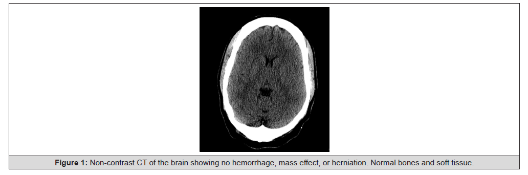
Figure 1:Non-contrast CT of the brain showing no hemorrhage, mass effect, or herniation. Normal bones and soft tissue.
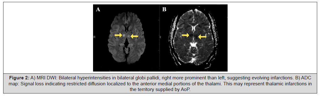
Figure 2:A) MRI DWI: Bilateral hyperintensities in bilateral globi pallidi, right more prominent than left, suggesting evolving infarctions. B) ADC map: Signal loss indicating restricted diffusion localized to the anterior medial portions of the thalami. This may represent thalamic infarctions in the territory supplied by AoP.
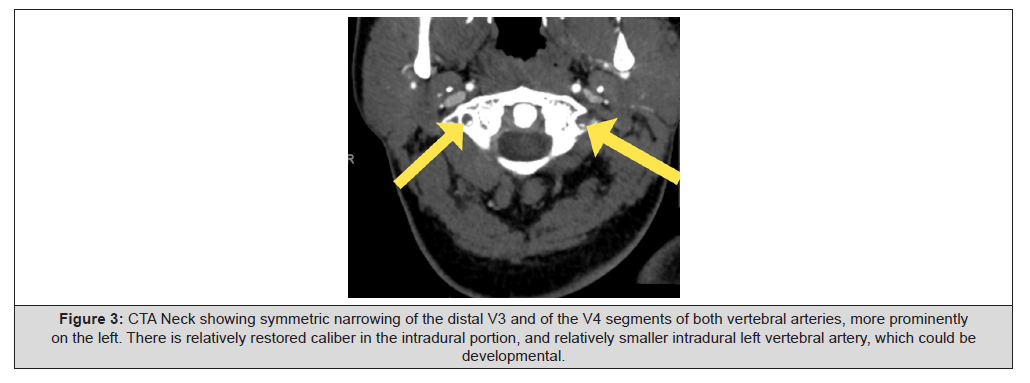
Figure 3:CTA Neck showing symmetric narrowing of the distal V3 and of the V4 segments of both vertebral arteries, more prominently on the left. There is relatively restored caliber in the intradural portion, and relatively smaller intradural left vertebral artery, which could be developmental.
Based on the patient’s clinical presentation and imaging findings, she was diagnosed with bilateral PPVAD and subsequent AoP stroke. Antiplatelet therapy was initiated with a one-time administration of aspirin 300-325 mg and clopidogrel 300 mg. The patient returned to baseline alertness and mentation within the following days. She continued dual antiplatelet therapy with aspirin 81 mg and clopidogrel 75 mg for 20 days and monotherapy with aspirin indefinitely thereafter.
Discussion
The blood supply of the thalami and midbrain can traditionally be distinguished into four common variations. Variant IIb, otherwise known as the variant involving the AoP, occurs when the AoP originates from a P1 segment of a unilateral PCA and then bifurcates to supply the perforating arteries [3,10]. These perforating arteries supply crucial blood flow to the rostral midbrain and bilateral paramedian thalami. Obstruction of these arteries can result in bilateral thalamic infarcts with variable mesencephalon involvement [2,3]. While AoP infarct only constitutes up to 2% of all ischemic strokes, Variant IIb is present in approximately 7-11% of the population [2,3,10].
Ischemia of the AoP presents primarily with symptoms reflecting bilateral paramedian thalamic infarction, such as: altered level of consciousness, memory impairment, oculomotor (predominantly vertical) gaze palsies, and akinetic mutism [2,10]. Delayed recognition of AoP infarction often results from a combination of a nonspecific, variable presentation of neurological deficits, difficulty in the detection of the miniscule infarct of the AoP on CT or MR imaging, sparsity of significant enhancement in early imaging, and a lack of awareness of AoP anatomical variants. These factors frequently lead to an initial misdiagnosis of viral infection or tumor [10].
This case was unique because our patient suffered a unique sequelae of spontaneous Postpartum Vertebral Artery Dissection (PPVAD), a rare complication of the puerperium period, and a subsequently incited AoP stroke. Diagnosis in this patient was made more complex by the combination of symptoms from the AoP stroke (vertical gaze palsy, memory loss) and bilateral PPVAD (bilateral posterior cervical headache). Similar to many AoP stroke work-ups, our initial recognition of the AoP involvement was superseded by considerations favoring toxic, metabolic, or hypoxicetiologies [7]. Additionally, early imaging showed motion artifacts and signal loss limiting MRA and MRV evaluation, which further complicated diagnosis.
Thorough analysis of neuroimaging was required to diagnose AoP stroke and rule out other etiologies of bilateral thalamic infarct. Bilateral posterior communicating arteries were evaluated to be patent with fetal contribution to the left PCA. A relatively smaller left P1 segment was noted which is consistent with the presence of the AoP. Areas of restricted diffusion localized to the anterior medial thalami were noted, suggestive of thalamic infarctions in the territory supplied by the AoP. Subsequent CTA revealed symmetric narrowing of distal V3 and V4 segments of bilateral vertebral arteries. These imaging findings and patient’s symptoms supported a diagnosis of bilateral thalamic infarcts secondary to spontaneous peri-partum bilateral vertebral artery dissection. Further imaging ruled out other causes of bilateral thalamic infarct including proximal large vessel occlusions at the Circle of Willis, thrombosis of dural venous sinuses, mass effects, ventricular and extra-axial cerebrospinal fluid space dilation, and paranasal sinus etiologies.
Conclusions
This case potentially represents one of the earliest documented occurrences of a PPVAD resulting in an AoP stroke. The bilateral nature of our patient’s dissection further underscores the exceptional rarity of this presentation. Our experience emphasizes the critical importance of a comprehensive understanding of cerebrovascular anatomical variations in effectively diagnosing and managing rare vascular conditions, particularly when such associations are not well-documented in literature.
Notably, clinicians should consider AoP involvement in cases with acute bilateral medial thalamic infarcts on diffusion-weighted MRI, especially after ruling out other toxic, metabolic, or hypoxic etiologies. Early recognition of this condition facilitates timely intervention with antiplatelet and/or thrombolytic therapy, potentially improving prognosis, as demonstrated in this case. This report underscores the value of maintaining a high index of suspicion for rare vascular anomalies in the face of atypical stroke presentations.
Disclosures
Human subjects: Informed consent for treatment and open access
publication was obtained or waived by all participants in this study.
Conflicts of interest: In compliance with the ICMJE uniform disclosure
form, all authors declare the following:
Payment/services info: All authors have declared that no financial
support was received from any organization for the submitted
work.
Financial relationships: All authors have declared that they have
no financial relationships at present or within the previous three
years with any organizations that might have an interest in the submitted
work.
Other relationships: All authors have declared that there are no
other relationships or activities that could appear to have influenced
the submitted work.
References
- Donkor ES (2018) Stroke in the 21st Century: A Snapshot of the Burden, Epidemiology, and Quality of Life. Stroke Res Treat 27: 3238165.
- Asim Kichloo, Shakeel M Jamal, El-Amir Zain, Farah Wani, Navya Vipparala, et al. (2019) Artery of Percheron Infarction: A Short Review. J Investig Med High Impact Case Rep.
- Ranasinghe K, Herath H, Dissanayake D, Seneviratne M (2020) Artery of Percheron infarction presenting as a nuclear third nerve palsy and transient loss of consciousness: a case report. BMC Neurology 20(1): 320.
- Nicholas A Lazzaro, B Wright, M Castillo, N J Fischbein, C M Glastonbury, et al. (2010) Artery of percheron infarction: imaging patterns and clinical spectrum. AJNR Am J Neuroradiol 31(7): 1283-1289.
- Rodriguez EG, Lee JA (2013) Bilateral thalamic infarcts due to occlusion of the Artery of Percheron and discussion of the differential diagnosis of bilateral thalamic lesions. J Radiol Case Rep 7(7): 7-14.
- Zhihua Xu, Lingling Sun, Yang Duan, Jinghua Zhang, Mengzhi Zhang, et al. (2017) Assessment of Percheron infarction in images and clinical findings. J Neurol Sci 383: 87-92.
- Pitts Tucker T, Small J (2018) Artery of Percheron: an unusual stroke presentation. BMJ Case Reports.
- Nathalie Zappella, Sybille Merceron, Chantal Nifle, Julia Hilly Ginoux, Fabrice Bruneel, et al. (2014) Artery of Percheron infarction as an unusual cause of coma: three cases and literature review. Neurocrit Care 20(3): 494-501.
- Marcel Arnold, Mathilde Camus Jacqmin, Christian Stapf, Anne Ducros, Anand Viswanathan, et al. (2018) Postpartum Cervicocephalic Artery Dissection. Stroke 39(8): 2377-2379.
- Neha Phate, Twinkle Pawar, Amol Andhale, Rohan Kumar Singh, Dhruv Talwar, et al. (2022) Artery of Percheron Stroke: A Case Report With a Diagnostic Challenge. Cureus 14(2): e21939.




 We use cookies to ensure you get the best experience on our website.