
Research Article 
 Creative Commons, CC-BY
Could Antimicrobial-Like Amphipathic Peptides Influence the Course of Alzheimer’s Disease? A Proposed Treatment Approach
*Corresponding author:GJ Mizejewski PhD, Division of Translational Medicine, Molecular Diagnostics Laboratory, Wadsworth Center, New York State Department of Health, Biggs Laboratory, Empire State Plaza, Albany, NY, USA.
Received:August 14, 2025; Published:August 26, 2025
DOI: 10.34297/AJBSR.2025.28.003671
Abstract
Alzheimer’s Disease (AD) is a common cause of the onset of dementia in the older population of the United States, accounting for 60-80% of all cases. Furthermore, AD affects 30 million people worldwide. In most people with AD, the loss of memory is the earlier manifestation of the disease. A recent advancement in the early detection of AD has been the development of a blood test for the ratio of two biomarkers in the vasculature of presumptive AD patients. The two biomarkers associated with the onset of AD include the Beta-amyloid peptides, and the hyper-phosphorylated microtubular p-Tau 217 protein. The Beta amyloid peptides are the cause of the plaque formations in brain cells, while the p-Tau 217 proteins are responsible for the diffuse presence of micro-fibrillary tangles widely distributed within the cytoplasm of brain cells. The present discourse describes the development of a proposed treatment regimen for AD involving the use of antimicrobial-like amphipathic peptides. An example of one such fetal peptide is described which may possibly be capable of addressing both the Beta-amyloid plaque formations and the Tau-induced microtubular tangle cell dispersements.
Keywords:Tau proteins, Beta amyloids, Peptides, Polymerization, Amphoteric, Anti-microbial-like peptide, Tangles, Plaques
Introduction
Alzheimer’s Disease (AD) is the most common cause of dementia in older populations of people in the United States 65 years and older [1]. The early memory loss of people with AD encompasses occurrences such as forgetting recent events and conversations which later developed into the inability to do every day common tasks. In advanced stages of AD, some people experience loss of familial face recognition, loss of certain brain functions, bodily dehydration, poor nutrition, susceptibility to infection, inflammatory disorders, and ultimately death [2]. AD has been uncovered to be causative in two biological processes as follows; first, the appearance of an increased amounts of circulating blood containing beta- amyloid peptides capable of transversing the blood-brain barrier and ultimately forming amyloid plaques within brain cells; and secondly, to the accumulation of neurofibrillary (microtubules) tangles widely distributed throughout the cytoplasm of brain cells [3]. These latter two processes result in an overall reduction of brain size, volume, and function.
One of the most prevalent symptoms in early AD affects a person’s memory retention in conjunction with multiple dysfunctional nerve networks [4]. Early in the onset of AD, individuals display mental problems which can include memory lapses and unclear thought processing. Frequently, it is the family members or friends that are most likely to first notice such mental issues. Moreover, the memory losses in AD can be long lasting, affecting the person’s ability to mentally function both at work and at home settings. AD in people with memory loss tend to: 1) repeatedly speak the same statements and questions; 2) forget conversations, appointments, and events; 3) misplace items and/or placing them in illogical locations; 4) getting lost in previously well-known locations; and 5) fumbling to find correct words to express themselves [5]. Additional memory-related symptoms can develop in situations involving highly detailed and focused thinking and reasoning, addressing judgements and decisions, and planning and performing familiar tasks. Such events occur together with obvious changes in personality, behavior, and memory retention.
Development of Blood Tests for Early Alzheimer’s Disease
In the last several years, improved blood tests for AD have been developed and reported in the medical literature involving the occurrence of two main blood biomarkers [6,7]. Such biomarkers include the 1) beta-amyloid peptides and 2) the hyperphosphorylated microtubular Tau protein. In the first instance, the overproduction of a beta-amyloid peptide can occur in which these peptides circulate in the bloodstream and subsequently transverse the blood brain barrier and enter into the brain. Secondly, the brain-derived microtubule-associated protein (Tau) has been found to be involved as a mutated hyperphosphorylated brain protein occurring at the amino acid 217 position of the Tau protein [8].
Tau-Induced Tangle Formation
Tau protein microtubules are crucial for maintaining the cell structural integrity of brain cells (neurons) by interacting with the microtubule system of the brain cellular cytoskeleton. Tau protein binding to the cell cytoskeleton proteins aid in stabilizing the cell microtubule network system, to ensure proper brain neuronal function. Tau proteins are quite soluble and abundant being localized throughout the central nervous system and in the cerebral cortex of the brain. In AD, Tau proteins become inappropriately hyper- phosphorylated at the protein’s amino acid position 217; the resultant aberrant protein then circulates throughout the cerebrospinal fluids and eventually accumulating within brain cells [9]. The hyperphosphorylated 217 Tau protein concentrations increase in AD patients and are causative in the formation of the microtubular tangles within brain cells causing disruptions in neural cell function. The hyper-phosphorylation at AA 217 prevents the Tau proteins from attaching to and supporting the microtubule network system, causing the microtubules to form insoluble aggregates (clusters) in brain cells collectively referred to as neurofibrillary tangles [10]. Thus, it is the accumulation of neurofibril tangles that disrupts neuronal activities, leading to brain cell dysfunction and ultimately resulting in cell death.
Beta Amyloid-Induced Plaque Formation
It is of interest that the formation of microtubular tangles correlate with the increased presence of beta-amyloid peptide plaques also localized within the cytoplasm of brain neurons. Thus, the presence of the amyloid plaques, together with the neurofibrillary tangles, are the two structural hallmarks of AD pathology and together play key roles in the progression of AD [10,11]. It is noteworthy that the increased presence of β-amyloid peptides are segments or fragments derived from a larger precursor protein called the “Amyloid Precursor Protein” (APP); “this protein is the original source from which the beta amyloid peptides are produced as fragments or segments. The APP is enzymatically cleaved by specific secretase enzymes; however, the dysfunctional APPs in AD can become irregularly and inappropriately cleaved leading to an overproduction of beta-amyloid derived peptide fragments [12]. This APP cleavage event results in an overabundance of blood clearance rate in the blood vasculature causing an accumulation of increased levels of beta-amyloid peptide fragments in the bloodstream. Such peptide fragments are known to circulate in the blood and are capable of crossing the blood-brain barrier due to the peptide’s smaller molecular size [13]. Once within the brain, the disorganized beta- amyloid fragments are able of enter neurons and become distributed throughout the brain neuronal cytoplasm; such peptides can eventually aggregate into larger oligomer segments, called protofibrils. These protofibrils later contribute to the insoluble amyloid plaque formations. The main classical peptide plaques display a dense accumulation core, while the diffuse plaques reveal a more scattered intracytoplasmic distribution in the cytoplasm of neurons [14]. Overall, the beta-amyloid plaques form clumps which become densely distributed throughout the brain neuronal cytoplasm of AD patients. Unfortunately, such plaques can occur simultaneously together with the formation of the neurofibrillary tangles mentioned above.
The presence of beta-amyloid plaques together with the tangles are the two known pathological neuronal dysfunctions leading to memory loss and cell death present in many AD patients brain cells [10]. The dual combination of such pathological events in the brain interferes with signal transduction communications and receptor crosstalk among and between neuronal pathways which further impact and impair cognitive brain function. Furthermore, the abnormal presence of plaques and tangles are known to attract and activate immune brain cells termed microglia and astrocytes, both of which can initiate inflammatory responses causing further damage to both neurons and other associated brain cells [15]. Thus, both the beta-amyloid peptide oligomers and aggregates becomes toxic to neurons resulting in multiple and varied brain cell malfunctions due to the multiple inflammatory reactions which trigger the programmed cell death (apoptosis) process.
Could the Presence and Use of Antimicrobial- Like Peptides Aid in Reducing the Damaging Effects of Beta Amyloid Plaques and Tangles Found in the Brain?s
Amyloids and Effects of Beta-Amyloid Induced Brain Cell Plaques
Amyloidosis (ALD) is a byproduct of cell cytoplasmic polypeptide assembly events, while amyloid bodies (A-bodies) consist of a highly organized form of protein aggregates that convert native- folded proteins into B-sheet rich molecular aggregates [16]. Interestingly, A-bodies consist of peptides that can bind to various heavy metals and function as cell-membrane disruptors and pore/ channel formers [17]. Such peptides not only disrupt cell surface membranes, but also function in cell membrane pore formation, binding to multiple receptors, and inducing the formation of endocytic vesicles formed within the brain cell cytoplasm.
Contrary to popular belief, ALD is a common occurrence in eukaryotic cells [16]. Previous reports have elucidated that ALD is a natural physiological process in mammalian cells which results from a response to multiple cell stress stimulations [17]. The ALD process enables cells to accumulate and aggregate proteins into organized fibril clusters for compartmentation and storage within the cell cytoplasm, thereby reflective of a cell adaptation stage resulting from multiple various stress stimulations [18]. In such events, ALD enables cells to safely store large quantities of fibrillary proteins in cytoplasmic compartments and permitting cells to enter into a dormant or resting stage [16]. Such events take place to allow cells to remain viable during periods of multiple extracellular stimulating stress agents. Eukaryotic cells frequently encounter environmental stress factors, such as inflammation, hypoxia, high temperatures, H2 O2 peroxidation, acidosis, pH extremes, oxidative stresses, and various other conditions that lead to abnormal cell growth regulations [19-21].
Structure Comparison of an Alpha-Fetoprotein (AFP) Derived Peptide to Abeta Peptides Which Cause Plaque Formation
Progressive neurodegenerative diseases such as Alzheimer’s Disorder (AD) and Parkinson’s disease employ the process of amyloidosis as described above, in which insoluble potentially toxic protein fibers are isolated and deposited in cellular compartments to guard against impairment of cell functions [16]. Aggregated amyloid fibers in AD are produced as intracellular, proteinaceous fiber deposits which display a combined-beta sheet and beta-turn secondary structure. Beta-amyloid peptides (Abeta) are normally cleaved by specific secretase enzymes (see above) from a larger transmembrane Amyloid Precursor Protein (APP) as described above [19]. The resultant Abeta peptides are ultimately found as the intracellular deposits of plaques found in the brain cells of AD patients resulting in cell neurotoxicity [22,23]. The transmembrane precursor proteins are cleaved by beta and gamma-secretase enzymes within the plasma membrane bilayer thus generating various forms of amyloid peptides most of which contain a 42 AA sequence composition (Table 1). However, some additional Abeta peptides can also include 40 AA residues and 25-35 AA residues in addition to the more numerous 42 AA peptides. It is the abnormal accumulation, aggregation, and presence of the multiple cleaved protein segments (fragments) within the cell membrane bilayer that eventually contributes to neural cell death [24] The Abeta peptide and cell membrane interactions are often accompanied by a cell membrane bilayer flip inversion of phosphatidylcholine and phosphatidylserine phospholipid head groups that result in the course of an inverted bilayer event [24-26]. The enhanced deposition of aggregated Abeta peptides is thought to involve a coordination of histidine metal ion binding because the Abeta peptide has a very high affinity for Cu2+ and Zn2+ binding. It is interesting that the AFP-derived antimicrobial-like GIP-34 peptide can also bind copper & zinc in a similar fashion [27].
Abeta Peptide Versus the GIP-34 Amino Acid Comparison
Abeta toxic peptides exhibit a 42-Amino Acid (AA) sequence composition comprised of the following Amino Acid (AA) single letter codes:
DAEFRHDSGYEVHHQKLVFFAEDVGSNKGAIIGLMVGGVVIA [14]. This 42-mer Abeta peptide contains 19 hydrophobic amino acids, 8 intermediate amino acids, and 15 hydrophilic amino acids making it similar in composition to many amphipathic peptides which also include multiple dipolar ions (Zwitterions) (Table 1). In comparison, the peptide AA sequence for an AFP-derived 34-mer (GIP) synthesized peptide which composes an amphipathic peptide with an AA sequence determined as:
LSEDKLLACGEGAASIIIGHLCIRHEMTPVNPGVGN. This GIP- 34 peptide is derived from AA-residues 446-480 found on the full-length alpha fetoprotein polypeptide [27]. In comparison to Abeta-42, the 34-mer GIP-peptide contains 16 hydrophobic amino acids, 7 intermediate amino acids, and 11 hydrophilic amino acids, thus fulfilling the descriptive composition of an amphipathic peptide [28] (Table 1). In comparison, the 42-AA Abeta peptide contains a few more hydrophobic and hydrophilic AAs than does the GIP-34 peptide; overall, the Abeta-42 peptide contains 8 more total AAs than the GIP-34 peptide [27,28]. In nature, the amphipathic peptides have the advantage that they bind either or both positive and negative charged surfaces of cell membranes. A structurally modeled minimal energy image comparison of Abeta-42 and GIP- 34 has further revealed that the two peptides show remarkably similar structure configurations as displayed in their non-solvent computer models [28].
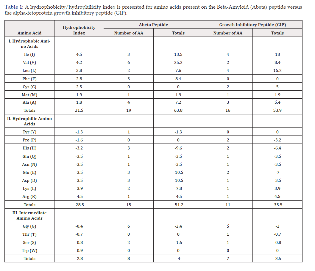
Table 1:A hydrophobicity/hydrophilicity index is presented for amino acids present on the Beta-Amyloid (Abeta) peptide versus the alpha-fetoprotein growth inhibitory peptide (GIP).
Note*: Single Letter Amino Acid Code, GIP=Growth inhibitory peptide
Amino Acid Sequence Matching in Abeta-42 versus GIP-34 peptides
An amino acid sequence match was performed to search for amyloid-associated peptide matches to the 34-mer AFP-derived GIP peptide (Table 2). Hence, the GIP-34 amino acid sequence was subjected to a FASTA search in the Genbank (GCG Wisconsin Program) database. FASTA employs Z-based statistic algorithms between comparison proteins and/or peptide amino acid sequences. The GCG search found identity/similarity sequence matches between GIP-34 and that of the Beta amyloid peptide Alzheimer’s associated protein. Such amino acid matches provide indirect evidence that the GIP-34 peptide contains short recognition AA-cassettes comparable to AA sequences of the AD derived Beta-amyloid peptides. As reported in a previous paper [21], the amino acid matching percent revealed that the 34-mer GIP and Beta-amyloid peptide had a 71% identity and a 29% similarity between the two peptides.
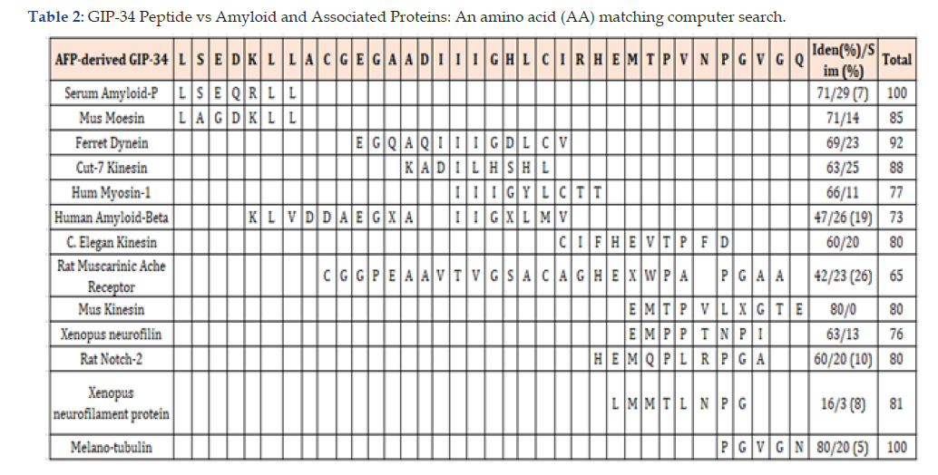
Table 2:GIP-34 Peptide vs Amyloid and Associated Proteins: An amino acid (AA) matching computer search.
Legend: The amino acid sequences of the growth inhibitory peptide (GIP) were compared and matched with proteins in the data bases using the GCG (Wisconsin program FASTA sequence described in Refs 17,18)
Abbreviations: Iden=identity; Sim=similarity; () =number of amino acids in sequence; Ache=acetyl cholinesterase; apolipoprot= apolipoprotein-E (APO-E); Ves-vesicular, AFP=alpha-fetoprotein.
Comparison of Microtubules, Microtubular- Associated Proteins, and the Cytoskeletal Proteins
From the data obtained to date (see above), GIP appears to function as a microtubular associated protein entity. Normally, the Microtubule Associated Proteins (MAPs) aid and function to regulate the assembly and stability of microtubules. The microtubules themselves are the largest of the cellular protein filaments and are mostly composed of a protein called Tubulin. The MAPs are proteins capable of binding to microtubules and as such can include the motor proteins entities such as dynein and kinesin (Table 2). Other microtubule proteins are comprised of Tubulin, Tau proteins, desmin, vimentin, ensconosin, katamin, and KIF2A. It is of interest that GIP shares amino acid sequence matching with many of the microtubule proteins as displayed in Table 2. Shared amino acid sequence matches between proteins and peptides imply and indicate possible docking/binding and amino acid pairing interactions between the two entities being matched. In comparison to the microtubules, the proteins of the cytoskeletal system comprise proteins such as: 1) Moesin; 2) Melsalin; 3) Diap-3; 4) Fascin; 5) Radixin; 6) Feinbrin; 7) Myosin-2; and 8) Doublecortin, some of which are displayed in Table 2. Overall, GIP shares AA sequence identities with many proteins of both the microtubular and cytoskeletal structural systems.
Microtubule Involvement in Tangle Formations
Microtubular Polymerization
Tubulin, the major protein comprising microtubules, is highly polymorphic and found in a variety of cell types especially neurons. In conjunction with basic elements of the cytoskeleton, microtubules are implicated in multiple cell functions, including chromosome segregation during cell division, distribution and transport of various organelles, and determination of cell morphology and cell polarity [29,30]. Overall, microtubules are thought to have structural, organizational and motility-related functions. Microtubules consist of protein polymers that form rod-like structures composed of multiple protofilaments. The protofilaments, in turn, are formed by the head-to-tail association of tubulin dimers. The microtubular association with the cytoskeleton reflects a scaffolding structure, which helps to define and maintain cell shape; in addition, microtubules mimic cell-like organelles performing as guidance tracks along which molecular complexes nutrients, and organelles are transported within cells. Thus, microtubules can serve as mobility- based structures, which promote mechanical forces which are assisted by motor proteins (see above and Table 2) that promote chromosome segregation during mitosis [31]. Plasma membrane- associated microtubules can also function in regulating cell surface receptor mobility and distribution, while cytoplasm-contained microtubules can mediate cytoplasmic endocytic/exocytic vesicle formation processes [32].
Microtubular polymerization experiments have been previously reported using GIP and employing α-tubulin brain extracts obtained from newborn chickens [29]. Studies of viscometrical measurements of polymerization using GIP peptides, titrated at varying concentrations, displayed a parallel rise in viscosity versus time. This rise in solution viscosity is consistent with the increase in the molecular size of the polymerized microtubule complex resulting in a more effective moment transect, manifested as an increase in solution viscosity. Thus, the solution viscosity increased with the amount of GIP added to the solution and this parallel rise event was found to correlate with increasing GIP concentrations. Thus, the addition of increasing levels of GIP to the reaction vessels resulted in specific viscosity readings that showed a steep rise peaking at a 4-fold increase and resulting in microtubule polymerization. Overall, the GIP peptide enhanced the ability of α-tubulin to polymerize in a cell-free solution, unlike control peptides that failed to demonstrate this same property [29].
The above data indicate that GIP shares some of the properties of microtubule-associated proteins, primarily capable of engaging in tubule polymerization. The highly soluble Tau proteins normally maintain the stability and integrity of brain neurons by binding to the microtubular system of the brain cell cytoskeletal system [8,9,33]. However, in AD, the Tau proteins become inappropriately hyper-phosphorylated at AA 217, leading to the formation of highly distributed microfibril tangles present within the brain cell microtubular system [9]. It is tempting to speculate that the GIP-34 peptide, by means of tubule polymerization, could compete and/or override the hyperphosphorylated Tau induced function of tangle formation. This GIP-induced polymerization could possibly result in formation of more uniform tubule distributions culminating in an even cytoplasmic neurofibril organized storage and compartmentalization within neuronal cells. In this proposed model, the GIP tubule induced polymerization might be capable of forestalling and/or reducing the start-up formation of the iwidely distributed cytoplasmic neurofibulary tangles [29]. Thus, it can be proposed that the prevention of the initial tangle formation might be enacted by means of tubule polymerization employing GIP-34. However, this GIP mode of action concept has yet to be performed and proven in experimental and clinical settings.
Summarizing Statements
It can be ascertained by the discussion of the above treatise, that a non-conventional hypothetical approach to a therapeutic treatment mode for early AD has been proposed. The suggested actions might have the potential to address the dual incidence of both brain cell inclusions of Abeta peptide plaques and those of microfibril tangles [34]. By means of employing GIP-derived amphipathic peptides, rogue circulating beta amyloid peptides could possibly be neutralized by binding interaction with the GIP molecule; secondly, the microfibril tangle formations might also be circumvented using a microtubule polymerization mode of action prior to formation of the tangles. Although not yet proven by direct experimental and clinical studies, the use of an AFP-derived amphipathic GIP-34 peptide has previously been reported to individually involve both such biological activities [28,29]. The binding and proposed inactivation/neutralization of Beta-amyloid peptide in blood circulation by GIP-34 has been further addressed using an “in silico” computer analysis. This procedure was accomplished by means of an amino acid pairing method first described and published by Root-Burnstein et al. [35]. These molecular docking and protein interaction sites on the Growth Inhibitory Peptide (GIP) have now been identified and mapped. Such sites were localized by means of a proprietary computer software program termed the “peptimer discovery platform.” This computer software was developed and generously provided to the author (GJM) by the Serometrix LLC Biotech CO, Syracuse, New York. In this computer program, it was demonstrated that the GIP sequence consisting of amino acid 462-GHLCIRH-470 contains molecular docking, binding, and molecular interactions with both Tubulin heterodimers and with the Glial Fibrillary acid protein. These latter proteins are expressed in brain cells such as astrocytes, obilgodendrites, and astroglia cells. As shown above, the process of tubule polymerization by GIP-34 has been experimentally studied and reported by Mizejewski et al. to prevent tangle formations [29]. Likewise, the amino acid pairing and computer docking of GIP with binding sites on Beta amyloid peptide has proved to be a promising venture. In summation, it has now been recognized that patho-physical changes may begin in AD patients many years prior to the clinical manifestations of AD. It is further known that the spectrum of AD activities spans from clinically asymptomatic to severely impaired patients [36]. Thus, it is crucial that early treatment of AD, as shown and proposed in this report, could possibly be implemented in the early stages of AD. In summation, GIP treatment has yet to be proven to be effective in AD patients and must await clinical testing [37].
Declarations and Acknowledgements
Funding: None; no US federal grants were used in the preparation
of this paper.
Conflicts of Interest: The author declares there are no known
conflicts of interest in the preparation of this manuscript.
Acknowledgements: The author extends his thanks and gratitude
to Ms. Sarah Andres for her commitment and time expenditure
in the skilled typing and processing of the manuscript,
references, and tables of this report.
References
- Scheltens P, De Strooper B, Kivipelto M, Holstege H, Van der Flier WM, et al. (2021) Alzheimer’s disease. Lancet397(10284): 1577-1590.
- Rostagno AA (2022) Pathogenesis of Alzheimer’s Disease. Int J Mol Sic 24(1): 107-108.
- Ogbodo J, Agbo C, Njoku U, Ogugofor M, Egba S, et al. (2022) Alzheimer’s Disease: Pathogenesis and Therapeutic Interventions. Curr Aging Sci: 15(1): 2-25.
- Eratne D, Loi S, Farrand S, Kelso W, Velakoulis D, Looi J, et al. (2018) Alzheimer’s disease: clinical update on epidemiology, pathophysiology and diagnosis. Australas Psychiatry (4): 347-357.
- Bocwinska Kiluk B, Wojiech J, Johannes K, Piotr L, Barbara M (2023) Alzheimer’s Disease-biochemical and psychological background for diagnosis and treatment. Int J Mol Sci 24(2): 1059.
- Mantzavinos V, Alexiou A (2017) Biomarkers for Alzheimer’s disease diagnosis. Curr Alzheimer’s Res 14(11): 1149-1154.
- Graff Radford J, Yong K, Apostolova LG, Bouwman FH, et al. (2021) New insights into atypical Alzheimer’s disease in the era of biomarkers. Lancet neurol 20(3): 222-234.
- Beharry C, Cohen LS, Di J, Ibrahim K, Briffa Mirabella S, et al. (2014) Tau-induced neurodegeneration: mechanisms and targets. Neuroscience bulletin 30(2): 346-358.
- Lee H, Perry G, Moreira PI, Garrett MR, Liu Q, Zhu XW, et al. (2005) Tau phosphorylation in Alzheimer’s disease: pathogen or protector? Trends in Molecular Medicine 11(4): 164-169.
- Braskie MN, Klunder AD, Hayashi KM, Protas H, Kepe V, Miller KJ, et al. (2010) Plague and tangle imaging and cognition in normal aging and Alzheimer’s disease. Neurobiology of aging 31(10): 1669-1678.
- Roberts GW (1988) Immunocytochemistry of neurofibrillary tangles in dementia pugilistica and Alzheimer’s disease: evidence for common genesis. Lancet 2(8626-8627): 1456-1458.
- Crescenzi O, Tomaselli S, Guerrini R, Salvadori S, D Ursi AM, et al. (2002) Solution structure of the Alzheiner amyloid beta-peptide (1-42) in an apolar microenvironment. Similarity with a virus fusion domain. Eur J Biochem 269(22): 5642-5648.
- Zhao H, Tuominen EK, Kinnunen PK (2004) Formation of amyloid fibers triggered by phosphatidylserine-containing membranes. Biochemistry 43(32): 10302-10307.
- Murakami K, Irie K, Morimoto A (2003) Neurotoxicity and physicochemical properties of Aβ mutant peptides from cerebral amyloid angiopathy. J Biol Chem 278(46): 46179-46187.
- Twarowski B, Herbet M (2023) Inflammatory Processes in Alzheimer’s Disease-Pathomechanism, diagnosis and treatment: A review. Int J Mol Sci 24(7): 6518.
- Mizejewski GJ (2017) Breast cancer and amyloid bodies: is there a role for amyloidsis in cancer-cell dormancy? Breast Cancer 26: 287-291.
- Schnued L, Raymick J, Tolleson W, Sarkar S, Zhang Yh, et al. (2012) Introducing Amylo-Glo, A novel flurescent amyloid specific histochemical tracer especially suited for multiple labeling and large-scale quantification studies. J Neurosci methods 209(1): 120-126.
- Audas Te, Audas DE, Jacob MD, JJ David Ho, Mireille Khacho, et al. (2016) Adaptation to stressors by systemic protein amyloidogenesis. Dev Cell 39(2): 155-168.
- Pinto M, Morange M, Bensaude O (1991) Denaturation of proteins during heat shock. In vivo recovery of solubility and activity or reporter enzymes. J Biol Chem 266(21): 13491-13946.
- Semenza GL (2012) Hypoxia-inducible factors in physiology and medicine. Cell 148(3): 399-408.
- Semenza GL, Wang GL (1992) A nuclear factor induced by hypoxia via de novo protein synthesis binds to the human erythropoietin gene enhancer at a site required for transcriptional activation. Mol Cell Biol (12): 5447-5454.
- Beard CM, Kokmen E, Sigler C, Smith GE, Petterson T (1996) Cause of death in Alzheimer’s Disease. Ann Epidemiol (3): 195-200.
- Buchsteiner A, Hauss T, Dante S, Dencher MA (2010) Alzheimer’s disease amyloid-beta peptide analogue alters the ps-dynamics of phospholipid membranes. Biochim Biphys Actra 1798(10): 1968-1976.
- Mingeot Liclercq MP, Lins L, Bensliman M, Van Bambeke F, Van Der Smissen P, et al. (2002) Membrane destabilization induced by beta-amyloid peptide 29-42: importance of the amino-terminus/Chem Phys Lipids 120(1-2): 57-74.
- Dante S, Hauss T, Brandt A, Dencher NA (2008) membrane fusogenic activity of the Alzheimer’s peptide A beta (1-42) Demonstrated by small-angle neutron scattering. J Mol Biol 376(2): 393-404.
- Terakawa MS, Yagi H, Adachi M, Lee YH, Goto Y (2015) Small liposomes accelerate the fibrillation of amyloid β (1-40). J Biol Chem 290(2): 815-826.
- Mizejewski GJ, MacColl R (2003) Alpha-fetoprotein growth inhibitory peptides: potential leads for cancer therapeutics. Mol Cancer Ther.2(11): 1243-1255.
- Mizejewski GJ (2019) The anti-estrogenic activity of Alzheimer’s amyloid-associated peptides: potential effects on hormone replacement therapy. A review and commentary. Journal of Cancer Research on Oncobiology 2(2): 1-8.
- Mizejewski G, Smith G, and Butterstein G (2004) Review and proposed action of alpha-fetoprotein growth inhibitory peptides as estrogen and cytoskeleton-associated factors. Cell Biol Int 28(12): 913-933.
- Audebert S, Desbruyers E, Koulakoff A (1993) Reversible polyglutamation of α and β-tubulin and microtubule dynamics in mouse brain neurons. Mol Biol Cell 4(6): 615-626.
- Mickey B, Howard J (1995) Rigidity of microtubules is increased by stabilizing agent s. J Cell Biol 130(4): 909-917.
- Hertzen KM, McCkung SCE, Walczak CE (2003) Kin I kinesins: insights into the mechanism of depolymerization. Crit Rev Biochem Mol Biol 38: 453-469.
- Alonso Ad, Li B, Grundke Iqbal I, Iqbal K (2008) Mechanism of tau-induced neurodegeneration in Alzheimer’s disease and related taupathies. Current Alzheimer Research (4): 375-384.
- Bondi MW, Edmonds EC, Salmon DP (2017) Alzheimer’s disease: past, present, and future. J Int Neuropsychol Soc 23(9-10): 818-831.
- Root Bernstein RS (1982) Amino acid pairing. J Theor Biol 94(4): 885-894.
- Aisen PS, Cummings J, Jack C, Morris JC, Sperling R, et al. (2017) On the path to 2025: understanding the Alzheimer’s disease continuum. Alzheimer’s Res Therap 9(1): 60-64.
- Thatte HS, Bridges KR, Golan DE (1994) Microtubule inhibitors differentially affect translational movement, cell surface express, and endocytosis of transferring receptors in K562 cells, J Cell Physil 160(2): 345-357.




 We use cookies to ensure you get the best experience on our website.