
Research Article 
 Creative Commons, CC-BY
Histological Analysis and Images of the Cardiac Fulcrum
*Corresponding author:Jorge Carlos Trainini MD PhD, Presidente Perón Hospital. National University of Avellaneda, Argentina.
Received:August 28, 2025; Published:September 08, 2025
DOI: 10.34297/AJBSR.2025.28.003687
Abstract
Objective: During myocardial dissection, the discovery of the cardiac fulcrum as the insertion point of the myocardium led us to further questions:
What are its characteristics? Where is it located? What is its histology? Is its presence analogous in different species? How is the myocardial muscle
inserted into this structure we have called the cardiac fulcrum? What are its properties? Does the fulcrums have any functional relationship with the
AV node, which is adjacent, located between it and the septal valve of the tricuspid valve?
Material and Methods: Seventy-one hearts from the morgue and slaughterhouses were used: a) 54 two-year-old cattle weighing 1300-1900g
(average 1650g); b) 17 humans (three at 8, 16 and 23 weeks of gestation respectively; four infants at 30 days, 36 days, 10 weeks and 27 weeks; one
4-year-old child; one 10-year-old child and eight adults with an average weight of 300g). Anatomical, histological, histochemical, and radiological
studies were performed. The hearts were fixed in 10% buffered formalin. Histology was performed with hematoxylin and eosin, Masson’s trichrome
staining, and 4-micron sections. 10% formalin was used as a buffer. Immunostaining (s100-neurofilaments) was also performed. All samples underwent
histochemical analysis using Alcian blue staining, a reliable marker for identifying the presence of hyaluronic acid, as an anti-friction mechanism,
and also providing a semiquantitative assessment.
Results: Anatomic, histological and radiological studies of the myocardium carried out in recent years in our research provide evidence that myocardial
fibers constitute a continuous muscle that describes a double helix to form both ventricles and that, to fulfill its muscular function, it needs a
support point that we have investigated, discovered, and called the cardiac fulcrum. Thus, the myocardium has the following characteristics derived
from the anatomical, histological and radiological analysis performed.
Conclusions: We have found in anatomy, histology and radiological studies of bovine and humans hearts (pregnant, pediatric, and adult) that the
cardiac fulcrum is a structure that grows in density toward a more solid center. In this scaling from lesser to greater density of the fulcrum, the fibers
are inserted just as in a tendon matrix (equivalent to the insertion of skeletal muscle tendons into bone).
Keywords:Cardiac anatomy, Cardiac fulcrum, Myocardial insertion, Cardiac images
Introduction
The existence of a bony formation called “os cordis” in mammals is a well-known fact in veterinary science. Beyond its mere mention, until our research, no function or meaning for its presence had ever been assigned to it, nor had it been described in humans [1,2]. The article we published in 2021 entitled “Myocardial torsion and cardiac fulcrum (Torsion myocardique et pivot cardiaque)” was the first human observation of the cardiac fulcrum and its functional importance [3]. During myocardial dissection, the discovery of the cardiac fulcrum as the insertion point of the myocardium led us to further questions: What are its characteristics? Where is it located? What is its histology? Is its presence analogous in different species? How is the myocardial muscle inserted into this structure we have called the cardiac fulcrum? What are its properties? Does the fulcrum have any functional relationship with the AV node, which is adjacent, located between it and the septal valve of the tricuspid valve? All of these requirements were investigated by us and disseminated in numerous publications [2-10]. The purpose of this presentation is to properly establish the histology of the cardiac fulcrum and its radiological images.
Material and Methods
Seventy-one hearts from the morgue and slaughterhouses were used: a) 54 two-year-old cattle weighing 1300-1900g (average 1650g); b) 17 humans (three at 8, 16 and 23 weeks of gestation respectively; four infants at 30 days, 36 days, 10 weeks and 27 weeks; one 4-year-old child; one 10-year-old child and eight adults with an average weight of 300g). Anatomical, histological, histochemical, and radiological studies were performed. The hearts were fixed in 10% buffered formalin. Histology was performed with hematoxylin and eosin, Masson’s trichrome staining, and 4-micron sections. 10% formalin was used as a buffer. Immunostaining (s100-neurofilaments) was also performed [11]. All samples underwent histochemical analysis using Alcian blue staining, a reliable marker for identifying the presence of hyaluronic acid, as an anti-friction mechanism, and also providing a semiquantitative assessment [12,13].
Results
Histological Analysis of the Cardiac Fulcrum. Myocardial Insertion
The consistency of the cardiac fulcrum in bovine hearts, osseous on palpation (Figure 1 and 2), has been confirmed by histology (Figures 3 and 4). The microscopic analysis of the bovine cardiac fulcrum shows a trabecular osteochondral matrix with segmental lines. Its general structure resembles the metaphyseal growth of long bones, and increased magnification reveals bone trabeculae with osteoblasts and segmental lines secondary to bone apposition. The same histological findings have been encountered in chimpanzees [14]. The sequential insertion of myocardial fibers in the bovine fulcrum can be seen in Figure 5 and 6 (Figures 1-6).
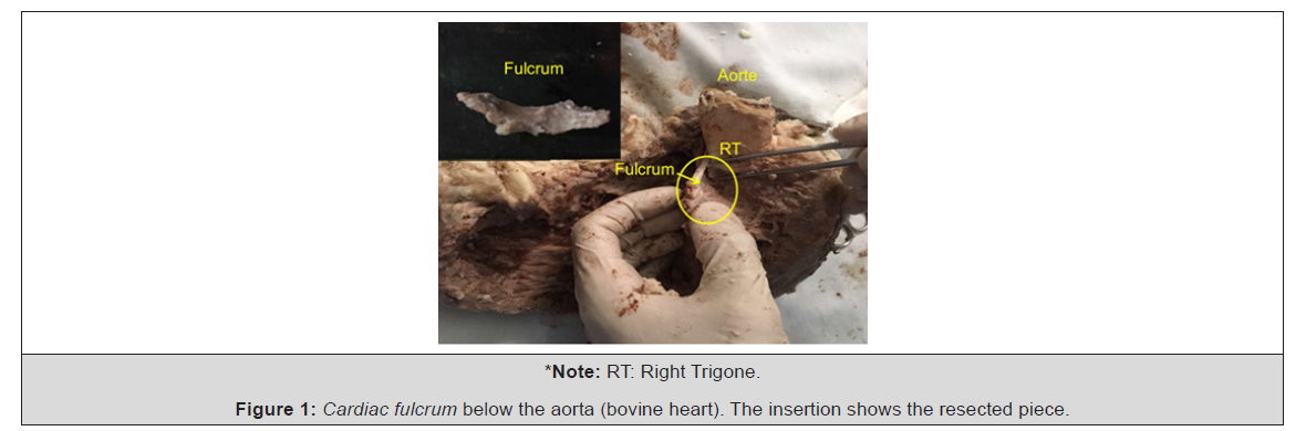
Figure 1:Cardiac fulcrum below the aorta (bovine heart). The insertion shows the resected piece.
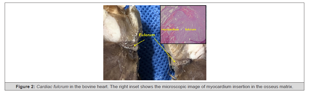
Figure 2:Cardiac fulcrum in the bovine heart. The right inset shows the microscopic image of myocardium insertion in the osseus matrix.
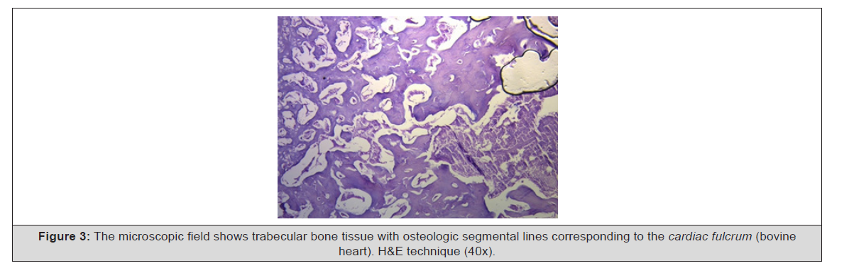
Figure 3:The microscopic field shows trabecular bone tissue with osteologic segmental lines corresponding to the cardiac fulcrum (bovine heart). H&E technique (40x).
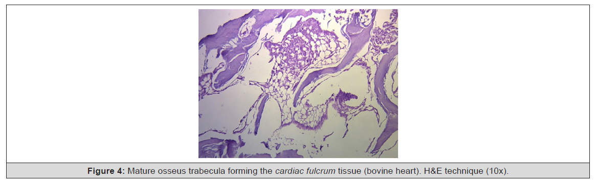
Figure 4:Myocardial insertion in the cardiac fulcrum (bovine heart). References.
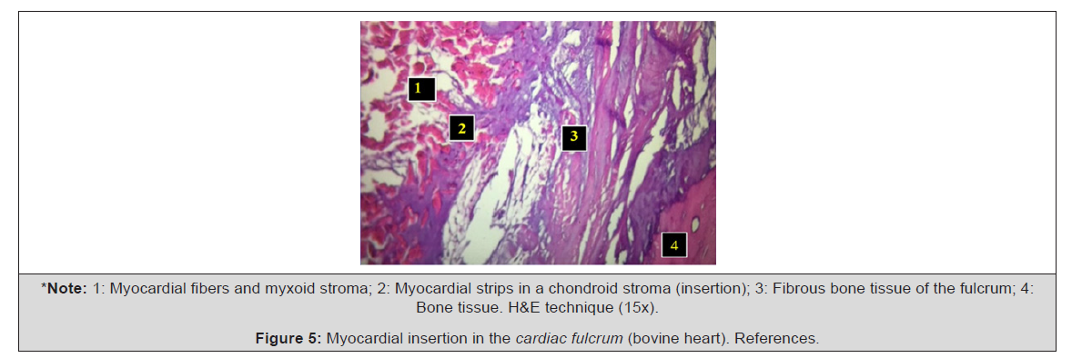
Figure 5:Myocardial insertion in the cardiac fulcrum (bovine heart). References.
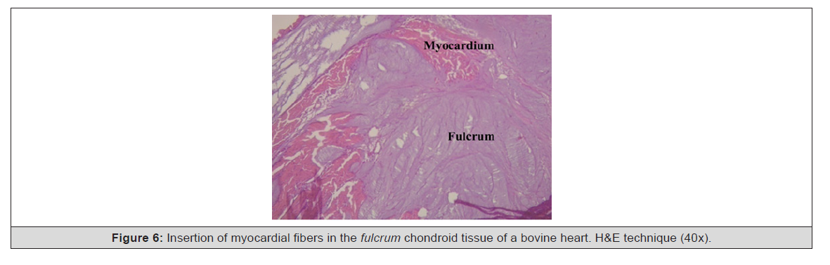
Figure 6:Insertion of myocardial fibers in the fulcrum chondroid tissue of a bovine heart. H&E technique (40x).
In the 10-year-old human heart, the histological description of the cardiac fulcrum is related with that early age, since the sample shows a central fulcrum zone formed by chondroid tissue. Given the age, its smaller size is logical, and characterized by more chondroid than osseus tissue (Figures 7,8). This finding was repeated in the 23-week-old human fetus with the characteristic prechondrial bluish areas in a myxoid stroma (Figures 9,10).

Figure 7:Cardiac fulcrum in a ten-year-old human explanted heart.
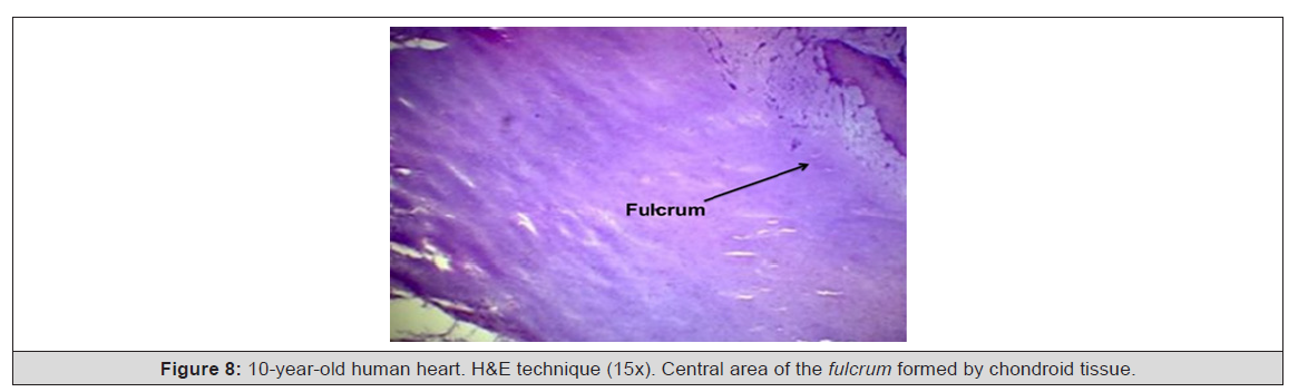
Figure 8:10-year-old human heart. H&E technique (15x). Central area of the fulcrum formed by chondroid tissue
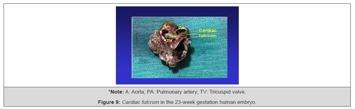
Figure 9:Cardiac fulcrum in the 23-week gestation human embryo
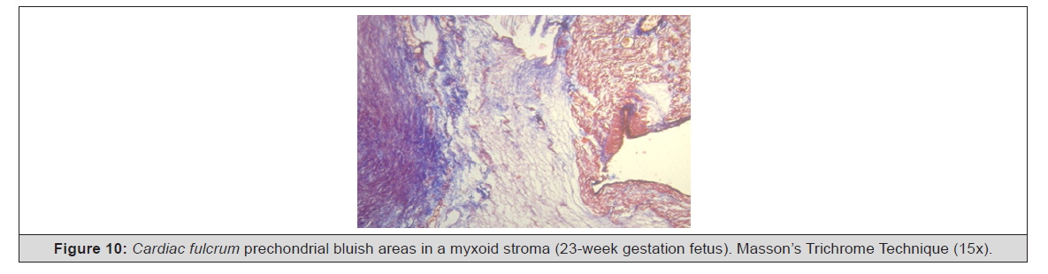
Figure 10:Cardiac fulcrum prechondrial bluish areas in a myxoid stroma (23-week gestation fetus). Masson’s Trichrome Technique (15x).
However, the histological analysis of the fulcrum in adult human hearts (approximate size 25mm long and 15mm wide) evidenced a chondroid-tendinous matrix, which needs additional clarification (Figure 11). As a rule, there is similar consistency in the detection, location and morphology of the fulcrum in all the hearts analyzed. They present myocardium insertion in the rigid fulcrum structure, forming a cardiomyocyte-matrix unit, independently of its osseous, cartilaginous or tendinous nature in the different specimens studied. As in any muscle, this point of attachment acts as a lever and also as a bearing, preventing the transfer of the ventricular rotation force, either by torque or torsion stress, to the great vessels, thus, dissipating the energy produced by the helical muscle movement. In this way, the energy of the myocardial torsion-detorsion movements is absorbed by the cardiac fulcrum, which is then shaped into the morphological characteristics of a helix (Figure 11).
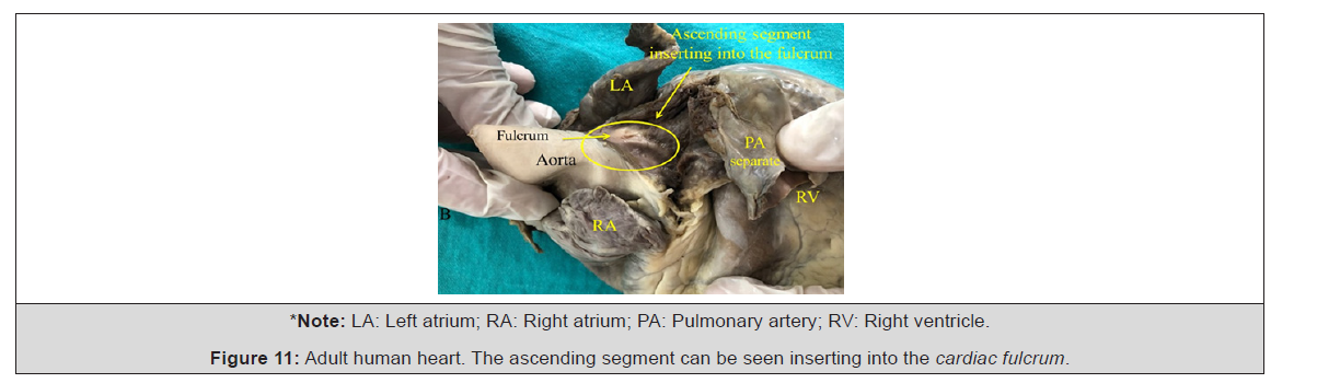
Figure 11:Adult human heart. The ascending segment can be seen inserting into the cardiac fulcrum
Having found an osseous structure in the bovine cardiac fulcrum and its relationship with the myxoid-chondroid texture in human hearts, even in embryos, is rational from the point of view of interpretation analysis. This disparity is associated with the different evolution given by age from chondroid to osseous material and with the greater power developed by bovine hearts, requiring a more rigid supporting point. In fact, tethering a heavy and powerful myocardium, such as that of a bovine heart, is not the same as tethering a myocardium of a human, which weighs only 270 grams. For the former, a very strong buoy, such as bone, is required. For humans, however, a cartilaginous-myxoid support is sufficient. Therefore, the histological analysis of the fulcrum in adult human hearts evidencing a chondroid-tendinous collagenous matrix, needs further clarification. The fact that it has been found in humans and different species implies that from a functional point of view its presence is synonym of myocardial insertion, as established in all histological analyses, becoming a solid point of reasoning to achieve its biomechanical function. And we find this demonstration when the histological examination is directed to the myocardial insertion in the cardiac fulcrum, be it of osseous, chondroid or tendinous nature. In all the hearts analyzed and according to the studies we have carried out in this investigation, this myocardial attachment was found to be as “ivy clinging to stone” in the rigid structure of the fulcrum, integrating an osseus, cartilaginous or tendinous cardiomyocyte- matrix unit, (Figure 12). In this concept there is analogy between skeletal and myocardial muscle. The former contracts between a fixed and a mobile supporting point, and this situation is found in the continuous myocardium, as there is greater solidity in the insertion between the fulcrum and the ascending segment compared with the attachment of the right segment in this support (Figure 12).
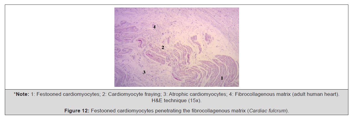
Figure 12:Festooned cardiomyocytes penetrating the fibrocollagenous matrix (Cardiac fulcrum).
At this point, fundamental questions arise: Why have we found that in adult human hearts the cardiac fulcrum has characteristics similar to a tendon despite it fulfills the same function of attaching the helical myocardium as in other species? Why does it not have the same structure as that found in the human embryo or child heart? Our interpretation is that perhaps the osseous-cartilaginous cardiac fulcrum is a vestigial organ specific of mammalian evolution. A vestigial structure must be understood as the preservation during the evolutionary process of genetically established attributes which have lost all or part of their ancestral function in a certain species [3]. As a result, it is found in the initial process of human gestation, but later loses its osteo-cartilaginous histology, remaining as a tendinous matrix sufficient to achieve myocardial insertion and accomplish a muscle power which is much lower than that of larger mammals. Let us recall that in bovines the fulcrum found in this investigation is of osseus nature. In Figure 13, a histological section of the fulcrum and its relationships can be seen in a porcine heart (Figure 13).
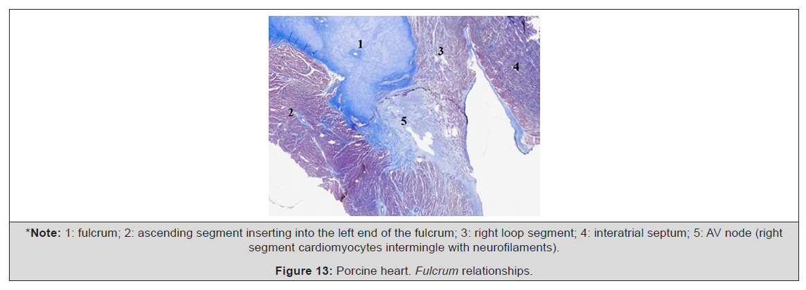
Figure 13:Porcine heart. Fulcrum relationships
To faithfully establish the identity of the cardiac fulcrum, a histological analysis has also been carried out on the trigones, trying to find cardiomyocytes as a probable cardiac muscle insertion in these structures. In our investigation, only collagenous tissue without cardiomyocytes was observed in the trigones, confirming that the fulcrum is the support of the myocardium, both at its origin and end.
Cardiac Fulcrum Images
Bovine hearts, studied with computed tomography (Figure 14,15), magnetic resonance imaging (Figure 16, 17) and x-rays (Figure 18) showed the osteochondral structure found in the dissections, evidencing the same morphology and analogous size. In computed tomography in humans, the analysis of the region where the cardiac fulcrum is located, through the dissections performed, revealed the presence of an intensity above 110HU (Hounsfield units), while the adjacent muscle has units below 80HU. Thus, in the image, the fulcrum structure reached an average of 132±4.5HU. In the adjacent areas, corresponding to the myocardial muscle, this value was between 47.96±12.5 and 77.59±21.64HU (Figure 19). Through echocardiography, recent and as yet unpublished works reveal the visualization of the cardiac fulcrum using the septum, the right coronary valve and the tricuspid septal valve as references for its location (Figure 20). The favoured views have been: modified 3-chamber parasternal long axis, modified apical 3-chamber and intermediate short axes parasternal and subxiphoid (between large vessels and short axis at the level of the AV valves). The intermediate apical 2-chamber view (between 4-chamber and pure 2-chamber) allows longitudinal insonation of the fulcrum. For their part, Sosa Olavarría, et al., [15] found the cardiac fulcrum using echocardiography, which they published under the title “Trainini’s cardiac fulcrum in the fetal heart” (2023). They studied 50 human pregnancies with fetuses between 18 and 37 weeks of gestation. Fetal cardiac ultrasound obtained 2D, Doppler, colour and three-dimensional, STIC, HD Flow and speckle tracking modalities (Figure 14-20).
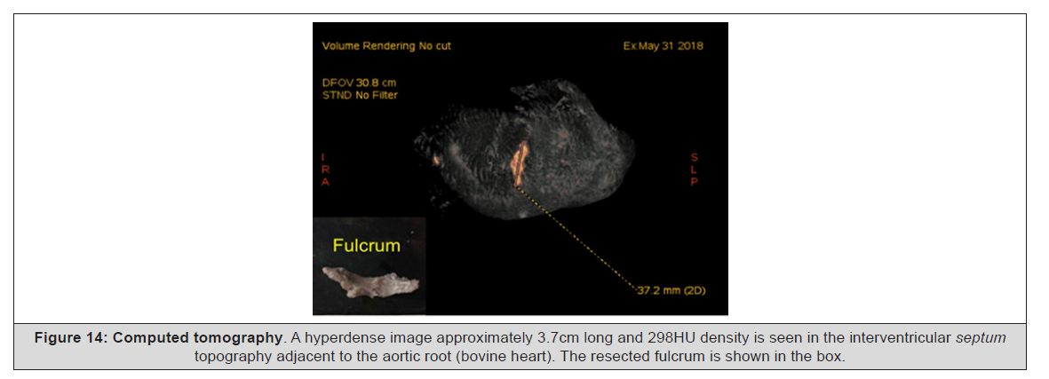
Figure 14:Computed tomography. A hyperdense image approximately 3.7cm long and 298HU density is seen in the interventricular septum topography adjacent to the aortic root (bovine heart). The resected fulcrum is shown in the box.
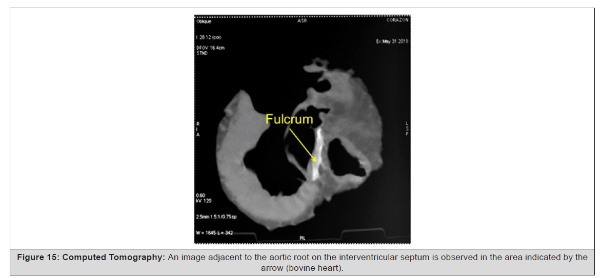
Figure 15:Computed Tomography: An image adjacent to the aortic root on the interventricular septum is observed in the area indicated by the arrow (bovine heart).
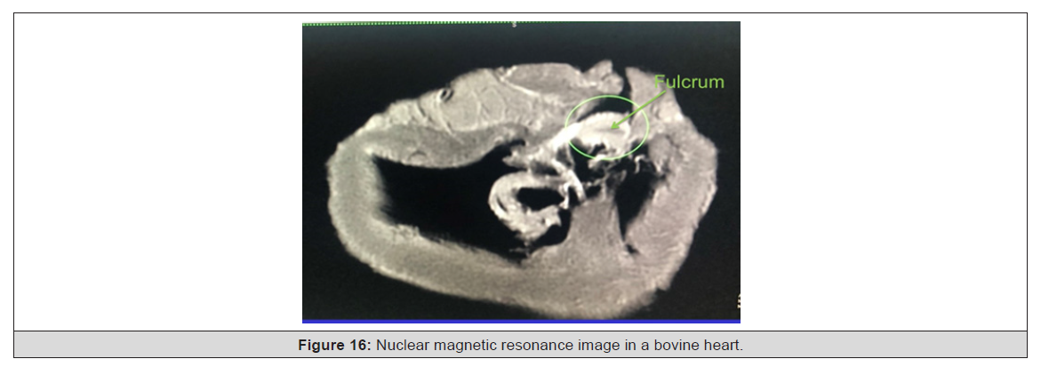
Figure 16:Nuclear magnetic resonance image in a bovine heart.
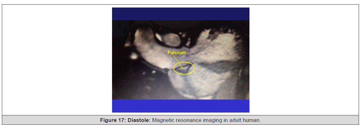
Figure 17:Diastole: Magnetic resonance imaging in adult human.
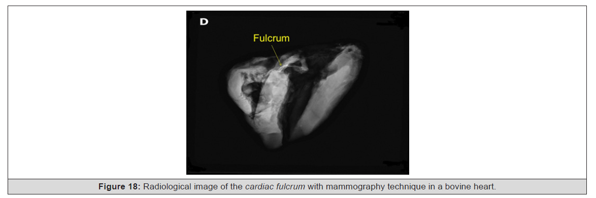
Figure 18:Radiological image of the cardiac fulcrum with mammography technique in a bovine heart
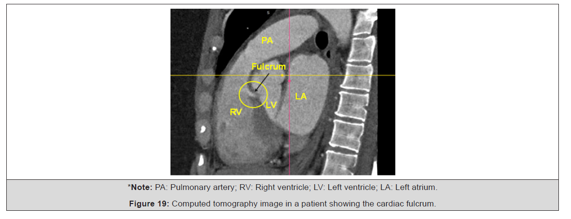
Figure 19:Computed tomography image in a patient showing the cardiac fulcrum.
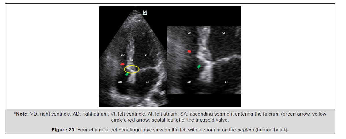
Figure 20:Four-chamber echocardiographic view on the left with a zoom in on the septum (human heart).
Discussion
The studies on the anatomy and histology of the myocardium carried out in recent years in our research [3,16-19] provide evidence that myocardial fibers constitute a continuous muscle that describes a double helix to form both ventricles and that, to fulfill its muscular function, it needs a support point that we have investigated, discovered, and called the cardiac fulcrum. Thus, the myocardium has the following characteristics derived from the anatomical and histological analysis performed:
1) It is a single, continuous and coiled muscle that forms a helix
with two spirals.
2) The myocardium is attached at its origin and end, as any muscle,
to a support that we have described and called the cardiac
fulcrum. The muscle fibers surrounding the atrioventricular
rings have no insertion into them.
3) The spatial helical arrangement forces the muscle to overlap
segments in its spatial configuration.
4) This anatomical condition has marked correspondence with
myocardial movements and with the stimulation that runs
throughout its segments.
5) The transverse interconnections between the muscle tracts do
not invalidate the concept of continuous myocardium, since
this compact arrangement is understood as the result of the
evolutionary development to obtain solidity of its structure in
strict relationship with function.
6) The fulcrum is contiguous to the AV node, which with its specialized
fibers surrounds and invades it.
7) In all the investigated hearts, we have found hyaluronic acid
in the cleavage planes between the myocardial bundles associated
with Thebesian and Langer venous conduits (Figure 21).
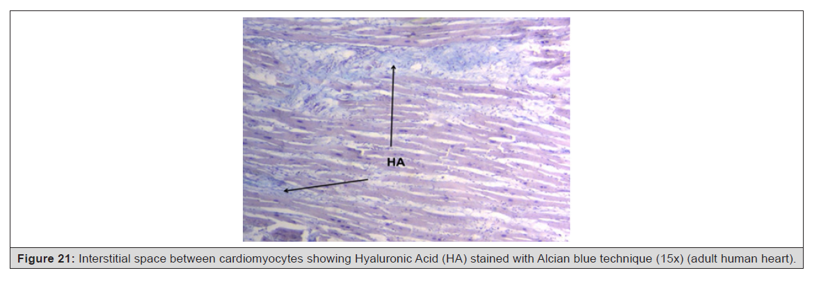
Figure 21:Interstitial space between cardiomyocytes showing Hyaluronic Acid (HA) stained with Alcian blue technique (15x) (adult human heart).
Therefore, the myocardium can be defined as a single muscle that in its longitudinal continuity adopts a spiral spatial conformation, inserted at its ends (origin and end) in an osteochondral-tendinous nucleus according to the specimens analyzed, called the cardiac fulcrum. This arrangement limits the two ventricular chambers [20]. The myocardium as a single, helical-coiled muscle is not accurately represented by the word “band,” a term that has generated discussion from an anatomical strictly academic point of view. The concept of “band” does not correspond to the etymology of the word and to the full individuality of its spiraling pathway where it is forced to superimpose the segments. In contrast to the band concept, some authors have proposed the concept of myocardial fiber arrangement as a mesh [3]. This term is not acceptable either, since this mesh structure is not related to the functional anatomical organization of the heart, as analyzed in this section. There are solid criteria that support the concept of cardiac myocardium continuity as a single, continuous and spiraling muscle:
1) Muscle homogenization conceals the real spiral continuity
of the fibers when its segments are overlapped. This implies
considering that its structural solidity is required in birds and
mammals to ensure that blood is ejected at a high velocity in
a limited time span, through an organ that must supply two
circulations (systemic and pulmonary). The anatomical investigation
of the heart through adequate dissection, histological
exploration, image analysis obtained through radiological and
echocardiographic studies, electrophysiological studies carried
out with 3D-EAM and diffusion tensor cardiac magnetic
resonance imaging show the continuous muscular pathway
that define the two ventricles.
2) Being able to unfold the myocardium and obtain a similar
thickness throughout the muscle length shows that its continuity
is real. When coiled, the thickness of the right ventricle is
less than that of the left ventricle, since the former is composed
of a single segment (right), while the latter presents attachment
two segments (descendent and ascendent).
3) Its function leads it to have a supporting point as any skeletal
muscle, both at its origin and end. If the myocardium did not
have this helical spatial anatomical conformation, with an insertion
at both ends in the cardiac base remaining free at the
apex, that is, pendant in the thorax; and if it did not present a
stimulation allowing torsion and detorsion, it would be unable
to fulfill its extraordinary muscular power. Echocardiography
with speckle-tracking techniques has demonstrated shortening
and lengthening movements during the systolic and suction
phases, respectively [21-24]. To calculate twist, the ultrasound
system algorithm performs an algebraic subtraction (it
adds the value of the positive apical rotation to the negative
basal rotation). Our experience in normal subjects shows that
this is around +19±9 degrees, always with a predominant apical
rotation [25,26].
4) The trigones do not show cardiomyocyte insertion, confirming
that the only myocardial attachment is the fulcrum.
5) Myocardial dissection, histological analysis, and cardiac function
do not correlate with a mesh conformation.
6) The contiguity of the cardiac fulcrum with the AV node, surrounded
by a rich plexus of neurofilaments, makes us consider
an anatomical electromechanical unit where stimulation energy
and muscle mechanics participate.
In this investigation, fresh bovine, porcine and human hearts were used to obtain detailed descriptions in order to elucidate the true spatial myocardial architecture.
Conclusion
To conclude, as previously reported, we find that the orientation and opposite rotational movement of the heart fibers, both at the base and at the apex, justify the continuous helical myocardium model. However, the question that arises from the logic of movement is that, to achieve torsion and the consequent detorsion, the muscle segments that in their continuity make up the ventricular chambers should do so on a supporting point as a skeletal muscle does on a firm insertion. It was found in our research and called the cardiac fulcrum. With the heart folded, we find the fulcrum embraced by the pulmotricuspid cord and the pulmonary artery on the right side of the aorta. The right ventricle is positioned anteriorly, so the fulcrum shows how the bundles emerging from it, when frontal, conceal the attachment of the ascending segment to said structure located below this view. When the pulmonary artery and the pulmotricuspid cord begin to unfold, the insertion of said ascending segment into the fulcrum is evident. The insertion of the myocardial fibers in the fibrous skeleton of the heart has been considered for three centuries. The development of the anatomical research reported in 1970 by Torrent Guasp [27-30] indicated that the myocardium originates and ends at the root of the great vessels, but that the fibers do not insert into the atrioventricular annuli, but merely attach to them, as confirmed by the images obtained by means of diffusion tensor magnetic resonance imaging.
In our anatomical investigations we found the nucleus called cardiac fulcrum, where the helical myocardium is inserted at both ends. Conversely, we have not found insertion of the cardiomyocytes in the collagenous matrix of the trigones; which confirms this finding. Our investigations have demonstrated that in the path of the aortic annulus septal segment, extending from the left to the right trigone, there is a solid structure that we have called cardiac fulcrum (below the origin of the right coronary artery) where the continuous myocardium is attached at its origin and end, since, as any muscle, it needs a supporting point to fulfill its function. Furthermore, we have not found any insertion of cardiomyocytes into the collagenous matrix of the trigones, a finding confirmed by histological studies. In all studies of animal and human hearts, the location of the fulcrum has been found to be contiguous but distinct from the classic fibrous core. The right and left trigones occupy the non-coronary sinus, the posterior half of the left coronary sinus, and the posterolateral part of the right coronary sinus. The fulcrum is located anteriorly, below the right coronary artery.
In our research, we always considered that there must be a fixation within the myocardium that would allow it to rotate in a helix to perform its fundamental movements and muscular power of shortening-twisting and lengthening-detwisting. This situation of investigating a continuous myocardial support correlates with an organic machine, such as the heart, which, without a solid anchor to a resistant nucleus, would lack the mechanical faculties necessary for its considerable power. This fixation point involves, as in any muscle, exercising the function of supporting the muscular lever and also allows it to act as a bearing or cushion, preventing the force of ventricular rotation, whether due to torque or torsional stress, from being transferred to the aorta. This way, it manages to dissipate the energy produced by the movement of the muscular helix and preventing the artery from becoming strangled or kinked during the systolic ejection period [2,18]. In the human hearts studied, the findings are surprising from the interpretation point of view, based on the fact that it is logical to consider its presence throughout the evolutionary chain of mammals. It should be considered that this structure, when analyzed in different specimens, has in common its function of supporting the helical myocardium to be able to generate the power required by any muscle, which is different in different mammals. Therefore, its presence is constant in all the hearts studied, both bovine and human, but its structural characteristics are distinct. And this difference in the intimate analysis of the cardiac fulcrum is undoubtedly related to the resistance it must oppose to the energetic action of the myocardium in hearts of different sizes.
It should be noted that what makes the concept of the fulcrum (support) important is the interweaving of the myocardial fibers and the chondroma. It is this functional element that gives value to this structure that supports and stabilizes myocardial movements. The fulcrum should not be considered a nucleus with sharp, rigid edges. The insertion of the myocardium into a structure with such characteristics would be inconvenient, as it would generate sudden tension at that point upon movement, causing tears at the insertion, given the force exerted by the myocardial band to eject the ventricular contents. Furthermore, its consistency decreases between bovines and humans, given that the force exerted is different depending on body weight. What we have found both in macroscopy and in the histology of bovine and human hearts (pregnant, pediatric, and adult) is a structure that grows in density toward a more solid center. In this scaling from lesser to greater density of the fulcrum, the fibers are inserted just as in a tendon matrix (equivalent to the insertion of skeletal muscle tendons into bone). This should be understood as a need to dissipate energy gradually with the least possible traction (bearing mechanism), avoiding a sudden and repetitive action-reaction principle, and also preventing the aorta from being dragged in the helical movement performed by the band.
We believe that a structure is only as good as its function. The fulcrum’s function as a support for the myocardium is important. Without this insertion, it is impossible to conjecture the movements and energy of the myocardial band to sustain the necessary circulatory physiology. Finally, regarding this important topic, we would like to transcribe what Best Adam, et al., [1] published in 2022 in “Anatomy, Histology, Embryology”, supporting the priority of our finding regarding the cardiac fulcrum:
“In human cardiac anatomy, in addition to trigones and atrioventricular rings, the heart has a cardiac fulcrum (Figure 1). Works by Trainini and coauthors elucidated the value of this structure in humans and proposed its function and importance once they had observed the attachment of the continuous myocardium to the fulcrum and naming the structure the ‘cardiac fulcrum’ [5,6]. They proposed that the fulcrum, a thickening at the base of the aorta made up of a collagen matrix, is essential for anchoring the myocardial band allowing the ban to contract and relax maintaining efficient cardiovascular blood flow” [5,6].
“Anatomy, histology, developmental functions of Ossa cordis. A review”.
Best Adam et al. University of Nottingham, UK; University of Tirol, Austria, University Colleague of London, UK; University of Montevideo, Uruguay. Anat, Histol, Embryol 2022; 00:1.13
Acknowledgment
None.
Conflict of Interest
None.
References
- Best A, Egerbacher M, Swaine S, Pérez W, Alibhai A, et al. (2022) Anatomy, histology, development and functions of Ossa cordis: A review. Anat Histol Embryol 51(6): 683-695.
- Trainini JC, Lowenstein J, Beraudo M, Wernicke M, Trainini A, et al. (2021) Myocardial torsion and cardiac fulcrum (Torsion myocardique et pivot cardiaque). Morphologie 105: 15-23.
- Trainini J, Lowenstein J, Beraudo M, Valle Cabezas J, Wernicke M, et al. (2023) “Anatomy and organization of the helical heart”. Undav Ed., Buenos Aires.
- Trainini J, Beraudo M, Wernicke M, Trainini A, Lowenstein J, et al. (2021) Cardiac Fulcrum. Cardiovasc Surg Int 2(1): 1011.
- Trainini JC, Beraudo M, Wernicke M, Trainini A, Haber Lowenstein D, et al. (2021) The myocardial support. Rev Argent Cardiol 89: 217-223.
- Trainini JC, Beraudo M, Wernicke M, Carreras Costa F, Trainini A, et al. (2022) Evidence that the myocardium is a continuous helical muscle with one insertion. REC: CardioClinics 57(3): 194-202.
- Trainini J, Beraudo M, Wernicke M, Herrero E, Trainini A (2024) The Cardiac Fulcrum Comments. International Journal of Cardiovascular Medicine 3(5).
- Trainini J, Lowenstein J, Beraudo M, Wernicke M, Mora Llabata V, et al. (2022) Cardiac Helical Function. Fulcrum and Torsion. Japan Journal of Clinical & Medical Research.
- Trainini J, Wernicke M, Beraudo M, Trainini A (2024) The fulcrum of the human heart (Cardiac fulcrum). World Journal of Biology Pharmacy and Health Sciences 17(01): 049–056.
- Trainini Jorge, Beraudo Mario, Wernicke Mario, Efraín Herrero, Trainini Alejandro (2024) The Cardiac Fulcrum-Comments. AJ Biomed Sci & Res 23(4): 420-425.
- Karamchandani JR, Nielsen TO, van de Rijn M, West RB (2012) Sox10 and S100 in the diagnosis of soft-tissue neoplasms. Appl Immunohistochem Mol Morphol 20(5): 445-450.
- Trainini J, Beraudo M, Wernicke M, Carreras Costa F, Trainini A, et al. (2023) The hyaluronic acid in intramyocardial sliding. REC: CardioClinics 58(2): 106-111.
- Trainini J, Wernicke M, Beraudo M, Trainini A, Valle Cabezas J, et al. (2025) Discovery of hyaluronic acid in the heart as a lubricating mechanism. Am J Biomed Sci & Res 27(6): 1012-1018.
- Moittié S, Baiker K, Strong V, Cousins E, White K, et al. (2020) Discovery of os cordis in the cardiac skeleton of chimpanzees (Pan troglodytes). Sci Rep 10(1): 9417.
- Sosa Olavarría A, Martí Peña A, Martínez A, Zambrana Camacho J, Ulloa Virgen J, et al. (2023) Fulcro cardíaco de Trainini en el corazón fetal. Rev Peru Ginecol Obstet 69(4): 1-8.
- Trainini JC, Herreros J, Elencwajg B, López-Cabanillas N, Lago N, et al. (2015) “Fundamentos de la Nueva Mecánica Cardí La Bomba de Succión”. Rev Argent Cardiol 84: 73
- Trainini JC, Lowenstein J, Beraudo M, Trainini A, Mora Llabata V, et al. (2019) “Myocardial Torsion”. Ed Biblos, Buenos Aires; Argentina.
- Trainini JC, Mora V, Lowenstein J, Beraudo M, Wernicke M, et al. (2020) La teoria de la banda miocárdica Nuevos descubrimientos que apoyan el complejo mecanismo de torsión miocá Revista de Ecocardiografía Práctica y Otras Técnicas de Imagen Cardíaca 3: 14-18.
- Trainini J, Lowenstein J, Beraudo M, Mora Llabata V, Carreras-Costa F, et al. (2022) “El corazón helicoidal. Fulcro y Torsion”. UNDAV Ed, Buenos Aires; Argentina.
- Trainini J, Lowenstein J, Elencwajg B, Beraudo M, Trainini A, et al. (2025) Fulcrum and Function of the Helical Heart: State of Research. Am J Biomed Sci & Res 27(2): 417-434.
- Beaumont A, Grace F, Richards J, Hough J, Oxborough D, et al. (2017) Left Ventricular Speckle Tracking-Derived Cardiac Strain and Cardiac Twist Mechanics in Athletes: A Systematic Review and Meta-Analysis of Controlled Studies. Sports Med 47(6): 1145-1170.
- Helle-Valle T, Crosby J, Edvardsen T, Lvseggen E, Amundsen BH, et al. (2005) New non-invasive method for assessment of left ventricular rotation: speckle tracking echocardiography. Circulation 112(20): 3149-3156.
- Maksuti E, Carlsson M, Arheden H, Kovács S, Broomé M, et al. (2017) Hydraulic forces contribute to left ventricular diastolic filling. Sci rep 7: 43505.
- Nakatani S (2011) Left ventricular rotation and twist: why should we learn? J Cardiovasc Ultrasound 19(1): 1-6.
- Mora Llabata V, Roldán Torresa I, Saurí Ortiza A, Fernández Galera R, Monteagudo Viana M, et al. (2016) Correspondence of myocardial strain with Torrent-Guasp’ s theory. Contributions of new echocardiographic parameters. Rev Arg de Cardiol 84(6): 541-549.
- Mora V, Roldán I Romero E, Saurí A, Romero D, Perez-Gozabo J, et al. (2018) Myocardial contraction during the diastolic isovolumetric period: analysis of longitudinal strain by means of speckle tracking echocardiography. J Cardiovasc Dev Dis 5(3): 41.
- Torrent Guasp F (1987) Nuevos conceptos sobre la estructura miocárdica ventricular. En Torrent Guasp F, editor. Estructura y mecánica del corazó Barcelona: Grass Ed, 35-97.
- Torrent Guasp F (1998) Estructura y función del corazó Rev Esp Cardiol 51(2): 91-102.
- Torrent Guasp F, Buckberg G, Carmine C, Cox J, Coghlan H, et al. (2001) The structure and function of the helical heart and its buttress wrapping. I. The normal macroscopic structure of the heart. Semin Thorac Cardiovasc Surg 13(4): 301-319.
- Trainini JC, Herreros J (2019) El explorador del corazó Biografía de Francisco Torrent Guasp. 87(2): 162-163.




 We use cookies to ensure you get the best experience on our website.