
Case Report 
 Creative Commons, CC-BY
IgG4-Related Bilateral Multiple Type III Renal Artery Aneurysms with Rupture: A Case Report
*Corresponding author:Ziyu Tang, Department of Interventional Medicine, Nanchong Central Hospital, China.
Received:October 13, 2025; Published:October 22, 2025
DOI: 10.34297/AJBSR.2025.28.003733
Abstract
Background: IgG4 - related disease (IgG4 - RD) is a systemic fibro - inflammatory disorder involving multiple organs. Renal aneurysms are life-threatening.
Only one case of IgG4-RD accompanied by a renal artery aneurysm has been reported, and this case was a single type II renal aneurysm. Here,
we report the first case of IgG4 - RD accompanied by bilateral multiple type III renal artery aneurysms with aneurysm rupture, which was relieved
after transcatheter arterial embolization and glucocorticoid treatment.
Case presentation: A 52-year-old Asian woman presented with restricted mouth opening and a palpable mass in the right parotid gland area.
During her hospitalization, she experienced abdominal pain and hemorrhagic shock. Multiple aneurysms were found in both renal arteries, and the
left renal aneurysm ruptured and bled. The patient underwent two transcatheter renal artery embolization surgeries due to bleeding from different
renal aneurysms at different times, and the hemostasis was successful. Through multidisciplinary consultation involving parotid gland pathology,
laboratory tests, and imaging examinations, the final diagnosis was igg4-rd accompanied by bilateral multiple renal aneurysms that had ruptured.
After treatment with glucocorticoids, the patient’s condition improved.
Conclusions: This case reports a rare case of IgG4-RD accompanied by rupture of multiple bilateral type III renal aneurysms. The patient achieved
remission after transcatheter renal artery embolization and glucocorticoid treatment.
Keywords:IgG4 - related disease, Renal artery aneurysms, Renal arterial embolization, Steroid treatment
Introduction
IgG4 - related disease (IgG4 - RD) is a systemic fibro - inflammatory disorder [1]. Characterized by elevated serum IgG4 levels, dense tissue infiltration by IgG4-positive plasma cells and fibrosis, it can affect multiple organ systems including the pancreas, bile ducts, salivary glands, lacrimal glands and lymph nodes [2-4]. Vascular involvement in IgG4-RD predominantly affects large and medium-sized arteries, such as the aorta and coronary arteries [5]. Only one prior report exists documenting IgG4-RD associated with a single Type II renal artery aneurysm [6]. Renal Artery Aneurysms (RAAs) represent potentially life-threatening vascular anomalies; their rupture carries an associated mortality rate of approximately 10% [7]. Here, we report a case of IgG4-RD accompanied by multiple type III renal aneurysms on both sides, with continuous rupture and hemorrhage of the left renal aneurysm. After transcatheter renal artery embolization and glucocorticoid treatment, the patient’s condition was relieved.
Case Presentation
A 52-year-old Asian female patient was admitted on December 23, 2024, due to restricted mouth opening for over two months. Physical examination revealed a 2×3 cm hard and poorly mobile mass in the right parotid region without redness or swelling. Laboratory tests showed normal blood routine, coagulation function, liver and kidney function, and tumor markers. Enhanced CT of the parotid gland showed a significantly enhanced and calcified mass (Figure 1). Partial parotidectomy was performed for biopsy due to negative fine-needle aspiration cytology. On December 30, 2024, the patient suddenly experienced severe pain in the left abdomen, accompanied by tachycardia, hypotension and other shock symptoms. Emergency chest and abdominal CT showed multiple patchy consolidations in both lungs (Figure 2), multiple nodular aneurysms in the distal segments of bilateral renal arteries, and a ruptured aneurysm in the lower pole of the left kidney with perirenal hematoma (Figure 3a). Urgent laboratory tests showed a hemoglobin level of 47 g/L. The patient received blood transfusion, anti-shock treatment and emergency transcatheter renal artery embolization. Intraoperative renal artery angiography revealed multiple nodular aneurysms in both kidneys (Figure 3b), with contrast extravasation in the lower pole aneurysm of the left kidney indicating rupture and bleeding (Figure 3c). Coils were used to embolize the responsible vessels of the ruptured aneurysm and another larger aneurysms, achieving complete occlusion of the target vessels (Figure 3d). Following embolization, the patient’s abdominal pain and fatigue improved with continuous fluid infusion and blood transfusion, vital signs gradually normalized, and hemoglobin levels increased to 80 g/L. On January 3, 2025, the patient experienced persistent severe left lower abdominal pain and shock again, with hemoglobin dropping to 37 g/L. Repeat enhanced abdominal CT showed an enlarged perirenal hematoma, extension of the left lower abdominal hematoma to the pelvis, significant enlargement and rupture of another aneurysm at the left renal hilum, and massive left pleural effusion (Figure 4A). Urgent transcatheter renal artery embolization was performed again (Figure 4B). Due to multiple renal artery aneurysms and consecutive ruptures of left renal artery aneurysms within a short period, the main trunk of the left renal artery was embolized during the procedure. The patient’s condition gradually improved following blood transfusion, anti-shock, and anti-infection treatments. On January 6, 2025, pathological examination of the parotid tissue revealed multiple hyaline nodules, abundant lymphoid cells, and lymphoid follicles. Immunohistochemistry showed CD138 positivity (plasma cells), IgG positivity (many cells), IgG4 positivity (approximately 25 cells per high-power field in hotspots), and an IgG4/IgG ratio <40% (Figure 5A), suggesting IgG4-related disease. Supplementary laboratory tests for connective tissue diseases, humoral immunity, and other autoimmune diseases showed a serum IgG4 level of 17.30 g/L (normal range <2.01 g/L), with other autoimmune markers (including antinuclear antibodies) within normal limits. Following multidisciplinary consultation involving the pathology, rheumatology, immunology, nephrology, and urology departments, the patient was finally diagnosed with bilateral multiple renal artery aneurysms with rupture caused by IgG4-RD. On January 9, 2025, the patient was initiated on methylprednisolone 30 mg/day for 4 weeks, followed by a weekly reduction of 5 mg until reaching a maintenance dose of 5 mg/day. At the 3-month follow- up, the parotid mass had significantly reduced, mouth opening returned to normal for eating, serum IgG4 was 3.07 g/L, renal function showed urea 24.02 mmol/L and creatinine 220.5 umol/L, and dialysis was not required.
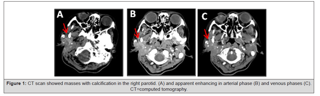
Figure 1:CT scan showed masses with calcification in the right parotid. (A) and apparent enhancing in arterial phase (B) and venous phases (C). CT=computed tomography.
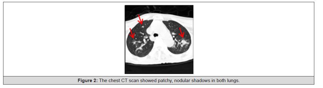
Figure 2:The chest CT scan showed patchy, nodular shadows in both lungs.
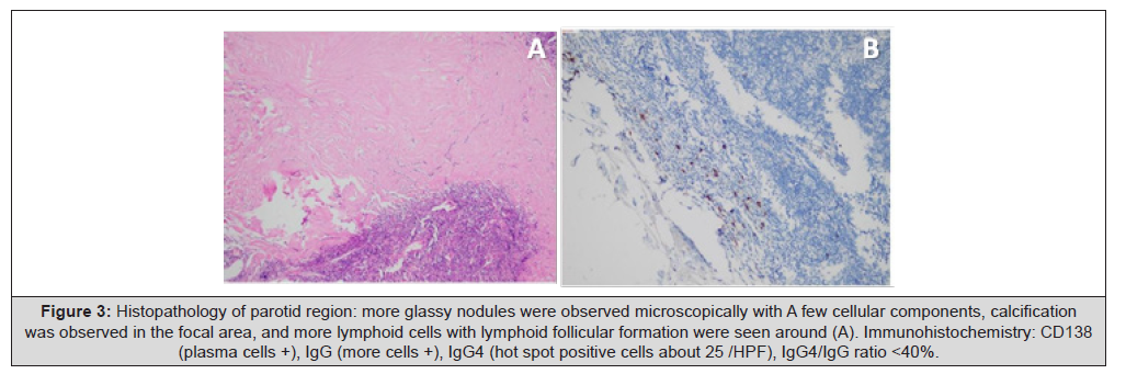
Figure 3:Histopathology of parotid region: more glassy nodules were observed microscopically with A few cellular components, calcification was observed in the focal area, and more lymphoid cells with lymphoid follicular formation were seen around (A). Immunohistochemistry: CD138 (plasma cells +), IgG (more cells +), IgG4 (hot spot positive cells about 25 /HPF), IgG4/IgG ratio <40%.
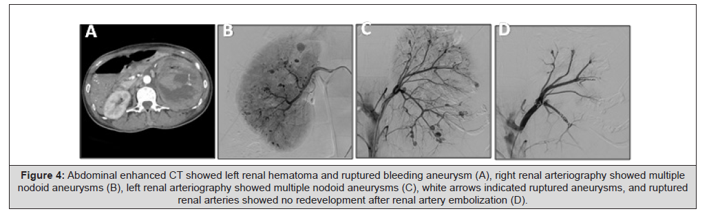
Figure 4:Abdominal enhanced CT showed left renal hematoma and ruptured bleeding aneurysm (A), right renal arteriography showed multiple nodoid aneurysms (B), left renal arteriography showed multiple nodoid aneurysms (C), white arrows indicated ruptured aneurysms, and ruptured renal arteries showed no redevelopment after renal artery embolization (D).
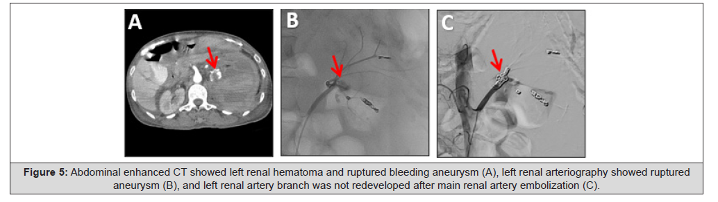
Figure 5:Abdominal enhanced CT showed left renal hematoma and ruptured bleeding aneurysm (A), left renal arteriography showed ruptured aneurysm (B), and left renal artery branch was not redeveloped after main renal artery embolization (C).
Discussion
IgG4-RD is an immune-mediated fibro-inflammatory disease that can affect various body systems [1], with an incidence of 0.28– 1.39 per 100,000 persons [8,9]. It predominantly involves organs such as the salivary glands, orbits, lacrimal glands, pancreas, biliary tract, lungs, kidneys, aorta, retroperitoneum, meninges, and thyroid [10,11]. IgG4-RD exhibits diverse manifestations, lacks clear diagnostic criteria, and overlaps with other diseases. Diagnosis requires integrating clinical, radiological, serological, and histopathological findings while excluding mimicking conditions. The diagnostic process involves three steps: (1) including patients with characteristic clinical and imaging manifestations of organ involvement or unexplained lymphoplasmacytic infiltration; (2) excluding patients with fever, no objective response to glucocorticoids, or specific autoimmune diseases; and (3)If the combined score of pathological examination, imaging examination and laboratory tests exceeds 20 points, it can be diagnosed as IgG4-related disease (IgG4-RD) [12]. In this case, the parotid mass was a typical manifestation of IgG4- RD. Normal tumor markers, biopsy results, and negative autoimmune, infectious disease tests ruled out tumors, infections, or other autoimmune diseases. The cumulative score of pathology, chest lesions, and laboratory tests was 32, confirming the diagnosis.
Renal artery aneurysms are defined as focal, isolated dilations of the three-layer arterial wall, with a diameter exceeding 1.5 times that of the adjacent normal proximal artery. They are rare and classified into three types: type I (saccular aneurysms of the renal aorta or proximal large branches, most common); type II (saccular or fusiform aneurysms of the renal aorta or proximal segmental branches); and type III (intralobar aneurysms originating from small segmental or accessory arteries). Type III aneurysms are primarily treated with embolization, using liquid embolic agents or coils for super selective embolization of renal segmental branches [11,13].
Vascular lesions in IgG4-RD mainly involve large and medium arteries, with rare renal artery involvement. The only previous report described a 3.2 cm type II renal artery aneurysm detected 12 years after IgG4-RD diagnosis, which enlarged to 4.4 cm after 1 year. Due to obstruction of the renal artery bifurcation, surgical repair was performed, with patent renal artery bypass at 2.5-year follow- up (IgG4 levels were not reported) [6]. This case differs in involving bilateral kidneys with multiple nodular type III aneurysms in the distal renal arteries. Recurrent left renal aneurysm ruptures precluded surgical resection or revascularization, leading to coil embolization with sacrifice of left renal function.
Steroid therapy is the cornerstone of IgG4-RD treatment [9]. but its use in IgG4-related arterial diseases is controversial [14]. No studies have evaluated steroid use after endovascular or surgical treatment for preventing new aneurysms. Previous reports of aortic or medium artery involvement described arterial wall thickening with severe perivascular fibrosis and late-phase contrast enhancement; steroids may weaken connective tissue, thinning the arterial wall [15]. In this case, steroid therapy was initiated after the second intervention due to rapid aneurysm enlargement and rupture, indicating active inflammation. Unlike previous cases of large/medium aneurysms, bilateral renal aneurysms in this patient lacked arterial wall thickening, consistent with peripheral aneurysms [14]. Therefore, steroid treatment did not make the aneurysm wall of this patient more fragile.
Follow-up for IgG4-RD primarily focuses on monitoring serum IgG4 concentration and assessing the function of affected organs. Serum IgG4 serves as an important biomarker for disease activity. Effective treatment typically induces a rapid decline in IgG4 levels, although normalization may not always occur. A rising titer may herald disease relapse. Organ-specific follow-up utilizes imaging (CT, MRI, ultrasound) and laboratory tests to evaluate structural and functional changes [1,4,16]. In this patient, post-treatment serum IgG4 decreased significantly and remained lower than baseline levels. Clinically, the parotid mass regressed and mouth opening normalized, indicating a positive response to steroid therapy. Nevertheless, long-term follow-up remains essential to promptly detect potential disease recurrence.
Conclusion
This is the first reported case of rupture of bilateral multiple renal aneurysms associated with IgG4-RD. This case enriches the understanding of IgG4-RD, especially its rare association with renal aneurysms. It also emphasizes the importance of multidisciplinary collaboration in the diagnosis and treatment of complex diseases such as IgG4-RD. The experience of using steroids for treatment in this case provides new insights into the management of IGG4-related arterial disease, although it remains controversial. To better understand the relationship between IgG4-RD and renal aneurysms and optimize treatment strategies, further research is needed.
Declarations
Ethics Approval and Consent to Participate
The patient’s treatment and data collection were conducted in accordance with the ethical standards of the institutional and national research committee. Ethical approval was obtained from Ethics Committee of Nanchong Central Hospital, and the patient provided written informed consent for participation in the study. This consent covered all aspects of the diagnostic and treatment procedures, as well as the use of her medical data for research purposes.
Consent for Publication
Written informed consent was obtained from the patient for publication of this case report and any accompanying images. A copy of the written consent is available for review by the Editor- in-Chief of this journal.
Availability of Data and Material
The data and materials used in this study are available upon reasonable request. The patient’s medical records, imaging studies, and laboratory results are stored securely at Nanchong Central Hospital. Interested researchers can contact the corresponding author to request access to these data, subject to the institution’s data access policies and relevant ethical regulations.
Competing Interests
The authors declare that they have no competing interests. There are no financial, commercial, or personal relationships that could potentially influence the objectivity or integrity of this case report. All authors have adhered to the highest standards of academic and professional conduct.
Funding
This research did not receive any specific grant from funding agencies in the public, commercial, or not - for - profit sectors. The study was conducted with the resources available within the institution, and no external funding was involved.
Authors’ Contributions
Ziyu Tang, Yunguo Liao, Jiaqi Pu contributed to the patient’s clinical management, data collection, and drafting of the initial manuscript. Hong Hu and Xin Wei supervised the overall study, reviewed the manuscript, and provided critical feedback. All authors have read and approved the final version of the manuscript.
Acknowledgements
The authors would like to thank the patient for her cooperation and participation in this study. We also express our gratitude to the medical staff at Nanchong Central Hospital who were involved in the patient’s care. Their dedication and expertise were crucial in the successful treatment of the patient and the compilation of this case report. Additionally, we acknowledge the support of the institutional research facilities that facilitated the data collection and analysis.
References
- Stone JH, Zen Y, Deshpande V (2012) IgG4-related disease. N Engl J Med 366(6): 539-551.
- Umehara H, Okazaki K, Masaki Y, Mitsuhiro Kawano, Motohisa Yamamoto, et al. (2012) Comprehensive diagnostic criteria for IgG4-related disease (IgG4-RD), 2011. Mod Rheumatol 22(1): 21-30.
- Deshpande V, Zen Y, Chan JK, Eunhee E Yi, Yasuharu Sato, et al. (2012) Consensus statement on the pathology of IgG4-related disease. Mod Pathol 25(9): 1181-1192.
- Glass LR, Freitag SK (2015) Management of orbital IgG4-related disease. Curr Opin Ophthalmol 26(6): 491-497.
- Coleman DM, Stanley JC (2015) Renal artery aneurysms. J Vasc Surg 62(3): 779-785.
- Meadors S, Modrall JG, Timaran CH, Malekpour F (2021) Case Report of a Renal Artery Aneurysm Due to IgG4-Related Disease. Ann Vasc Surg 73: 515-520.
- Ghosh S, Dutta SK (2021) Endovascular interventions in management of renal artery aneurysm. Br J Radiol 94(1124): 20201151.
- Uchida K, Masamune A, Shimosegawa T, Okazaki K (2012) Prevalence of IgG4-Related Disease in Japan Based on Nationwide Survey in 2009. Int J Rheumatol 2012: 358371.
- Wallace ZS, Miles G, Smolkina E, Natalia Petruski Ivleva, Duane Madziva, et al. (2023) Incidence, prevalence and mortality of IgG4-related disease in the USA: a claims-based analysis of commercially insured adults. Ann Rheum Dis 82(7): 957-962.
- Katz G, Stone JH (2022) Clinical Perspectives on IgG4-Related Disease and Its Classification. Annu Rev Med 73: 545-562.
- Wallace ZS, Deshpande V, Mattoo H, Vinay S Mahajan, Maria Kulikova, et al. (2015) IgG4-Related Disease: Clinical and Laboratory Features in One Hundred Twenty-Five Patients. Arthritis Rheumatol 67(9): 2466-2475.
- Wallace ZS, Naden RP, Chari S, Hyon K Choi, Emanuel Della Torre, et al. (2020) The 2019 American College of Rheumatology/European League Against Rheumatism classification criteria for IgG4-related disease. Ann Rheum Dis 79(1): 77-87.
- Rundback JH, Rizvi A, Rozenblit GN, M Poplausky, S Maddineni, et al. (2000) Percutaneous stent-graft management of renal artery aneurysms. J Vasc Interv Radiol 11(9): 1189-1193.
- Kasashima F, Kawakami K, Matsumoto Y, Endo M, Kasashima S, et al. (2018) IgG4-Related Arterial Disease. Ann Vasc Dis 11(1): 72-77.
- Kasa K, Ohki T, Ito E, Fukasawa N, Shukuzawa K, et al. (2024) Immunoglobulin G4-related hepatic artery aneurysm. J Vasc Surg Cases Innov Tech 10(1): 101377.
- Kamisawa T, Zen Y, Pillai S, Stone JH (2015) IgG4-related disease. Lancet 385(9976): 1460-1471.




 We use cookies to ensure you get the best experience on our website.