
Research Article 
 Creative Commons, CC-BY
Intravitreal Bevacizumab Injection Combined with Ex-Press Mini Glaucoma Shunt Implantation in The Treatment of Neovascular Glaucoma: 2 Year Experience
*Corresponding author:Avtandil Gagnidze, East-West University, Tbilisi, Georgia.
Received:October 13, 2025; Published:October 17, 2025
DOI: 10.34297/AJBSR.2025.28.003729
Summary
Aims and Methods: Neovascular Glaucoma (NVG) remains one of the most challenging ophthalmic diseases due to its ischemic pathogenesis. Surgical
treatment alone is often inadequate; thus, combining anti-VEGF therapy with surgery enhances outcomes. This prospective, non-randomized
study evaluated the effectiveness of dual (pre- and post-operative) Intravitreal Bevacizumab (IVB) injections combined with Ex-Press Glaucoma Mini
Shunt implantation in 36 patients (42 eyes) with NVG. The study group received IVB two weeks before and after surgery, while the control group
received a single intraoperative injection. Parameters compared included surgical success, intraocular pressure (IOP), visual acuity, anti-glaucoma
medication use, and retinal nerve fiber layer (RNFL) thickness.
Results and Conclusion: Surgical success was higher in the dual-injection group (96.8% vs. 72.7% at 12 months; 93.5% vs. 81.8% at 24 months).
Mean IOP significantly decreased in the injection group (baseline 35.4±4.9mmHg to 18.3±1.7mmHg at 24 months) compared with the control group
(baseline 36.5±5.8mmHg to 22.0±8.0mmHg). Postoperative medication use was also markedly reduced in the injection group. No significant differences
were found in visual acuity or RNFL thickness between groups at 12 or 24 months. Dual IVB administration before and after Ex-Press shunt
implantation proved to be an effective and safe NVG management strategy. The approach minimized intraoperative complications and maintained
stable IOP and ocular function over two years.
Keywords:Neovascular glaucoma, Bevacizumab, Anti-VEGF, Ex-Press Mini Shunt, Intraocular pressure, Glaucoma surgery, Intravitreal injection, Ocular ischemia
Introduction and Background
Neovascular Glaucoma (NVG) results from retinal ischemia, which triggers overexpression of Vascular Endothelial Growth Factor (VEGF) and other angiogenic mediators. These diffuse into the anterior segment, inducing abnormal vessel growth on the iris and trabecular meshwork. NVG progresses through three phases: rubeosis iridis (new iris vessels, open angle), secondary open-angle glaucoma (fibrovascular membrane obstructing outflow), and secondary angle-closure glaucoma (membrane contraction and synechial closure).
Effective management targets three priorities: eliminating ischemia, controlling IOP, and reducing inflammation and pain. Anti-VEGF agents (bevacizumab, ranibizumab, aflibercept) have revolutionized early treatment by inducing rapid regression of neovascularization and facilitating Panretinal Photocoagulation (PRP). However, their benefit is temporary without subsequent PRP. IOP-lowering drugs, including beta-blockers and carbonic anhydrase inhibitors, provide short-term relief, while steroids and cycloplegics control inflammation and discomfort.
PRP remains the gold-standard definitive therapy, destroying ischemic retina and reducing VEGF production. When the view is obscured, anti-VEGF injections, Pars Plana Vitrectomy (PPV) with endolaser, or Cyclophotocoagulation (CPC) may be necessary
If IOP remains uncontrolled, Glaucoma Drainage Devices (GDDs) offer the best surgical outcome for eyes with vision. Trabeculectomy is less favored due to fibrosis, while CPC is reserved for painful or blind eyes.
Modern NVG management relies on early recognition, timely anti-VEGF and PRP, and a staged, individualized approach. This strategy has transformed NVG from a once hopeless condition into a controllable, sight-preserving disease.
Neovascular Glaucoma (NVG) is a form of secondary glaucoma characterized by new vessels on the iris and angle of the Anterior Chamber (AC). It is usually associated with a poor visual prognosis [1,2]. The mechanism of anterior segment neovascularization is ischemia of the posterior segment of the eye resulting from a number of ophthalmic and systemic etiologies. The main causes include Diabetic Retinopathy (DR), Retinal Vein Occlusion (RVO), Retinal Artery Occlusion (RAO) and ocular ischemic syndrome [3]. Trabeculectomy, the most common type of glaucoma filtration surgery, is considered the mainstay of incisional glaucoma surgeries [4]. However, this technique is still associated with some postoperative complications, including hyphema, Vitreous Hemorrhage (VH), choroidal detachment, transient bleb leak, and endophthalmitis [5]. In recent years, glaucoma drainage devices have gained popularity in the surgical treatment of NVG because their success is thought to be less dependent on control of intraocular inflammation and the failure of a filtering bleb [6]. Of the various filtration surgeries, the Ex- PRESS® device is unique in terms of being short and plateless and being made of stainless steel. Its surgical procedure is more similar to trabeculectomy, involving a filtering bleb around the scleral flap. The wound healing process and surgical results of Ex-PRESS® mini shunt surgery have been comparable to those of trabeculectomy in randomized controlled trials, mainly involving Primary Open Angle Glaucoma (POAG) patients [7]. However, the efficacy and safety of Ex-PRESS® mini shunt surgery for other types of glaucoma, Including Neovascular Glaucoma (NVG), remain unclear. A study found that Vascular Endothelial Growth Factor (VEGF) is a key factor causing NVG, as demonstrated by significantly higher VEGF levels in the aqueous humor of NVG patients [8]. Because of their role in inhibiting intraocular neovascularization and mitigating damage to the blood ocular barrier due to leakage from new vessels. Inhibition of VEGF-A by bevacizumab has been shown to be successful in causing short-term regression of retinal neovascularization. Intravitreal injection of this agent has demonstrated rapid regression of Neovascularization of the Iris (NVI). Small case series have shown regression of NVI for 4-10 weeks after a single intravitreal injection of bevacizumab [9].
Recently, anti-VEGF factors have been used alone or in combination for the treatment of neovascular glaucoma. This study was to determine the efficacy of intravitreal bevacizumab combined with Ex-Press Mini Glaucoma Shunt implantation for the treatment of neovascular glaucoma.
Methods
This prospective, non-randomized, open-label, controlled clinical study was conducted at the Department of Ophthalmology, New Vision University Hospital, from December 2019 to November 2021. It included patients diagnosed with Neovascular Glaucoma (NVG), characterized by active neovascularization of the iris and/ or the anterior chamber angle, and elevated intraocular pressure (IOP >21mmHg) associated with ischemic retinal conditions. Eligible participants were aged between 18 and 85 years and had either received Pan-Retinal Photocoagulation (PRP) or showed resistance to medical treatment. All participants provided informed consent after being fully briefed on the treatment options.
Patients self-selected into one of two groups based on personal preference after receiving detailed information regarding both treatment protocols, including efficacy, risks, and costs.
In the intervention group, patients received an intravitreal injection of bevacizumab (1.25mg/0.05mL) 14 days prior to Ex-Press Mini Glaucoma Shunt surgery. Injections were performed under topical anesthesia (tetracaine), with the injection site located 3-3.5 mm from the limbus. Post-injection compression was applied using a cotton swab for 5-10 seconds, followed by monitoring of IOP and light perception. Patients returned for evaluation two days post-injection.
In the control group, bevacizumab was injected directly into the anterior chamber during the Ex-Press shunt implantation surgery.
The surgical technique was consistent across both groups. Procedures were carried out under topical tetracaine anesthesia, occasionally supplemented with retrobulbar anesthesia based on pain assessment. A conjunctival incision was made from the 11 to 1 o’clock position, forming a rectangular flap directed toward the superior fornix. A partial-thickness 4x4 mm scleral flap was created toward the limbus. Mitomycin-C (0.04%) was applied for two minutes to reduce fibrosis. A paracentesis was made at the 10 o’clock meridian, and viscoelastic material was injected into the anterior chamber to improve visualization. A 25-gauge needle was used to enter the anterior chamber parallel to the iris, and the Ex-Press shunt was inserted using an injector. The scleral flap was sutured with 8-0 nylon, and the conjunctiva was closed with 9-0 nylon. A subconjunctival injection of antibiotics and steroids concluded the procedure.
Preoperatively, all patients used Levofloxacin eye drops. Postoperatively, a tapering regimen of Levofloxacin and Dexamethasone was administered (six times daily for one week, then five times daily the second week). Antiglaucoma medications were prescribed as needed.
Baseline IOP was defined as the average of three preoperative ambulatory measurements. Follow-up visits occurred at designated intervals: 1-3 days, 2 weeks ±1 day, 1 month ±3 days, 3 months ±5 days, 6 months ±7 days, 9 months ±14 days, 12 months ±14 days, 18 months ±14 days, and 24 months ±14 days post-surgery. Evaluations included Best Corrected Visual Acuity (BCVA), IOP measurements, gonioscopy, ultrasound biomicroscopy, computer perimetry, and retinal nerve fiber layer analysis.
Outcome measures included IOP, BCVA, surgical success rate, use of antiglaucoma medications, and postoperative complications. Surgical success was defined as maintaining IOP between 6 and 21 mmHg, with or without antiglaucoma medication, and without severe complications or need for reoperation. Surgical failure was defined as IOP persistently outside this range for over two weeks, loss of light perception, or development of serious complications such as endophthalmitis, malignant glaucoma, or displacement/exposure of the drainage implant.
Results
36 neovascular glaucoma patients (42 eyes) were assigned to receive either 1.25mg/0.05ml intravitreal bevacizumab for 14 days before Ex-Press Mini Glaucoma Shunt implantation and repeated IVB injection 14 days after surgery (injection group) or Ex-Press Mini Glaucoma Shunt implantation with intraoperative intracameral bevacizumab injection (control group). Baseline data for the two groups are shown in Table 1, with no notable differences observed in initial clinical parameters (Table 1).
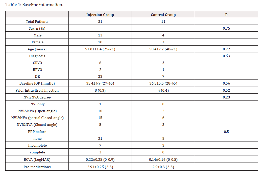
Table 1:Baseline information.
Of the 36 patients enrolled, 33 completed the 2-year follow-up period, while the remaining 3 were followed for 1 year. Specifically, one patient from the injection group and two patients from the control group completed only the 1-year follow-up.
A reduction in Intraocular Pressure (IOP) was observed in both the study and control groups; however, the absolute IOP values were consistently lower in the study cohort, aligning with the established gold standard in glaucoma management.
A notable asymmetry was also identified in the stability of postoperative outcomes. In the study group, the reduction in IOP remained stable throughout the follow-up period, without significant fluctuations. Conversely, in the control group, attainment of target IOP required, on average, three months, whereas in the study group, a markedly lower IOP was achieved as early as the immediate postoperative period.
The observed intergroup differences were predominantly attributable to a substantial disparity in the incidence of intraoperative and early postoperative complications, with a clear advantage in favor of the study group. Detailed IOP measurements at each time point are presented in (Table 2). Additionally, a significant difference in surgical success rates was noted between the groups, as shown in (Table 3).
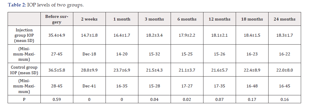
Table 2:IOP levels of two groups.
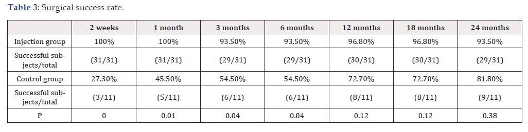
Table 3:Surgical success rate.
BCVA demonstrated stability over the 2-year follow-up in both study groups. The corresponding data are summarized in (Table 4).

Table 4:BCVA of two groups.
The number of antiglaucoma medications used at various follow- up visits was significantly reduced compared to baseline in both groups. However, medication use gradually increased over time in both cohorts. No significant differences were found between the groups at any time point (Table 5).

Table 5:Number of antiglaucoma medication.
According to the Retinal Nerve Fiber Layer (RNFL) thickness analysis, the data remained largely stable in both groups, with only a mild tendency toward thinning. This subtle decline is consistent with the progressive nature of the disease, despite the sustained normalization of intraocular pressure achieved through treatment (Table 6).

Table 6:RFNL thickness.
Discussion
Based on the statistical analysis of our results, we conclude that despite the challenges in successfully treating Neovascular Glaucoma (NVG), the treatment protocol we employed combining bevacizumab with Ex-Press shunt implantation represents a highly effective and reliable therapeutic approach.
This study demonstrated that intravitreal injection of anti-VEGF agents prior to glaucoma surgery promotes rapid regression of neovascularization in the anterior chamber angle and significantly reduces the risk of intraoperative hemorrhage. Bevacizumab, an anti-VEGF drug, inhibits both angiogenesis and fibroblast proliferation, both of which are critical in neovascularization and wound healing modulation. A key factor in managing NVG is mitigating ischemic processes in ocular tissues, which subsequently decreases VEGF production.
Standard NVG treatment typically includes Panretinal Photocoagulation (PRP), retinal cryotherapy, or endolaser therapy to reduce retinal ischemia, though these methods act slowly and with limited efficacy. Intravitreal anti-VEGF injections have emerged as an effective adjunct therapy, dramatically and rapidly reducing retinal and anterior segment neovascularization and improving NVG outcomes. Although bevacizumab is approved for antiangiogenic use in metastatic colorectal cancer combined with chemotherapy, it is used off-label in ophthalmology.
Multiple reports have documented successful bevacizumab use alongside glaucoma drainage devices and trabeculotomy in NVG management. Combining intravitreal anti-VEGF injections with glaucoma surgery is theoretically ideal: the anti-VEGF drug transiently suppresses neovascularization early, while PRP provides longer-term control of retinal ischemia.
We observed that early administration of bevacizumab minimizes ocular tissue damage, particularly in patients who have not previously undergone PRP. While some case series have noted only transient Intraocular Pressure (IOP) lowering with anti-VEGF monotherapy, the effect depends on disease stage and the drug’s short duration of action. In advanced NVG or cases with extensive peripheral anterior synechiae, prognosis for IOP control is poor, often necessitating additional interventions such as PRP and implantation of drainage devices. Despite maximal medical therapy, surgical intervention remains necessary but is frequently complicated by intraoperative difficulties and suboptimal outcomes.
The core challenge in managing glaucoma is preserving central and peripheral visual function, which depends on stable IOP control. Since NVG pathogenesis involves severe chronic vascular disease in our study, primarily proliferative diabetic retinopathy and postthrombotic retinopathy the effective IOP-lowering achieved by filtration surgery in primary glaucoma often lacks durability and relevance in NVG. Thus, adjunctive therapy targeting neovascularization is essential.
Several authors, including Min Tang and Nina Asrini Noor, have explored combinations of anti-VEGF agents (such as ranibizumab) with glaucoma drainage devices, reporting benefits but also a high rate of intraoperative complications, notably hyphema and hemophthalmos (~40-42%), complicating postoperative IOP control [10,11].
Our treatment protocol differs by administering anti-VEGF injections two weeks prior to surgery rather than during the procedure, aiming to minimize intraoperative complications. The anti- VEGF effect peaks approximately two weeks post-injection and lasts at least two weeks. Using this approach, we achieved a critically low rate of intraoperative complications, enabling Ex-Press shunt implantation even in patients with severe neovascularization (rubeosis) of the anterior chamber angle and iris cases previously considered unsuitable for this surgery.
Conclusions
A preoperative intravitreal injection of bevacizumab may serve as an effective adjunctive therapy in the management of neovascular glaucoma. This approach promotes rapid regression of anterior chamber angle neovascularization and facilitates subsequent surgical intervention. Implantation of the Ex-Press Mini Glaucoma Shunt in eyes pretreated with anti-VEGF agents where neovascularization has already regressed provides a sustained intraocular pressure-lowering effect in patients with otherwise refractory neovascular glaucoma.
A repeat bevacizumab injection administered two weeks after shunt implantation further enhances and prolongs the regression of neovascularization, thereby maintaining the long-term hypotensive efficacy achieved through Ex-Press Mini Glaucoma Shunt implantation.
In all observed eyes, visual function was preserved with stable and optimal intraocular pressure values throughout the follow-up period.
Further long-term studies are warranted to confirm the longterm safety and efficacy of this combined treatment protocol over extended observation periods.
Ethics approval and consent to participate
This study was conducted in accordance with the Declaration of Helsinki and was approved by the Medical Ethics Committee of New Vision University. All participants provided written informed consent before enrollment. Clinical trial number: not applicable.
Consent for publication
Not applicable. This study does not contain any individual person’s data in any form (including individual details, images, or videos).
Availability of data and materials
The datasets generated and/or analyzed during the current study are not publicly available due to patient confidentiality and institutional policies.
Competing interests
The authors declare that they have no competing interests.
Funding
The funding body had no role in study design, data collection, analysis, or manuscript preparation.
Authors’ contributions
Dr. Lela Nemsadze designed the study and supervised the project. Dr. Elene Bugashvili collected the clinical data and performed the initial statistical analysis and drafted the initial manuscript. Dr. Avtandil Gagnidze performed the statistical analysis and contributed to Background. All authors contributed to manuscript editing and final revisions. All authors read and approved the final version of the manuscript.
Acknowledgements
The authors would like to thank the staff of the Department of Ophthalmology at New Vision Univeristy Hospital for their assistance with patient care and data collection.
Authors’ information
Dr. Lela Nemsadze is head of the Department of Ophthalmology, New Vision Univeristy Hospital, with a special interest in glaucoma surgery. Dr. Elene Bugashvili is an ophthalmologist at New Vision University hospital. Avtandil Gagnidze is Professor and Vice-President for Research and Development at the East-West University.
References
- Senthil S, Dada T, Das T, Kaushik S, Puthuran GV, et al. (2021) Neovascular glaucoma-A review. Indian J Ophthalmol 69(3): 525-534.
- Yang H, Yu X, Sun X (2018) Neovascular glaucoma: Handling in the future. Taiwan J Ophthalmol 8(2): 60-66.
- Vasudev D, Blair MP, Galasso J, Kapur R, Vajaranant T (2009) Intravitreal bevacizumab for neovascular glaucoma. J Ocul Pharmacol Ther 25: 453-458.
- Olmos LC, Lee RK (2011) Medical and surgical treatment of neovascular glaucoma. Int Ophthalmol Clin 51: 27-36.
- Takihara Y, Inatani M, Fukushima M, Keiichiro Iwao, Minako Iwao, et al. (2009) Trabeculectomy with mitomycin C for neovascular glaucoma: prognostic factors for surgical failure. Am J Ophthalmol 147(5): 912–918.
- Eid TM, Radwan A, el Manawy W, Ihab el-Hawary, et al. (2009) Intravitreal bevacizumab and aqueous shunting surgery for neovascular glaucoma: safety and efficacy. Can J Ophthalmol 44(4): 451-456.
- de Jong LA (2009) The ex-PRESS glaucoma shunt versus trabeculectomy in open-angle glaucoma: a prospective randomized study. Adv Ther 26(3): 336-345.
- Lim TM, Bae SH, Cho YJ, Lee JH, Kim HK, et al. (2009) Concentration of Vascular Endothelial Growth Factor after Intracameral Bevacizumab Injection in Eyes with Neovascular Glaucoma. Korean J Ophthalmol 23(3): 188-192.
- Silva Paula J, Jorge R, Alves Costa R, Maria de Lourdes VR, Ingrid U Scott, et al. (2006) Short-term results of intravitreal bevacizumab (Avastin) on anterior segment neovascularization in neovascular glaucoma. Acta Ophthalmol. Scand. 84(4): 556-557.
- Tang M, Fu Y, Wang Y, Zhi Zheng, Ying Fan, et al. (2016) Efficacy of intravitreal ranibizumab combined with Ahmed glaucoma valve implantation for the treatment of neovascular glaucoma. BMC Ophthalmol 16: 7.
- Noor NA, Mustafa S, Artini W (2017) Glaucoma drainage device implantation with adjunctive intravitreal bevacizumab in neovascular glaucoma: 3-year experience. Clin Ophthalmol 11: 1417-1422.




 We use cookies to ensure you get the best experience on our website.