
Review Article 
 Creative Commons, CC-BY
Investigations of Glycoconjugates from Human Erythrocyte- and Lymphocyte Membranes with Polarisation Optical-Histochemical (Topo-Optical) Staining Reactions
*Corresponding author:Josef Makovitzky, Univ Heidelberg, Dep of Neuropathology and Univ Freiburg i.Br. Institue for Forensic Medicine, Germany.
Received:September 17, 2025; Published:October 30, 2025
DOI: 10.34297/AJBSR.2025.28.003742
Summary
Topo-optical staining reactions are suitable for the selective detection of glycoconjugates located on the membrane surface. In this paper, we demonstrated the glycoconjugate components located on the surface of red blood cell membranes and lymphocyte membranes. With the help of the topo- optical reaction, we demonstrated the differences between human red blood cell and lymphocyte membranes. We selectively demonstrated the O-acetyl-sialic acid, which occurs only on the surface of human B lymphocytes and is a monitoring marker of children with acute lymphoblastic leukemia.
Keywords:Human red cell membrane, Human lymphocyte cell membrane, Sialic acid, O-acetyl-sialic acid, Topo-optical staining reaction, Polarization microscopy
Introduction
The beginning of modern carbohydrate histochemistry is dated with the sensitive reaction “Periodic acid-Schiff reaction” (PAS), [1]. Nevertheless, carbohydrate research has long been pushed into the background by intensive protein and nucleic acid structure research in the second half of the 20th century. This also applies to membrane research: the function and structure of various proteins and lipids were rapidly elucidated, but that of membrane glycoproteins was slow to emerge.
After the immunological revolution of the 1970s, interest grew in learning about the structure of the various carbohydrate components of the membrane in order to better understand their biological function [2-4] In addition to numerous ultrastructural [5] and immunohistochemical methods [6-9], it was only with the help of polarisation microscopy that it became possible to precisely determine the spatial arrangement of the carbohydrate components of the biomembrane at the molecular level.
The “topo-optical reactions” used for this purpose are characterised by the fact that they amplify the original anisotropic effect of dye molecules or primarily colourless compounds on repetitive (micellar) structures several times by means of oriented addition. This creates a possibility to make latent or weakly anisotropic structures visible or to increase the anisotropic effect and thus to carry out a polarisationoptical analysis on repetitive biological structures.
Originally, these reactions were called “topochemical” by Schmidt [10]. In modern histochemical literature, however, this term was used for quite different reactions without an anisotropic effect, which is why Romhányi [3,4,11] suggested the term “topooptical” reaction instead. A topo-optical reaction is, for example, the toluidine blue precipitation method, which has been widely used in the study of sub microscopic structures (Typ 1 acc. to Romhányi [3,4,11,12] and Makovitzky [13-15], Modis [16,17].
The principle of the method is based on the direct binding of toluidine blue dye molecules to the chromotrope. The oriented binding induces birefringence (=anisotropic effect), which is additionally stabilised by subsequent precipitation of the bound dye with simultaneous multiple amplification of the anisotropic effect.
Acc. to Romhanyi [11,18,19,20], the product obtained by a
topo-optical reaction is an oriented dye aggregate consisting of
three components:
I. the substrate responsible for the oriented dye binding.
II. the bound dye
III. the precipitant, which reacts with the oriented bound dye to
give an oriented aggregate.
The effect of the precipitant in stabilising the topo-optical reaction is explained by the fact that the dissolved precipitant, e.g. potassium hexacyanoferrate (III), forms an oriented complex with the associated toluidine blue molecules without destroying the primary oriented dye attachment to the structure. At the same time, the intensity of the anisotropic effect is increased.
Acc. to Hebenstreit and Keller [21], one mole of potassium hexacyano-ferrate(III) binds three moles of toluidine blue. By means of the toluidine blue precipitation method, it was possible to selectively display the original directionally (repetitively) arranged acidic groups at pH values between 1 and 7 in the polarisationoptical analysis (type I of the Romhány topo-optical reactions). At pH1-3 SO- - 4 and SO- -3 at pH 3-5 next to these groups, and the PO- -4, at pH 5-7 next to these groups and the COOH groups are responsible for the oriented dye binding, in this case for the oriented binding of the toluidine blue dyestuff molecules [Makovitzky 14,15] and Scheven and Jurke [22].
III.1. Type II of the Topo-Optical Reactions Includes e.g. Mild Sulphation,
the Aldehyde-Bisulphite-Toluidine Blue Reaction (ABT), the Permanganate-Bisulphite-Toluidine Blue Reaction (PBT) as well as the sialic acid and O-acylsialic acid specific reactions [6]. In all topo-optical reactions of type 2, the new groups aligned by chemical reactions are responsible for the oriented dye binding (Romhányi et al., [23,24,25] Makovitzky [14]. The toluidine blue precipitation method was also introduced into electron microscopic histochemistry [26,27] as was later the ABT reaction [28].
III.2. The Polarisation-Optical Analysis of Glycoconjugates with the Toluidine Blue Precipitation Method
The cell membrane carbohydrate components are divided into two groups acc.to Roseman [29].
I. the membrane glycoproteins, which are located on the surface
in the outer half of the membrane (not on the cytoplasmic
side), are the N-terminal region of the protein chain and have
branched oligosaccharid chains.
II. the so-called free glycoproteins, which are also localised in the
outer half of the membrane, but are only loosely connected to
it.
Structure of the Human Erythrocyte Membrane
Bio membranes reveal, as a rule, only weak intrinsic und extrinsic -form birefringences, owing to their lipid bilayer und thei protein framework, respectively. The erythrocyte membrane consists of a bimolecular lipid layer. This is the basic unit in other cell types as well. Proteins are embedded in the bimolecular lipid layer. The polar (hydrophilic) head groups of the lipids are directed outwards, the apolar (hydrophobic) lipid sections make up the hydrophobic membrane interior [30,31].
The proteins accessible from the outer environment and part of the lipids of the outer lamella carry the oligosaccharide chains. Structurally and functionally, a number of proteins especially attached to the membrane interior also belong to the membrane [32,33]. The lipid matrix contains mainly phospholipids. The total lipid content of the human erythrocyte membrane is 5x10 - 10 mg / cell and is divided between the main lipid components: Phosphatydilcholine, physphatidylethanolamine, sphingomyelin, phosphatydilserine, lysolecithin, phosphatidic acid, phosphatidylinositides, polyglycerol phospholipids [34]. Other lipid components are glycolipids and cholesterol. The phosphlipids are unequally distributed in the two lipid lamellae, resulting in lipid asymmetry [35]. The outer and inner lamellae also differ in their fluidity [36,37].
Numerous proteins (membrane proteins) are associated with or even embedded in the lipid matrix. Ectoproteins of the outer lipid lamella include acetylcholinesterase and so-called lesser (minor) glycoproteins.
The following ectoproteins are localised in the inner lipid lamella: spectrin, ankyrin and erythroactin, which essentially build the membrane skeleton of the erythrocyte [33,38]. The membrane skeleton interacts with the cytoplasmic side of the membrane (membrane inner side) in that the main component spectrin is directly and indirectly bound to the inner lipid lamella via ankyrin/syndein and protein of band 4.1. Glycophorins belong to the transmembrane proteins. They determine the immunological behaviour of erythrocytes [39,40] and are available as transport proteins (anion and glucose transport proteins) [33]. The transmembrane proteins owe their name to the fact that they are embedded in the lipid matrix with the hydrophobic segments and project beyond the two lipid lamellae with the hydrophilic C- and N-terminal segments, i.e. the C-terminal segments of the glycophorins lie on the inside of the membrane (cytoplasmic), whereas the N-terminal segments lie on the outside of the membrane. Together with the other transmebrane proteins, ectoproteins and the sugar chains of glycolipids, they form the glycocalyx.
Results on the Erythrocyte Membrane with Topo-Optical Reactions
The sign of polarisation of the biomembrane is linearly negative/radier positive in the unstained state, caused by the lipid components (Romhányi 3, 24, Romhányi: personal communication 41) and [14,15,24,42,43]. The retardation of the unstained erythrocyte ghost membrane is 0.4 nm (Schmitt et al., [48].
According to the toluidine blue precipitation method (0.1% toluidine blue solution at pH 7.4, the retardation is 41-42 nm, so a 100-fold increase in intensity can be achieved with this method. Based on the optical analysis, there is a positive polarisation cross on the membranes of the erythrocytes. The dye molecules are aligned radially (vertically) with their light retarding bands oriented towards the surface [14,24,33,42,44,45, 46,47]. Based on the optical analysis, the sign of the human erythrocytes stained with toluidine blue topo-optical reaction is: linear negative/radial positive. After various enzymatic and chemical degradation reactions at the erythrocyte membrane, such as trypsin and sialidase digestion, alkaline and acid hydrolysis, digitonin treatment, phospholipase C digestion or sialic acid extraction, the role of the glycocalyx or its glycoprotein stock could be determined [14,33,42,43,45]. The role of the glycocalyx or its glycoprotein components in the formation of the topo-optical reaction with toluidine blue has been clearly demonstrated [42,43].
(Table 1)
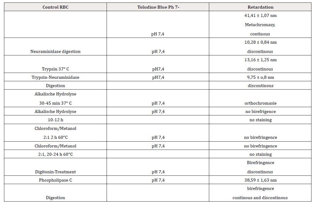
Table 1:
Based on extensive polarisation-optical-histochemical studies [42], it is assumed that the toluidine blue-induced anisotropy of the human erythrocyte membrane is related to the spatial state of the glycocalyx, i.e. depends on the density and distribution of the negatively charged groups on the outside of the cell membrane. The glycoproteins (terminal sialysed glycophorins) are responsible not only for the binding but also for the simultaneous optimal orientation of the toluidine blue dye molecules. This statement has been confirmed by several authors [33,42-47]. The optical behaviour of human erythrocytes changes at different pH-values, ionic strengths, temperatures and salt concentrations.
Between pH5.0-pH7.4 the surface appears in a deep green polarisation colour. This is due to the fact that the sialic acid molecules, which are terminally localised on oligosaccharide chains, are oriented membrane-parallel and the binding sites - the COOH groups of the sialic acid molecules - are located on the sugar side chains of the glycophorins at a distance of 0.4-0.5 nm [32]. One must hypothetically also think of the free COOH groups of glutamic acid and aspartic acid, because the toluidine blue-induced anisotropy does not disappear even after combined digestion attempts [14,46,47]. The optimal preparation of erythrocytes for polarisation optical studies is in PBS pH 7.4 and isoosmolarity (300 mOsm) (Figures 1-4).
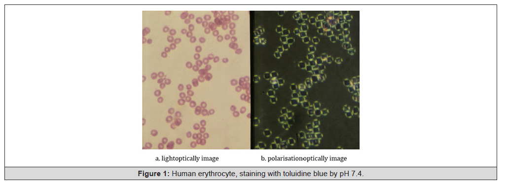
Figure 1:Human erythrocyte, staining with toluidine blue by pH 7.4.
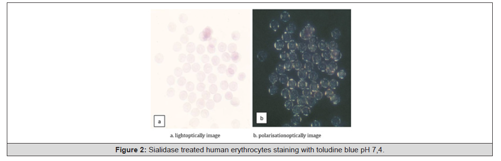
Figure 2:Sialidase treated human erythrocytes staining with toludine blue pH 7,4.
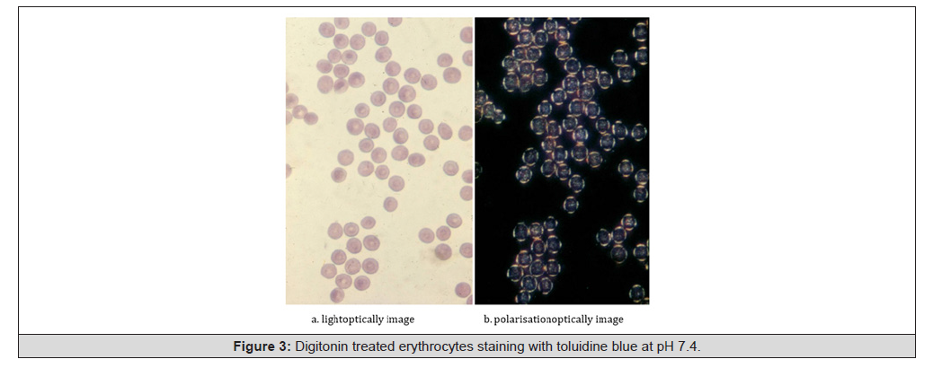
Figure 3:Digitonin treated erythrocytes staining with toluidine blue at pH 7.4.
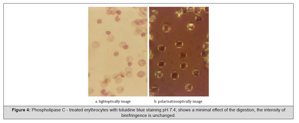
Figure 4:Phospholipase C - treated erythrocytes with toluidine blue staining pH 7.4, shows a minimal effect of the digestion, the intensity of birefringence is unchanged.
Phospholipase C digestion and digitonin treatment clearly demonstrate the membrane-building role of lipid molecules responsible for the optimal arrangement of sialic acid-bearing glycophorins and thus for the optimal topo-optical response [14,42,43]. Phospholipase D digestion was ineffektive. Erythrocytes from 12-, 21- and 42-day preservation at 4°C show a marked decrease in anisotropy and exhibit the phenomenon of “vesiculation”. Cryopreservation is optimal at -25°C or -30°C. The erythrocytes show intense anisotropy and metachromasia even after 180 days [42,43,45]. The findings reflect a spatially structurally unchanged erythrocyte membrane surface or glycocalyx. This series of experiments demonstrates the effect of temperature on the toluidine blue topo-optical staining reaction of human erythrocyte membrane [49]. Dried human red blood cell smears were fixed in 5% GA at pH 7.4 in phosphate buffer for 15 min between 0°C and 45°C and stained with 0.1% toluidine blue at pH 7.4.
Optimum staining and birefringence were found at room temperature. Membrane of red blood cells stained with toluidine blue at temperatures below 10°C was 20-30% of optimal Retardation. Between 10 and 28°C, the retardation was measured up to 40-42 nm. After maximum values between 28 and 31°C, between 31 and 37°C, they were only 50-60% of the optimum. Between 37-45°C, with toluidine blue topo-optical reaction stained the birefringence oft he human red blood cells is granular, indicating a breakdown of the membrane order [49]. The electrophoretic mobility of human erythrocytes treated with procaine hydrochloride was unchanged, the amount of bound toluidine blue molecules was not changed, because the red blood cells bind a part of the toluidine blue dyestuff molecules in an unoriented manner [43].
Acc. to Stibenz and Geyer (1980), the length of the extracellular segment of gylkphorin ANN in the calculated secondary structure is a maximum of 16.8 nm (168 AU). The N-terminal segments carry the sialic acid molecules, and their orientation, or spatial position, is no longer as optimal after procaine hydrochloride treatment. In the stretched state, the length of glycophorin A is 23 nm. (230 AU, Makovitzky and Geyer 1980, Stibenz and Geyer 1980,1982). Due to the topo-optical reactions with thiazine dyes (toluidine blue, methylene blue, 1,9 di methyl methylene blue, azure a,b, c and thionin) the spatial extent of the glycocalyx is uniform, as is the distribution of sialic acid on the surface (no capping phenomena).
We examined the different ghosts polarisation-optically with
toluidine blue staining reaction by pH 7.0 -7.4.
I. reconstructed Ghost (Schwoch and Passow 1973)
II. non-reconstructed Ghost (Schwoch and Passow 1973)
III. haemoglobin depleted (white) Ghost (Dodge 1963)
IV. dialysed white Ghost
V. freeze-thaw Ghost
(Table 2)

Table 2:
Whether the ghosts exhibit the phenomenon of metachromasia was difficult to judge, even in polychrome light. The ghosts, Hbfree erythrocytes show anisotropy reduced to about half with the toluidine blue stain, the measured duct differences are between 20- 22 nm. The reconstructed ghost show a stronger anisotropy and higher differences in gait, but do not reach the values of control erythrocytes. In different ghost preparations we cannot expect sialic acid loss (Stibenz personal communication: [32,33], thus it can be assumed that structurally (spatially) altered ghost glycocalyces are present.
The toluidine blue dye molecule-binding glycocalyx components of the ghosts, i.e. the N-terminal extracellular glycophorin A segments or their sialic acid residues, are located in a different spatial environment. With the toluidine blue topo-optical staining reaction at pH 7.0 - 7.4, we can make exact statements about the spatial arrangement of the terminal sialic acid residues and thus indirectly about the structure of the glycocalyx [33, 42,43]. First investigations of control as well as 3 and 6weeks liquid preserved erythrocytes with the toluidine blue topo-optical staining reaction showed a decrease of metachromasia as well as anisotropy of the erythrocytes with increasing storage time [50].
Halbhuber et al., 1983 a,b). The measured anisotropy in polychromatic light is 41-42 nm of the control erythrocytes and for 6 weeks preserved erythrocytes with unchanged cell diameter 13 to 15 nm. From these results it must be concluded (Stibenz and Makovitzky 1982, unpublished, 50) that in the course of storage both the oriented toluidine blue binding and the spatial order of the toluidine blue oriented binding sialic acid residues decrease. The conditions thus support a structural change of the glycocalyx (change of steric localisation) during the preservation of the erythrocytes. In addition, there is the phenomenon of vesiculation in blood conserv.
The vesicles can be stained by pH 7.4 and show up light-optically with the mPAS reaction, with the sialic acid-specific topo-optical reaction, they show a positive polarisation-optical reaction [50,51]. For the in vivo ageing process of erythrocytes, the involvement of lipid peroxidation processes was made probable, in which, among other things, malondialdehyde is formed by oxidation of Cn /Cn+3 - positioned double bonds of unsaturated fatty acids. My idea was the basis for the incubation of human erythrocytes with dextran (Correspondence with D. Lerche 1980, [53].
Cell surface properties are involved in the aggregation process of red blood cells. Using the topo-optical toluidine blue reaction, conformational changes of the glycocalyx (main component glycophorin A) were found when red blood cells were incubated and fixed in the presence of dextran. Relative differences in optical path as a measure of red blood cell membrane anisotropy decreased in relation to dextran concentration during fixation. These conformational changes could not be detected by electrophoretic measurements. When incubating, fixing and staining red blood cells in the presence of dextran, anisotropy decreased only at low dextran concentrations and increased at rising dextran concentrations. This biphasic course of differences in optical path seems to be due to different effects of dextran superimposing upon each other: (i) a disturbing influence on the spatial order of sialic acid carrying oligosaccharide side chains due to H-bond interaction, and (ii) an increase in the size of dye aggregates and suppression of the thermal motion of macromolecules at higher dextran concentrations [33,54].
We investigated the influence of hypo or hypertonic stress, ionic strength, pH and procaine hydrocloride on toluidine blue induced birefringence. In isotonic buffered salt medium at pH 7.4, the induced anisotropy is about 40 nm. It decreases with hypotonic stress. Extensive removal of ions leads to the extinction of the anisotropy. Similarly, the spatial arrangement of the sialic acid residues must decrease. Subsequent second incubation of sucrosewashed erythrocytes with PBS leads to a partial reappearance of the birefringence and thus to a partial restoration of the original arrangement/orientation of the terminal sialic acid residues [43, ,33,44,46,47, Makovitzky and Geyer, unpublished,50] (Table 3).
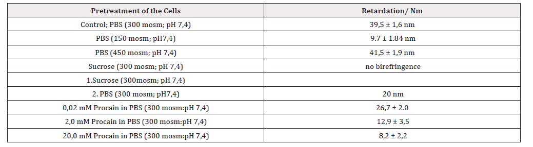
Table 3:
Summarized: acc. to Makovitzky and Geyer (1977 42) Geyer and Makovitzky 1980 [43] Makovitzky 1984,1991 [14,15], Halbhuber et al., 1984 1984 a [55,56] Stibenz 1985 [33] the toluidine blue topooptical reaction is the most sensitive reaction of the erythrocyte membrane. The structural investigation of the glycocalyx of RBC results light optical in metachromasia of the basic toluidine blue molecules to the sialic acid residues of the glycocalyx, under crossed polar birefringence. The extent of metachromasia is proportional to the packing density of the bound dye molecules and thus also to the packing density of the binding sialic acid residues.
The induced birefringence is an indirect measure of the spatial arrangement of the bound toludine blue dyestuff molecules and thus also of the binding sialic acid residues, i.e. in other words the decrease in the induced birefringence (the pathway difference) therefore reflects the increasing arrangement of the dyestuffbinding sialic acid residues of the glycocalyx, or the extracellular glycophorin A segment, which carries the main part of the sialic acid residues.
The glycocalyx elements and thus the extracellular glycophorin-A segment are densely packed close to the membrane (average density of the glycocalyx is 50-70 Å. The sialic acid-bearing sugar side chains show a high similar spatial order, as evidenced by the toluidine blue topo-optical reaction at pH 7.4 and the sialic acid specific topo-optical reaction: continuous birefringence [14,33,43]. We cannot support the intercalation of toluidine dye molecules into the lipid layer of the red blood cell membrane [3,12,14,24,33,42,43].

Figure 5:The Aldehyde-bisulfite toluidine reaction (ABT-r) acc. Romhányi et al., 1975 (25) and Fischer 1978 (6).
During the ABT reaction, the sign of the birefringence changes [6,25] so that one speaks of an inverse topo-optical reaction. Lightoptically, the reaction is characterised by basophilic metachromasia and polarisation-optically green polarisation colour. Based on the polarisation-optical analysis: The linearly oriented OH groups of the sugars are arranged perpendicularly on the surface, parallel to the surface. Similarly, the α-hydroxysulphonate groups and the oriented bound dye molecules.
The PAS reaction with post-precipitation shows anisotropy (birefringence, between two crossed polarisation filters in linearly polarised light), but the new complex is not stable, the anisotropy is extinguished after 10-15 minutes (Makovitzky and Richter 2009 [60]. Therefore, after the PAS reaction (oxidation of vicinal OH groups to aldehyde groups and conversion to sulfonic acid groups with Schiff’s reagent), the addition of toluidine blue at pH 1-2 and precipitation with potassium hexacyanoferrate (III) was inserted. Only with this multi-step procedure a stable anisotropic (substratedye- precipitation complex) complex is formed.
The topo-optical “anisotropic PAS reaction” the Aldehyde- Toluidine Blue Reaction (ABT-R) is suitable for the selective presentation of the linearly oriented OH groups of mono-, di- or oligosaccharides. It is also called aldehyde-bisulphite-toluidine blue reaction (ABT-reaction). The basis is the reaction first described by Malinin (1970): After periodic acid oxidation of vicinal OH groups, CHO groups are formed. These become SO--3 groups of the α hydroxysulphonates by (bi-)sulphite addition. These in turn can be selectively visualised with toluidine blue and/or thiazine dyes at pH 1-2 [14,24,25,60]. If the sign of the birefringence is linearly negative with respect to the length (radial positive spherite), then the sugar chains are parallel to the surface. If the sign of the birefringence is linearly positive with respect to the length (radially negative spherite, Fischer 1978 [6], then the sugar chains lie perpendicular to the surface (Figure 6).
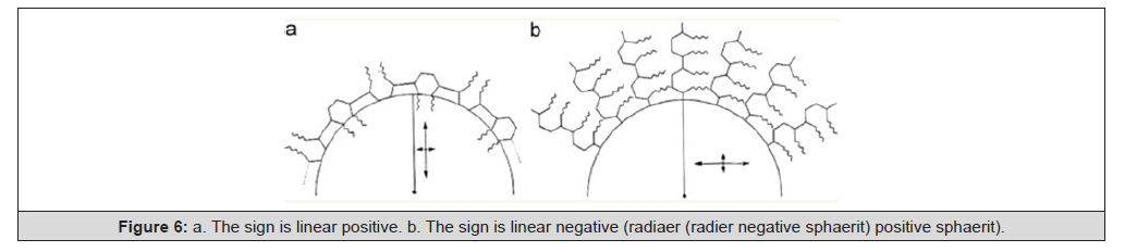
Figure 6:a. The sign is linear positive. b. The sign is linear negative (radiaer (radier negative sphaerit) positive sphaerit).
Acc. to Fischer 1978 [6].
We could repeat the results with the thiazine dyes azure A, B, C, methylene blue and 1:9-dimethylmethylene blue(1.9DmMb) at pH 1-2. Except for methylene blue, the reaction results are stable over time. The prerequisite for the metachromasia and birefringence (anisotropy) produced in the reaction is that the vicinal, linearly oriented OH groups are 4-5 Å away from each other. If the distance is greater than 0.5 nm, there is neither metachromasia nor anisotropy (Sylvén 1954 [61]. Erythrocytes do not give a positive ABT reaction, although surface-oriented oligosaccharide chains are present [24,25,42].
We tried all thiazine dyes for the selective visualisation of the sugar chains on the surface and found 1,9 dimethylmethylene blue and azure B suitable for selective visualisation. The reaction was also positive with methylene blue, but under the influence of light the metachromasia and anisotropy disappeared and we saw a transformation into orthochromasia and isotropy, the dyes and the precipitaion complex have broken up. The sign of the birefringence is linear negative (radial positive spherite), i.e. the linearly oriented OH groups and the dye molecules are bound oriented perpendicular to the surface, the sugar chains are membrane parallel. A control reaction with 1.9 DmMb at pH 1 turned out negative.
The lymphoid cells show a positive ABT reaction, but only in the pH range 3-4 acc. to Romhányi 1974 [24]. The two populations of T and B lymphocytes show a clearly positive reaction with the ABD reaction (1,9 dimethylmethylene blue instead of toluidine blue). Birefringence is coherent with deep green polarisation colour, metachromasia is seen light-optically. The sign of the birefringence with respect to length is linearly negative (radial positive spherite), the linearly oriented OH groups are oriented perpendicular to the surface, the sugar chains are oriented parallel to the membrane surface.
The control reaction on the lymphocytes at pH 1 and 2 with 1.9 DmMb shows a positive reaction. This means that glycosaminoglycans are present at the lymphocyte surface. After hyaluronidase or chondroitinase AC and -B digestion, the reaction at pH 1-2 with 1.9 DmMb was negative, i.e. the surface contains sulphated glycosaminoglycans (hyaluronic acid, chondroitin-4- and chondroitin-6-sulphate as well as keratan sulphate, Makovitzky 1984 [14].
The ABD reaction at pH 1 and 2 is positive, i.e. the T and B lymphocytes show a positive topo-optical reaction with green polarisation colour and anisotropy, light-optical metachromasia. In contrast, the human erythrocytes and thrombocytes with 1.9 DmMb show no reaction in this pH range (Makovitzky 1984 [14]. Culling et al. (1971,1974,1980, [62, 63, 64] found that after treatment with 0.5% KOH alcohol solution, the subsequent PAS reaction is specific for O acyliered carbohydrate derivatives. Fischer [6] further developed the KOH-PAS reaction into a polarisationoptical- histochemical reaction (KOH-ABT reaction). The principle of the KOH-ABT reaction consists in the cleavage of interfering O-acyl substituents from potential vicinal diols caused by alkaline pretreatment.
To specify the KOH effect, a blocking with sodium borohydride can be carried out after a primary periodic acid oxidation. This is followed by a KOH-ABT reaction [6,60]. The comparable periodic acid/borohydride (PB)-KOH-ABD reaction showed in a mixed population (T+B), positively and negatively reacting lymphocytes. Human erythrocytes were negative with this reaction. The mild PAS (mPAS) reaction is suitable for the selective preparation of N acetyl neuraminic acid (sialic acid). Based on chemical and biochemical studies, it was found that the periodic acid in low concentration (0.01%) at 4° C for 10 min. reacts only with the linearly oriented OH groups of sialic acid. The mild PAS reaction (mPAS) is well detectable polarisation-optically, but shows a low intensity lightoptically.
The specificity of the mPAS reaction was largely confirmed by Klessen [65-67] With the mPAS reaction, human erythrocytes, thrombocytes and lymphocytes show a positive reaction only with 1.9 DmMb and azure B. All cell types show unison light-optically a metachromasia, polarisation-optically a deep green polarisation colour. The sign is linear negative (radial positive spherite), the reaction indicates that the linear oriented OH groups are spatially in an optimal position for the dye molecules. The dye molecules are bound perpendicular to the membrane surface. The reaction specificity was tested with sialidase digestion and chemical sialic acid extraction [14].
The sialic acid specific topo-optical reaction is a modified ABT/
ABD reaction, which differs from this one by a mild periodic acid
oxidation and use of 1,9-dimethylmethylene blue or azure B at pH
1 and pH 1.2. [13,14].
The prerequisite of the reaction are:
i. the selective oxidation of the vicinal OH groups at C8-C9 and
C7,
ii. the groups are not substituted, C7-aldehyde-neuraminic acid
residues are formed, and
iii. a selective sulphite addition of the newly formed CHO group at
C7 with the formation of hydroxyl sulphonate groups (Figure
7).
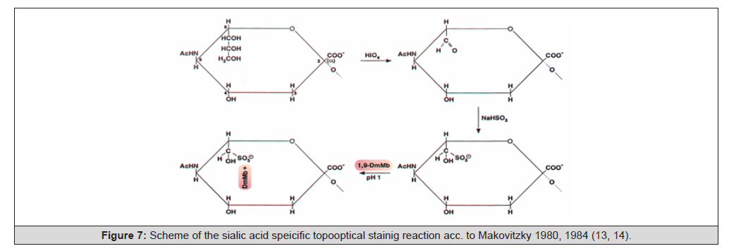
Figure 7:Scheme of the sialic acid speicific topooptical stainig reaction acc. to Makovitzky 1980, 1984 (13, 14).
The mild PAS reaction is a specific reaction for the histochemical preparation of sialic acids, proves also after an oxidation time of 10 min at 4°C measured higher periodic acid consumption of N-acetylneuraminic acids with 1.15 mol H5IO6 in contrast to other carbohydrates, which together consumed less than 0.07 mol periodic acid [68] (Figure 8).
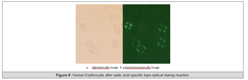
Figure 8:Human Erythrocyte after sialic acid specific topo-optical stainig reaction.
The distribution or extension of sialic acid on the surface of erythrocytes, lymphocytes is uniform and contiguous (no capping phenomenon). The periodic acid/borohydride-KOH-mPAS-sialic acid specific reaction was simultaneously +/- in lympohycyte suspension (T+B). We observed the same phenomenon in chicken and goose lymphocyte suspensions (Makovitzky and Rudas 1979 unpublished, [69]. The differences between T and B lymphocytes were clearly demonstrated by mass spectrometry, liquid chromatography and thin-layer chromatography [70,71]. Thus, human B lymphocytes contain glycosaminoglycans, N-acetylneuraminic acid and in position C9 O-acylated sialic acid on their surface. Since the beginning of 2000, this finding has been used in diagnostics: 9-O-acylsialic acid is a biomarker for acute lymphoblastic leukaemia (patent EP1083181 B1, priority date 1999 [72-75] (Figure 9).

Figure 9:Scheme of the KOH-sialic acid specific reactions acc. to Makovitzky (1984, 14).
At the same time, I was able to selectively visualise membranebound RNA on the surface of T and B lymphocytes after a permanganate-sulphite-toluidine blue or permanganate-sulphite- 1,9-dimethylmethylene blue reaction. RNAse digestion (120-150 min) removed the RNA and thus the anisotropy. Alkaline hydrolysis according to Geyer and Scheibner also gave the same result. Alarcon- Segovia et al. (1979, [76] suggested that the lymphocyte membrane should contain membrane-bound RNA at the surface, the function of which is that the antibody does not enter the cell by phagocytosis or endocytosis, but binds to the surface RNA components first.
Acknowledgement
None.
Conflict of Interest
None.
References
- McManus JFA (1976) Histological demonstration of mucin after periodic acid. Nature 158: 202.
- Schauer R (1988) Sialic acids as antigenic determinants of complex carbohydrates. Adv Exp Med Biol 228: 47-72.
- Romhányi Gy (1978) Ultrastructure of biomembranes as shown by topo-optical reactions. In: Acta Biol Acad Sci Hung 29(4): 311-365.
- Romhányi Gy (1989) Ultrastructural and cell biological aspects of membrane glycoproteins In: A biomembránok strukture és toimiködése / The structure and function of biomembranes / ed/ed: János Somogyi – Budapest: Akadémiai Kiadó 1: 73-91.
- Geyer G (1977) Electron Microscopic Histochemistry. Part 1: Detection and contrasting methods for carbohydrates, proteins and amino acids, nucleic acids, lipids and minerals. In: Handbuch der Histochemie / ed v Walther Graumann, Karlheinz Neumann– Stuttgart G Fischer Verlag.
- Fischer J (1978) Polarization optical analysis of the orientation of polyaldehyde macromolecules in complex biological structures. Habilitationsschrift Pécs, (hungarian)
- Makovitzky J (1986) The distribution and localization of the monoclonal-defined antigen 19-9 (CA19-9) in chronic pancreatitis and pancreatic carcinoma. Virchows Arch (Cell Pathol) 51(6): 535-544.
- Schwenk J, Makovitzky J (1989) Comparative study on expression of the blood- group antigens Le a, Le b, Le x, Le y and the carbohydrate antigens CA 19-9 and CA 50 in chronic pancreatitis and pancreatic carcinoma. Virchows Archiv A pathol Anat 414(6): 465-476.
- Schwenk J, Makovitzky (1990) The occurence of carbohydrate antigens (CA19-9, CA 50) in chronic pancreatitis and pancreatic carcinoma. Acta histochemica Suppl-Bd, XXXVIII 183-187.
- Schmidt W J (1947) The change of optical anisotropy in topochemical reactions of histological structures. About Oberhess Ges Nature and Medicine Giessen NF Naturwiss Abt 23(1): 56-85.
- Romhányi Gy (1966) On the ultrastructure of the intercellular substance of connective tissue based on top optical reactions and polarization optical studies: Doctor of Science thesis. (On the ultrastructure of the intercellular substance established on the basis of polarization optical examination of topo-optical reactions) DSc. Thesis) Pé
- Romhányi G (1988) The topo-optical reactions and their roles in biological ultrastructure research. In: Tolnai M editor Lecture at the Hungarian Academy of Sciences (25th March 1983) Akadémia Kiadó
- Makovitzky J (1980) A topo-optical detection of C8-C9-unsubstituted neuraminic acid residues in the glycokalix of erythrocytes. Acta Histochem 66(2): 192-6.
- Makovitzky J (1984) Polarization optical analysis of blood cell membranes. In: Progress in Histochem Cytochem 15: 1-100. In memoriam to Günther Geyer Gustav Fischer Verlag. Stuttgart New York. ISBN 3-437 10922-7 (Stuttgart), ISBN 0.89574-194-6 (New York).
- Makovitzky J (1991) The topo-optical Analysis of Blood Cell membranes : 89-92. In: Neue Methoden in der Histochemie und ihre Anwendung. Gedenksymposium aus Anlaß des 10. Todestages von Prof Dr med habil. Günther Geyer. Jena 18 Bis 20 Juni 1990 Friedrich Schiller Universität Jena.
- Módis L (1974) Topo-optical investigations of mucopolysaccharides (acid glycosaminoglycans). In Handbuch der Histochemie / hrsg v Walther Graumann, Karlheinz Neumann-Stuttgart G Fischer Verl II (4): 1-170.
- Módis L (1991) Organization of the extracellular matrix: a polarization microscopic approach. – Boca Raton: CRC Pr - (The author had dedicated this book to George Romhányi, em Professor of pathology, Pécs, Hungary, one of the pioneers of the polarization microscopy).
- Romhányi G (1963) On the submicroscopic basis of the metachromatic reaction. Acta Histochem 15: 201-33.
- Romhányi G (1970) On top optic reactions and their significance in submicroscopic structural research. The current position of morphology in biology and medicine (About topo-optic reactions and the significance of submicroscopic structure research) Ed Kettler LH, Berlin, Akademischer Verlag: 537–545.
- Romhányi G (1988) The topo-optical reactions and their roles in biological ultrastructure research. In Tolnai M, editor Lecture at the Hungarian Academy of Sciences (25th March 1983) Akadémia Kiadó
- Hebenstreit D, Keller G (1968) Investigations on the influence of complex cyanides and monovalent or polyvalent anions on the metachromasia of acidic mucopolysaccharides and toluidine blue. Inaug- Diss Erfurt.
- Scheven Chr, Jurke A (1991) On the pH dependence og the binding of toluidine blue to fixed erythrocytes: 127-129. In New Methods in Histochemistry and their Application. Commemorative symposium on the occasion of the 10th anniversary of the death of Prof Dr med habil Günther Geyer Jena, 18 to 20 June 1990, Friedrich Schiller University of Jena.
- Romhányi Gy, Bukovinszky A, Deák Gy (1973) Sulfation as a collagen-specific reaction. The ultrastructure of sulfate collagen, basement membranes and reticulin fibers as shown by topo-optical reactions. Histochemistry 36: 123-138.
- Romhányi Gy, Molnár L, Németh À (1974) Ultrastructure differences in cell membranes of erythrocytes, myeloid and lymphoid cells as shown by topo-optical reactions. Histochemistry 39: 261-276.
- Romhányi Gy, Deák Gy, Fischer J (1975) Aldehyde-bisulfite-toluidine blue (ABT) staining as topo-optical reaction for demonstration of linear order of vicinal OH groups in biological structures. Histochemistry 43(4): 333-348.
- Geyer G, Linss W (1978) Toulidine blue staining of cartilage subunits. Acta Histochem 61(1): 127-134.
- Geyer G, Linss W, Makovitzky J (1978) Electron microscopic toluidine blue staining of erythrocyte membrane. Anat Anz 143: 291-295.
- Sótonyi P, Tátrai E (2009) Electronmicroscopic demonstration of Romhányi aldehyde-bisulfite-toluidine blue (ABT)- topo-optical staining reaction. Acta Histochem 111: 326-328.
- Roseman S (1975) Sugars of the cell membrane. In: Weissmann G, Clairborn E, editors Cell membranes. Biochemistry, Cell Biology, Pathology H P Publ Co New York : 55-64.
- Singer S J (1973) The molecular organization of membranes. Ann Rev Biochem 43: 805-826.
- Marchesi VT (1979) Functional proteins of human red blood cell membrane. Seminars in Hematology 16(1): 3-19.
- Stibenz D (1982) personal communication.
- Stibenz D (1985) Milieu- and conservation-related structural changes of the membrane of human erythrocytes: Habilitation thesis- Jena.
- Op den Kamp JA (1979) Lipid Asymmetry in Membranes. Review Article. Annual Review of Biochemistry 48: 47-71.
- Op den Kamp JA, de Gier J, van Deenen LL (1974) Hydrolysis of phosphatidylcholine liposomes by pancreatic phospholipase A2 at the transition temperature. Biochim Biophys Acta 345(2): 253-256.
- Tanaka KI, Ohnishi S (1976) Heterogeneity in the fluidity of intact erythrocyte membrane and its homogenization upon hemolysis. Biochim Biophys Acta 426(2): 218-231.
- Cogan U, Schachter D (1981) Asymmetry of lipid dynamics in human erythrocyte membranes studied with impermeant fluorophores. Biochemistry 20(22): 6396-403.
- Branton Daniel, COHEN Carl M, TYLER Jonathan (1981) Interaction of cytoskeletal proteins on the human erythrocyte membrane. Cell 24(1): 24-32.
- Furthmayr H (1978) Structural comparison of glycophorins and immunochemical analysis of genetic variants. Nature (London) 271(5645): 519-524.
- Furthmayr H (1978) Glycophorins A, B and C: A family of sialoglycoproteins. Isolation and preliminary characterrization of trypsin derived peptides J supramol Struct 9: 79-95.
- Romhányi (1982) personal communication.
- Makovitzky J, Geyer G (1977) Investigations on the toluidine blue colouring of the glycocalyx. Histochemistry 50: 261-270.
- Geyer G, Makovitzky, J (1980) Erythrocyte membrane topo-optical staining reflects glycoprotein conformational changes. J of Microscopy 119(3): 407-414.
- Gliesing M (1987) Topo-optical investigations on the membrane of intact and altered erythrocytes: PhD thesis- Jena.
- Oehring H (1987) Elimination mechanisms of altered erythrocytes. Doctoral Thesis-Jena.
- Halbhuber K-J, Oehring H, Gliesing M, Stibenz D (1990) Polarization optical investigation (topo-optical analysis) of the structure of the human erythrocyte glycocalyx. Influence of pH, ionic strength and diamide-induced spectrin cross linking. Cell Mol. Biol 36(6): 643-57.
- Halbhuber K-J, Klinger M, Oehring H, Linss W, Gliesing M (1992) Structural alteration of the membrane skeleton and of the glycocalyx-preconditions for the IgG-receptor expression in the red blood cell (RBC) membrane. Recent Advances in Cellular and Molecular Biology / eds R J Wegmann, M A Wegmann- Leuven: Peeters: 227-242.
- Schmitt FO, Bear RS, Ponder E (1936) Optical properties of the red cell membrane. J cell comp Physiol 9: 89-92.
- Németh À (1980) personal communication.
- (1982) Stibenz and Makovitzky unpublished.
- (1979) Makovitzky and Geyer unpublished.
- (1980) Correspondence with D Lerche.
- Gawrisch K, Stibenz D, Möps A, Arnold K, Linss W, et al. (1986) The rate of lateral diffusion of phospholipids in erythrocyte microvesicles Biochimica et Biophysica Acta (BBA) – Biomembranes 856(3): 443-447.
- Halbhuber KJ, Gliesing M, Stibenz D, Makovitzky J (1984) Histochemical Methods for the detection of alterations in erythrocyte membrane. Histochemical J 16: 351-353.
- Halbhuber KJ, Gliesing M, Stibenz D, Makovitzky J (1984) Topooptical investigations of human erythrocyte glycocalx age-related changes. Histochemistry 81: 187-193.
- Ruhenstroth Bauer G (1961) Experiences with electrophoresis of blood cells. Bibl Haematol 12: 5-19.
- Ruhenstroth Bauer G, Lücke Huhle C (1968) Electrophoretic investigations on cells of the lymphatic organs. Z Naturforsch B 23: 115.
- Criegee R (1937) The Oxidative Cleavage of the C-C Bond. Angew Chem 8: 153-55.
- Makovitzky J, Richter S (2009) The relevance of Aldehyde-bisulfite toluidine blue reaction and its variants in the submicroscopic carbohydrate research. Acta histochem 111(4): 273-291.
- Sylvén B (1954) Metachromatic dye-substrate interactions. Q J Microsc Sci 95(3): 327-358.
- Culling CF, Reid PE (1977) The apparent failure of sodium borohydride reduction to block further PAS reactivity in rat epithelial mucins. Histochem J 9(6): 781-795.
- Culling CF, Reid PE, Clay MG, Dunn WL (1974) The histochemical demonstration of O-acetylated sialic acid in gastrointestinal mucins. Their association with the potassium hydroxide-periodic acid-schiff effect. J Histochem Cytochem 22(8): 826-831.
- Culling CF, Reid PE, Dunn WL (1971) The effect of saponification upon certain histochemical reactions of the epithelial mucins of the gastrointestinal tract. J Histochem Cytochem 19(11): 654-662.
- Klessen Ch (1977) Specificity and control reactions in carbohydrate histochemistry. Acta Histochem (Jena) Supl XVIII 45-58.
- Klessen Ch (1978) Demonstration of an alkali PAS effect when using periodic acid in low concentrations. Histochemistry 56(3-4): 299-305.
- Klessen Ch (1978) Graumann W: Histochemical investigations on the KOH-PAS effect. Acta Histochem (Jena) Supl XX 319-323.
- Roberts GP (1977) Histochemical detection of sialic acid residues using periodate oxidation. Histochem J 9(1): 97-102.
- (1979) Makovitzky and Rudas unpublished.
- Kamerling JP, Makovitzky J, Schauer R, Vliegenhart JFG, Wember M, et al. (1980) Histochemistry and nature of sialic acids in human lymphocytes. 13th FEBS Meeting 180.C4-P3.
- Kamerling JP, Makovitzky J, Schauer R, Vliegenhart JFG, Wember M, et al. (1982) The nature of sialic acids in human lymphocytes. BBA (Amst) 714: 351-355.
- (1999) patent EP1083181 B1, priority date.
- Pal S, Ghosh S, Bandyopadhyay S, Mandal C, Bandyopadhyay S, et al. 2004) Differential expression of 9-O-acetylated sialoglyco-conjugates on leukemic blats: a potential tool for long term monitoring of children with acute lymphoblastic leukemia. Int J Cancer 111(2): 270-277.
- Pal S, Ghosh S, Mandal C, Kohla G, Brossmer R, et al. (2004) Purification and characterization of 9-O-acetylated sialoglycoproteins from leucemic cells and their potential as immunological tool for monitoring childhood acute lymphoblastic leucemia. Glycobiology 14: 859-870.
- Bandyopadhyay S, Mukherjee K, Chatterjee M, Bhattacharya DK, Mandal C, et al. (2005) Detection of immune-complexed 9-O-acetylated sialoglycoconjugates in the sera of patients with pediatric acute lymphoblastic leukemia. J Immunol Methods 297: 13-26.
- Alarcón Segovia D, Ruiz Argüelles A (1979) Antibody penetration into living cells. A new meschanism of immunologically mediated damage. Res Invest Clin 31: 111-120.
- J Makovitzky (2008) Investigation of glycoconjugates by polarization microscope and topo-optical staining reactions. Inaugural Lecture at the Hungarian Academy of Sciences, Budapest 2008 Investigation of glycoconjugates using polarization microscopy and topo-optical staining reactions. Inaugural lecture at the Hungarian Academy of Sciences Budapest.




 We use cookies to ensure you get the best experience on our website.