
Case Report 
 Creative Commons, CC-BY
Post-Colonoscopy Splenic Injury: A Rare Complication in a Patient with Prior Abdominal Surgery and Tobacco Use
*Corresponding author:Leonard Ranasinghe, MD, PhD, California Northstate University College of Medicine, 9700 West Taron Drive, Elk Grove, CA, USA
Received:October 27, 2025; Published:October 30, 2025
DOI: 10.34297/AJBSR.2025.28.003745
Abstract
Although colonoscopy is a commonly performed and generally low-risk procedure, it can occasionally lead to fatal complications such as post-colonoscopy splenic injury (PCSI). Due to its nonspecific symptoms and delayed onset, PCSI should remain a diagnosis of high clinical suspicion. This case report details a 53-year-old man with a history of hypertension, long-term tobacco use, and prior abdominal surgery who presented with acute left-sided chest and upper abdominal pain two days after a screening colonoscopy with polypectomy. Initial laboratory tests and imaging were inconclusive. However, subsequent contrast-enhanced computed tomography revealed a subcapsular hematoma with active extravasation. He was successfully treated with coil embolization following transfusion and stabilization. This case underscores how prior intra-abdominal surgery may predispose patients to splenic trauma through adhesions, and how smoking-related vascular fragility may represent an underappreciated risk factor. In addition, pharmacologic factors such as concurrent antiplatelet therapy or chronic NSAID use may exacerbate bleeding, turning a mechanically induced injury into a rapidly progressive hemorrhage. The patient’s delayed presentation and symptom overlap with cardiopulmonary conditions underscore the need for heightened clinical vigilance post-colonoscopy. This case report aims to raise awareness of PCSI among endoscopists, emergency physicians, and primary care providers to promote earlier recognition and timely intervention. It also calls attention to the need for further research to better define patient-specific risk factors and inform procedural strategies to mitigate the risk of splenic injury following colonoscopy.
Keywords:Post-colonoscopy splenic injury, Colonoscopy complications, Contrast-enhanced computed tomography, Endoscopic adverse events, Abdominal adhesions, Vascular fragility, Iatrogenic splenic injury
Introduction
Colonoscopy is a widely performed procedure that plays a critical role in colorectal cancer screening and surveillance. In the United States alone, approximately 13 million colonoscopies are performed annually, with 57% conducted for screening purposes [1]. Between 2005 and 2021, utilization among individuals aged 50- 75 increased by 22.2%, reflecting growing reliance on this tool in preventive care [2]. Despite its widespread use, colonoscopy is not without risk. Adverse events occur in approximately 2.8 per 1,000 colonoscopies and may include hemorrhage, intestinal perforation, combustion of intestinal gases, and cardiopulmonary complications related to sedation [3]. Among these complications, splenic injury remains an underrecognized yet potentially fatal consequence. Its reported mortality rate has increased from 5.4% to 10% in recent years [4,5]. However, post-colonoscopy splenic injury (PCSI) is frequently underreported and often misattributed to post-procedural discomfort, particularly in hemodynamically stable patients [3-6]. Although the incidence of PCSI is low at approximately 14.1 cases per 100,000 colonoscopies, it represents a significant clinical concern given the sheer volume of procedures performed annually [7].
Up to 85% of patients with PCSI present within 24 hours of colonoscopy with left upper quadrant abdominal pain, Kehr’s sign (referred left shoulder pain), dizziness, pallor, and hemodynamic instability [3,4,5,8]. These nonspecific symptoms often overlap with more common gastrointestinal or cardiovascular complaints, which may delay diagnosis and management. However, the concurrence of abdominal and left shoulder pain following a colonoscopy should raise suspicion for splenic injury, alongside other critical complications such as perforation or hemorrhage [5]. With an aging population and growing reliance on endoscopic procedures for gastrointestinal diseases, recognizing rare but serious complications like PCSI is essential. Early identification and prompt imaging, particularly with contrast-enhanced computed tomography (CT), are critical for accurate diagnosis. Management depends on the severity of injury and may range from conservative approaches to surgical intervention, such as embolization or splenectomy, respectively [3,5,8]. As colonoscopy utilization continues to rise, heightened clinical vigilance is vital to reduce PCSI-related morbidity and mortality.
Case Presentation
Our patient is a 53-year-old male with a past medical history of hypertension, 30 pack-year smoking history, and a surgical history of abdominal laparotomy with bowel section following a stab wound to the right abdomen approximately ten years prior. He presented to the Emergency Department (ED) alert and oriented x3 with two hours of left-sided chest discomfort and moderate left-sided abdominal pain with associated nausea. The pain began two days after undergoing a screening colonoscopy during which four polyps were removed. Following the procedure, he noted poor appetite but denied pain until shortly before his ED visit. He described the discomfort as a pinching sensation radiating from the left chest to the upper abdomen but was unable to quantify its severity. He denied recent trauma, anticoagulant use, or antiplatelet therapy prior to admission. The patient’s outpatient medication list included albuterol, hydrochlorothiazide, ibuprofen, acetaminophen, tramadol, colchicine, ferrous sulfate, megestrol acetate, naproxen, and oxycodone/acetaminophen. Although both ibuprofen and naproxen appear on his medication list, it is possible that he was not taking them simultaneously. Given his complaints of both chest and left-sided abdominal pain, the patient was administered 30 mg of intravenous ketorolac tromethamine and morphine for analgesia. Additional medications included 325 mg of chewable aspirin, 5 mg diazepam PO, 0.4 mg nitroglycerin IVP, and 4 mg ondansetron HCl IVP.
Initial vital signs showed a temperature of 97.6°F, heart rate of 71 beats per minute (bpm), respiratory rate of 20 breaths per minute, blood pressure of 111/83 mmHg, and oxygen saturation of 98% on room air. Over the next hour, he developed worsening hypotension and tachycardia, with vitals at 18:32 revealing a heart rate of 120 bpm. By 19:12, his heart rate was 106 bpm, respiratory rate was 24 breaths per minute, and blood pressure had dropped to 83/39 mmHg. Laboratory findings showed no leukocytosis, normal hemoglobin, normal coagulation parameters, and an unremarkable chemistry panel (Table 1). Electrocardiogram revealed normal sinus rhythm with a QT interval of 404 ms and a corrected QT (QTC) of 461 ms. A left axis deviation was noted, but this was deemed stable and not indicative of acute pathology (Figure 1). Chest X-ray demonstrated aortic atherosclerotic calcification and a large right-sided bulla over the mid upper lateral hemithorax, likely secondary to the patient’s smoking history (Figure 2).
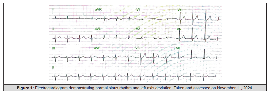
Figure 1:Electrocardiogram demonstrating normal sinus rhythm and left axis deviation. Taken and assessed on November 11, 2024.
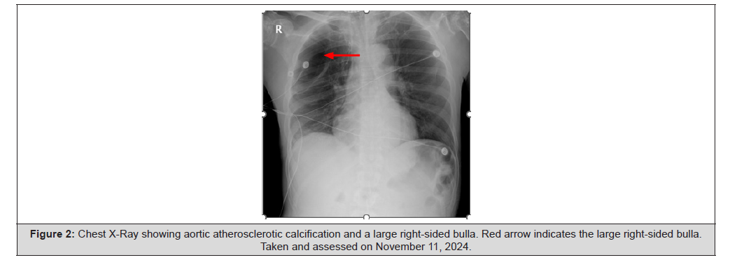
Figure 2:Chest X-Ray showing aortic atherosclerotic calcification and a large right-sided bulla. Red arrow indicates the large right-sided bulla. Taken and assessed on November 11, 2024.
Three hours later, contrast-enhanced CT imaging of the chest, abdomen, and pelvis showed a subscapular splenic hematoma with active extravasation from the posterior periphery of the spleen (Figure 3). Furthermore, there was moderate, diffuse intraperitoneal hemorrhage predominantly in the left upper quadrant, extending along the anterior left peritoneal cavity, anterior to the stomach, and posterior margin of the left hepatic lobe. Other hemorrhagic collections were noted in the perihepatic space, bilateral paracolic gutters, and pelvis. No intraperitoneal free air or bowel obstruction was identified. Additional findings included retained surgical staples from prior abdominal surgery and bilateral apical bullous changes, more prominent on the right. He was also noted to be hypotensive and anemic with a hemoglobin of 6.8 g/dL, which was corrected with administration of normal saline bolus, adjustment to Trendelenburg position, transfusion of two units of red blood cells, administration of tranexamic acid, and fresh frozen plasma. Interventional radiology was consulted, and the patient underwent successful coil embolization of the ruptured branch of the splenic artery without complications. Subsequently, his hemoglobin stabilized at 10.0 g/dL. After three days in the ICU, he was transferred in stable condition.
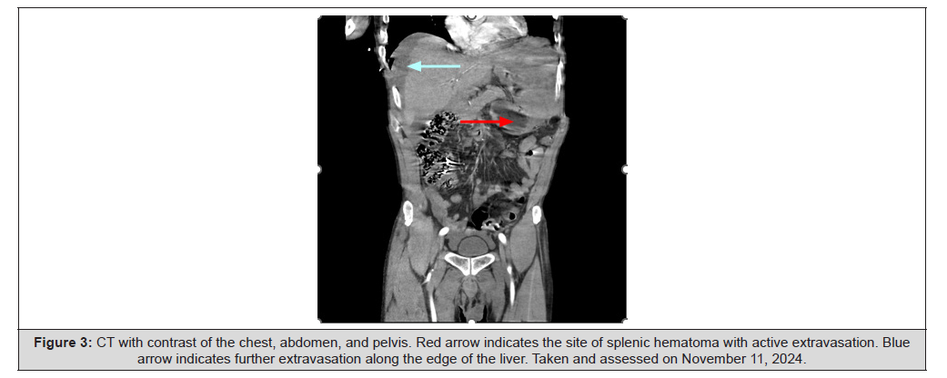
Figure 3:CT with contrast of the chest, abdomen, and pelvis. Red arrow indicates the site of splenic hematoma with active extravasation. Blue arrow indicates further extravasation along the edge of the liver. Taken and assessed on November 11, 2024.
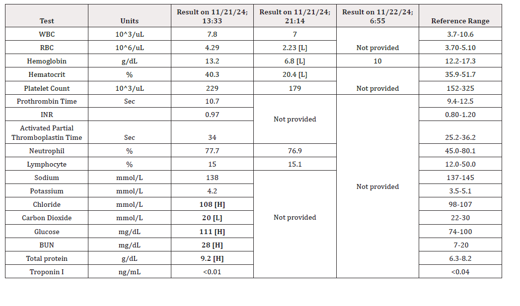
Table 1:Laboratory values taken on November 21, 2024 and November 22, 2024.
Note*: [H = high]; [L = low].
Discussion
Our case report joins a small yet growing body of evidence highlighting the potential adverse effects of colonoscopies, with the aim of increasing awareness and reducing associated mortality. While most reports emphasize mechanical injury mechanisms, our case uniquely illustrates how overlapping mechanical, vascular, and pharmacologic factors can interact to both precipitate and worsen splenic hemorrhage. Although the exact mechanism of PCSI remains unknown, prior studies suggest that excessive traction on the splenocolic ligament, tension on splenocolic adhesions, or direct trauma to the spleen while navigating the colonoscope through the splenic flexure can cause splenic injury [3-6]. In our patient, a history of abdominal laparotomy likely led to splenocolic adhesions that restricted splenic mobility, rendering the spleen more susceptible to traction injury as the colonoscope advanced through the splenic flexure. Compounding this, his chronic 30 pack-year smoking history may have weakened splenic vascular integrity through endothelial dysfunction and chronic inflammation [9]. Thus, even minimal traction could have placed disproportionate stress on fragile arterial branches, priming the spleen for rupture under relatively mild procedural strain.
The situation was further aggravated by pharmacologic factors. Our patient’s left-sided chest and abdominal pain initially raised concern for myocardial infarction, leading to the administration of 325 mg of aspirin before the diagnosis of splenic injury was established. However, in the setting of chronic NSAID use, this combination likely amplified platelet dysfunction through additive COX-1 inhibition. Together, these agents may have exacerbated ongoing splenic hemorrhage, accelerating his hemoglobin decline and worsening hemodynamic instability. Therefore, it is crucial to obtain a thorough medication history in post-colonoscopy patients presenting with chest pain, as certain interventions intended for alternative diagnoses may inadvertently worsen hemorrhage.
Timely recognition of PCSI is critical, as delays in diagnosis can allow hemorrhage to progress to significant hemodynamic compromise. The majority of PCSI cases (74%) develop symptoms within 24 hours of the colonoscopy [10], though some patients exhibit delayed symptom presentation up to three days post-procedure [11]. Our patient had a moderately delayed symptom presentation two days after his colonoscopy, which may be attributed to his splenocolic adhesions hindering the onset of signs of peritoneal irritation [12]. This case underscores the importance of maintaining a high index of suspicion for PCSI even when symptom onset is atypical, particularly in patients with prior abdominal surgery or other predisposing factors.
If splenic trauma is suspected, immediate contrast-enhanced CT imaging is essential for assessing the extent of injury and guiding management. CT not only confirms the diagnosis but also facilitates injury classification using the American Association for the Surgery of Trauma (AAST) grading scale, which evaluates factors such as laceration depth, hematoma size, and evidence of active extravasation. A recent systematic review reported CT sensitivity for splenic trauma to be as high as 98.5% [5], making it the most reliable tool for evaluation. For our patient, imaging revealed a subcapsular hematoma involving over 50% of the splenic surface with active extravasation, consistent with a Grade III splenic injury. This finding was critical in expediting interventional radiology consultation and guiding successful coil embolization, thereby preventing further hemodynamic decline and possible splenectomy. Ultimately, this case reinforces several key clinical lessons. First, prior abdominal surgery with resultant adhesions can predispose patients to splenic traction injury and delay symptom onset. Second, coexisting vascular risk factors such as chronic smoking may compound susceptibility to rupture. Third, administration of antiplatelet agents before PCSI diagnosis can aggravate bleeding and worsen clinical outcomes, particularly in patients with chronic NSAID use. Together, these insights expand the current understanding of PCSI by illustrating how mechanical predisposition, vascular fragility, and pharmacologic factors can converge to influence bothpresentation and outcome. Heightened awareness of these overlapping risks is essential for diagnosis and appropriate management in post-colonoscopy patients.
Conclusion
Post-colonoscopy splenic injury, though rare, is a potentially life-threatening complication that requires heightened clinical awareness. A combination of abdominal and left shoulder pain following a colonoscopy should prompt immediate consideration of PCSI, particularly in the presence of hemodynamic instability. Early recognition and timely imaging with CT are critical to confirm the diagnosis, guide management, and reduce morbidity and mortality. While prior abdominal surgery and excessive traction on the splenocolic ligament are established risk factors, additional contributors, including chronic smoking and pharmacologic interactions, may exacerbate splenic injury. Further research is needed to clarify this association and to inform risk mitigation strategies during colonoscopy procedures.
Acknowledgement
None.
Conflict of Interest
None.
References
- Joseph D, Meester R, Zauber A, Manninen D, Winges L, et al. (2016) Colorectal cancer screening: estimated future colonoscopy need and current volume and capacity. Cancer 122(16): 2479-2486.
- Ebner D, Rutten L, Miller Wilson L, Markwat N, Vahdat V, et al. (2024) Trends in colorectal cancer screening from National Health Interview Survey: analysis of modalities impact on overall screening Rates. Cancer Prev Res (Phila) 17(6): 275-280.
- Li S, Gupta N, Kumar Y, Mele F (2017) Splenic laceration after routine colonoscopy, a case report of a rare iatrogenic complication. Transl Gastroenterol Hepatol 2: 49.
- Shankar S, Rowe S (2025) Splenic injury after colonoscopy: case report and review of literature. Ochsner J 11(3): 276-281.
- Ullah W, Rashid M, Mehmood A, Zafar Y, Hussain I, et al. (2020) Splenic injuries secondary to colonoscopy: rare but serious complication. World J Gastrointest Surg 12(2): 55-67.
- Skipworth J, Raptis D, Rawal J, Damink S, Shankar A, et al. (2009) Splenic injury following colonoscopy—an underdiagnosed, but soon to increase, phenomenon? Ann R Coll Surg Engl 91(4): W6-11.
- Koop H, Skupnik C, Schnoor T, Horenkamp Sonntag D (2024) Splenic injury associated with colonoscopy in hospitalized patients: incidence, risk factors, management, and outcome. Endosc Int Open 12(12): E1453-E1457.
- Touati M, Bouzid A, Khefacha F, Othmane M, Belhadj A, et al. (2024) Post-colonoscopy splenic injury: a case report on diagnostic challenges and treatment strategies. Int J Surg Case Rep 123: 110243.
- Rao K, Beri G, Sterling M, Salen G (2009) Splenic injury as a complication of colonoscopy: a case series. Am J Gastroenterol 104(6): 1604-1605.
- Ha J, Minchin D (2009) Splenic injury in colonoscopy: a review. Int J Surg 7(5): 424-427.
- Prowda J, Trevisan S, Lev Toaff A (2005) Splenic injury after colonoscopy: conservative management using CT. Am J Roentgenol 185(3): 708-710.
- Cappellani A, Di Vita M, Zanghì A, Cavallaro A, Alfano G, et al. (2008) Splenic rupture after colonoscopy: report of a case and review of literature. World J Emerg Surg 3(1): 8.




 We use cookies to ensure you get the best experience on our website.