
Review Article 
 Creative Commons, CC-BY
Recalibrating Retinal Immunity: Peptide-Based Strategies for Durable Vision Preservation in Retinitis Pigmentosa
*Corresponding author:Jonathan RT Lakey, University of California, Irvine- Department of Surgery and Biomedical Engineering, 4University of California, Irvine, Department of Biomedical Engineering, Irvine, CA, USA.
Received:August 04, 2025; Published:August 11, 2025
DOI: 10.34297/AJBSR.2025.28.003638
Abstract
Retinitis pigmentosa (RP) is a heterogeneous group of inherited retinal degenerative diseases characterized by progressive photoreceptor loss, chronic inflammation, and immune dysregulation that ultimately lead to irreversible vision impairment. Current treatments largely focus on symptomatic management, with limited options to halt or reverse disease progression. Emerging regenerative immunotherapies and peptide-based biologics offer promising avenues to address the multifactorial pathogenesis of RP by targeting key mechanisms such as oxidative stress, neuroinflammation, and aberrant immune activation. Peptide therapeutics possess unique advantages including high specificity, low immunogenicity, and the ability to modulate immune responses and promote retinal repair. Bioactive peptides like thymosin β4 and melanocortin-derived KPV exhibit immunomodulatory and neuroprotective effects by restoring epithelial integrity, reducing oxidative damage, and recalibrating local immune responses. Advances in delivery systems are enhancing peptide stability, targeted retinal delivery, and therapeutic durability. Autologous Active Specific Immunotherapy (AASI) represents a transformative immunologic approach aimed at inducing antigen-specific tolerance by selectively reprogramming maladaptive T cell responses against retinal autoantigens. Preclinical studies in autoimmune and inflammatory diseases highlight the potential of AASI to suppress pathogenic effector T cells while expanding regulatory T cells, thereby restoring immune homeostasis without broad immunosuppression. Combining AASI with stem cell therapies or peptide biologics may synergistically promote retinal regeneration and durable immune quiescence. Challenges remain in clinical translation, including manufacturing standardization, regulatory approval pathways, long-term safety, and trial design complexity. Integration of multi-omic biomarker profiling and artificial intelligence-driven patient stratification promises to optimize personalized treatment regimens. Together, these advances herald a paradigm shift toward precision regenerative immunotherapy in RP, with the potential to arrest degeneration, restore vision, and improve quality of life for affected individuals.
Keywords:Retina, Peptides, Immunity
Introduction
Retinitis pigmentosa (RP) refers to a clinically and genetically heterogeneous group of inherited retinal dystrophies [1]. RP is characterized by progressive photoreceptor degeneration and retinal pigment epithelium (RPE) dysfunction. RP affects approximately 1 in 4,000 individuals worldwide, representing one of the leading causes of inherited blindness [2]. Unlike age-related macular degeneration (AMD), which typically affects central vision later in life [3], RP often presents with night blindness and peripheral vision loss in adolescence or early adulthood, ultimately progressing to complete blindness in severe cases [4]. The etiology of RP is multifactorial, involving over 80 identified genes associated with rod and cone photoreceptor structure and function, RPE integrity, ciliary trafficking, and retinal metabolism [5]. However, despite the substantial genetic underpinnings, many cases exhibit variable penetrance and disease progression, implicating additional layers of environmental, immunological, and metabolic influence [1,2,5].
Recent evidence suggests that RP pathogenesis extends beyond intrinsic photoreceptor dysfunction [6]. Chronic inflammation, oxidative stress, microglial activation, and secondary immune-mediated tissue damage are increasingly recognized contributors to disease progression, particularly in later stages [7]. Studies have identified persistent activation of retinal microglia, complement deposition, and pro-inflammatory cytokine expression (e.g., IL-1β, TNF-α, IL-6) in both animal models and postmortem RP retinas [8]. This inflammatory milieu exacerbates photoreceptor apoptosis, disrupts the blood-retinal barrier, and may hinder regenerative responses [5,7,8]. Despite advances in gene therapy for specific monogenic subtypes (e.g., RPE65-Luxturna) [9], current treatments remain limited in scope, and most patients lack effective interventions that halt or reverse vision loss. Importantly, no approved therapies directly target the inflammatory or immune components of RP pathobiology [10].
Emerging concepts in retinal disease now emphasize the dual necessity of controlling neuroinflammation and promoting tissue regeneration. Studies in degenerative retinal diseases have demonstrated that targeting innate immune modulators, such as TLRs and microglial polarization pathways, may attenuate photoreceptor degeneration and support retinal survival [4-8]. Additionally, advances in peptide-based therapeutics offer novel tools to modulate retinal immune responses, promote cellular repair, and even restore immune tolerance to self-antigens implicated in secondary autoimmune retinopathies [11,12]. For example, peptides derived from ocular immunosuppressive environments, such as α-MSH, SOCS1-mimetic peptides, or thymosin β4, show promise in preclinical models for preserving retinal structure and function through anti-inflammatory and pro-repair mechanisms [12,13].
More recently, the concept of Antigen-Adaptive Selective Immunotherapy (AASI) has emerged, leveraging immune-tolerizing peptides to reprogram pathogenic T cell responses implicated in autoimmunity and post-injury retinal degeneration [14,15]. These precision immunotherapies hold potential to prevent secondary immune-mediated damage in RP and complement gene-based interventions by restoring immunological homeostasis. In parallel, organ-specific targeting strategies using homing peptides capable of crossing ocular barriers and binding selectively to degenerating retinal tissues have opened new avenues for tissue-specific delivery of bioactive peptides and regenerative cargos [16]. This review will explore emerging translational strategies in RP through three interrelated domains:
1. Retina-targeted stem/progenitor cell and peptide-based delivery
systems – including retinal homing peptides for organ-specific
targeting of therapeutic agents to degenerating photoreceptor
and RPE cells [17].
2. Peptide-based immunoregenerative strategies, using therapeutic
peptides (e.g., α-MSH analogs, SOCS1-mimetics, KPV,
thymosin β4) to modulate neuroinflammation, promote RPE
regeneration, and attenuate fibrosis [18].
3. Antigen-Specific Tolerogenic Peptide Therapy leveraging retinal
autoantigen-derived peptides (e.g., recoverin, arrestin)
[19] to restore immune tolerance in autoimmune retinopathies
and RP with inflammatory overlap [20].
Together, these emerging strategies represent a paradigm shift from symptomatic management to targeted immunoregulation and tissue preservation, offering a promising future for halting progression and restoring vision in individuals with RP.
Immunopathogenesis of Retinitis Pigmentosa
Genetic and Environmental Contributions. RP arises from a complex interaction between inherited mutations and secondary modulators such as oxidative stress, immune activation, and metabolic disturbances, which together undermine retinal immune privilege and tissue integrity [21]. Over 80 genes have been implicated in non-syndromic RP, encompassing critical roles in phototransduction, ciliary trafficking, RPE phagocytosis, and mitochondrial homeostasiss [22,23]. Among the most frequently mutated genes are RHO, RPGR, USH2A, and EYS, each associated with distinct patterns of photoreceptor loss and disease progression [1]. Mutations in genes such as PDE6B, NR2E3, and PRPF31 have been linked to disrupted retinal metabolism, DNA repair, or RNA splicing, resulting in accelerated apoptosis of rod photoreceptors and secondary cone degeneration [1].
In In addition to genetic defects intrinsic to photoreceptor cells, the pathogenesis of RP is increasingly recognized as being influenced by environmental and microenvironmental stressors [24]. Chronic oxidative stress, arising from high metabolic demand, continuous light exposure, and mitochondrial respiration, disproportionately affects metabolically active rod outer segments, leading to mitochondrial dysfunction and lipid peroxidation [21]. This oxidative milieu promotes protein misfolding, DNA damage, and cellular debris accumulation, each of which can act as danger-associated molecular patterns (DAMPs) that trigger innate immune responses [25]. Furthermore, inflammation-inducing factors such as infections, trauma, or even gut dysbiosis have been shown to influence microglial activation and immune cell infiltration in the retina [26]. Emerging data also implicate retinal dysbiosis and systemic immune priming as modulators of retinal inflammation, particularly in autoimmune retinopathies and syndromic forms of RP. Disruption of the outer blood-retinal barrier, characterized by altered expression and organization of tight junction proteins in the retinal pigment epithelium (RPE), may permit immune surveillance of normally sequestered retinal antigens (e.g., recoverin, α-enolase) [27]. This immune exposure can initiate a pathological feedforward loop involving autoreactive immune responses and progressive photoreceptor degeneration. Thus, therapeutic strategies aimed at modulating extrinsic stressors and restoring metabolic homeostasis may offer promising avenues for intervention in both monogenic and complex forms of retinal degenerative disease.
Dysregulated Immune Pathways
The chronic progression of RP reflects not only intrinsic photoreceptor degeneration but also a failure of neuroimmune regulation and tissue repair [8]. Central to this process are the maladaptive responses of microglia and Müller glia [8]. In early stages, microglia exhibit a homeostatic, neuroprotective phenotype [28]; however, sustained photoreceptor loss and oxidative stress promote their shift toward a pro-inflammatory, neurotoxic state characterized by secretion of IL-1β, TNF-α, and reactive oxygen/nitrogen species [29]. These activated microglia not only exert direct cytotoxic effects but also serve as key amplifiers of innate immune signaling, initiating a cascade of molecular events driven by pattern recognition receptors such as Toll-like receptors (TLRs), NOD-like receptors (NLRs), and RIG-I-like helicases [30], which detect cellular stress signals including misfolded proteins, mitochondrial debris, and nucleic acid fragments. This culminates in inflammasome activation. NLRP3, in particular induces caspase-1-mediated cleavage and release of IL-1β and IL-18, further amplifying inflammation and photoreceptor apoptosis [31]. Concurrently, dysregulation of autophagy, a process essential for cellular homeostasis and clearance of photoreceptor debris, results in toxic aggregate accumulation and sustained immune activation [31]. This chronic innate immune stimulation may, in turn, facilitate recruitment and activation of the adaptive immune system-a process increasingly evident in RP subtypes with autoimmune or para-infectious features.
Aberrant T cell responses, characterized by retinal antigen-specific Th1 and Th17 populations and impaired Treg function, have been observed in both animal models and patient cohorts [32]. Chronic exposure to pro-inflammatory cytokines such as IFN-γ, IL-17, and GM-CSF drives Müller cell gliosis, RPE dedifferentiation, and vascular dysfunction, contributing to tissue remodeling and progressive vision loss [7,8,21,29]. Crucially, resolution pathways are often inadequate. Deficits in pro-resolving mediators (e.g., neuroprotectin D1, lipoxins), insufficient Treg activity, and persistent antigen presentation prevent the restoration of immune homeostasis, even after the inciting genetic or environmental insult subsides. Collectively, RP represents a convergence of genetic vulnerability, metabolic dysregulation, and chronic immune activation. This evolving immunopathogenic model supports therapeutic strategies that go beyond gene correction to include targeted immunomodulation and regenerative, peptide-based approaches designed to interrupt destructive immune cascades, re-establish tolerance, and preserve retinal architecture.
Limitations of Current Therapeutics in Retinitis Pigmentosa
The therapeutic landscape for RP has seen major advances over the past decade. However, despite innovations, effective treatments remain unavailable for the vast majority of individuals with RP. The only FDA-approved gene therapy, voretigene neparvovec-rzyl (Luxturna), targets RPE65-associated disease and benefits a limited subset of patients with confirmed biallelic mutations and sufficient viable retina9. Numerous gene-specific strategies are under development, including antisense oligonucleotides, base editing, and optogenetics, yet these remain restricted to select genotypes and early disease stages [33].
Pharmacologic interventions have demonstrated limited efficacy and are not universally recommended due to inconsistent benefits and potential toxicity [33]. Neuroprotective agents including ciliary neurotrophic factor (CNTF), brimonidine, and anti-apoptotic peptides have been tested in clinical trials, yet none have yielded robust, sustained improvements in visual function. Cell-based therapies offer promise but face hurdles related to cell survival, integration, immunogenicity, and host inflammation as well as the added challenge of vector choice and engineering methods. Moreover, retinal prosthetics provide rudimentary vision restoration but are limited by resolution, invasiveness, and patient eligibility. Despite mechanistic advances, real-world effectiveness of these approaches remains constrained by the progressive and multifactorial nature of RP, particularly in mid- to late-stage disease. Crucially, most current therapies do not address secondary drivers of disease progression such as chronic retinal inflammation, microglial activation, or immune- mediated degeneration [34]. Persistent innate immune activation and failed resolution mechanisms have been implicated in bystander photoreceptor loss, RPE dysfunction, and retinal remodeling, regardless of the initiating mutation [35]. Collectively, limited capacity to incorporate strategies to modulate immune responses or prevent subsequent degeneration of spared retinal cells, has impaired long-term effectiveness.
Unmet Needs in Disease Management
Although preservation of structural retina is a prerequisite for many emerging therapies, patients frequently experience functional symptoms (e.g., photosensitivity, contrast loss, delayed dark adaptation, visual field constriction) despite apparent anatomic stability [36]. These deficits often arise from non-cell autonomous mechanisms including oxidative stress, cytokine-mediated synaptic dysfunction, and glial remodeling that are not resolved by mutation correction alone. RP also has systemic associations, particularly in syndromic forms such as Usher and Bardet-Biedl syndromes, which involve multi-organ dysfunction including hearing loss, obesity, diabetes, and cognitive impairment [37]. These manifestations may reflect shared pathogenic mechanisms (e.g., ciliary dysfunction, mitochondrial stress) that are not targeted by ocular therapies. Importantly, chronic retinal degeneration itself may trigger systemic immune priming, blood-retinal barrier breakdown, and secondary autoimmune responses, compounding disease progression. Moreover, none of the currently available or investigational RP therapies directly promote tissue regeneration or restore immune tolerance. Photoreceptor or RPE replacement is technically challenging and carries risks of immune rejection and inflammation. Immunomodulatory agents that target retinal microglia or restore regulatory T cell activity are not yet integrated into standard treatment paradigms [38]. This therapeutic void is especially problematic in advanced disease stages, where photoreceptor loss, gliosis, and immune remodeling dominate the retinal landscape. Additionally, repeated therapeutic interventions including subretinal injections and chronic inflammation may sensitize the retina to immune activation, raising concerns about long-term tolerance and safety. Together, these limitations highlight the need for next-generation therapies that extend beyond genetic correction to encompass immune recalibration, neuroregeneration, and tissue-specific delivery. Organ-targeted peptides and antigen-specific tolerogenic therapies represent promising strategies to modulate the retinal immune microenvironment, halt secondary degeneration, and promote long-lasting retinal preservation regardless of genotype or stage of disease [39] (Table 1).
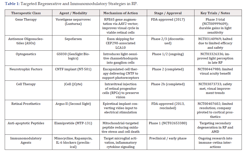
Table 1:Targeted Regenerative and Immunomodulatory Strategies in RP.
Targeted Regenerative and Immunomodulatory Strategies in RP
Conventional therapies are typically mutation-specific, temporizing, and insufficient to restore functional vision once significant retinal loss has occurred. Regenerative and immunomodulatory strategies, including stem cell transplantation, RPE replacement, Müller glia reprogramming, and peptide therapeutics are being investigated to address these critical gaps.
Retinal Progenitor Cells (RPCs) offer a potential source of photoreceptor and glial replacement. Preclinical studies demonstrate the ability of transplanted RPCs to integrate into the outer nuclear layer, secrete neuroprotective factors, and partially restore visual function in animal models of RP [40]. Several early-phase human trials using fetal- or hESC-derived RPCs have shown safety and signals of efficacy when delivered subretinally (e.g., NCT02464436, NCT03073733) [41]. However, challenges such as immune rejection, surgical delivery limitations, and inconsistent photoreceptor differentiation limit widespread application [42,43].
Mesenchymal Stem Cells (MSCs) are also under investigation for RP due to their paracrine secretion of neurotrophic and anti-inflammatory cytokines (e.g., BDNF, CNTF, TGF-β). Intravitreal injection of MSCs has been shown to delay photoreceptor degeneration in animal models, and preliminary clinical trials report structural preservation and improved visual acuity in some patients [44]. Nonetheless, the therapeutic benefits are often transient, and repeated administration may be needed due to the short-lived nature of secreted factors. Moreover, the risk of proliferative vitreoretinopathy or ectopic tissue formation persists, particularly with non-encapsulated delivery [45].
Induced Pluripotent Stem Cells (iPSCs) have revolutionized regenerative medicine with their capacity to generate patient-specific retinal cell types. iPSC-derived RPE or photoreceptor precursors have been shown to engraft and preserve retinal structure and function in RP models [46]. However, the risk of tumorigenicity, immune recognition, and manufacturing variability remain significant translational hurdles. Moreover, retinal integration of photoreceptors without synaptic connectivity has proven insufficient to restore complex visual processing [47].
Müller Glia Reprogramming represents an emerging endogenous regenerative strategy. In lower vertebrates, Müller cells can dedifferentiate into retinal progenitors and regenerate photoreceptors, but this capacity is minimal in humans [48]. Peptides such as HB-EGF and Notch inhibitors have been tested in combination with transcription factors (e.g., Ascl1) to induce reprogramming in mammalian retina, with early signs of neurogenesis [49]. Further work is needed to improve lineage fidelity, mitigate reactive gliosis, and optimize delivery to activate Müller glia repair pathways in human RP. However, reprogramming retinal cells remains a promising strategy [49]. Realizing its full therapeutic potential will require overcoming delivery challenges, safety concerns, and integration barriers. In parallel, peptide therapeutics represent a compelling complementary approach, scalable, modulable, and mutation-agnostic, capable of promoting endogenous repair, immune recalibration, and mitochondrial stabilization, all hallmarks of neuroprotective intervention in RP.
Peptide-Based Therapeutics for Retinitis Pigmentosa
Peptide-based therapeutics offer a promising avenue for the treatment of RP by modulating degenerative pathways, restoring mitochondrial function, mitigating oxidative stress, and preserving photoreceptor survival. Melanocortin peptides such as α-MSH and its synthetic analog NDP-α-MSH exert potent anti-inflammatory and neuroprotective effects in retinal tissue via melanocortin receptor 1 (MC1R)-dependent mechanisms. These peptides inhibit NF-κB activation and suppress retinal microglial activation, thereby reducing photoreceptor apoptosis in rd10 and light-induced degeneration models [50,51]. Similarly, the tripeptide KPV (Lys-Pro-Val), derived from α-MSH, attenuates retinal inflammation and preserves barrier function in diabetic retinopathy models by modulating IL-1β and tight junction protein expression [52].
The mitochondria-targeted tetrapeptide SS-31 (elamipretide) is among the most advanced candidates for RP, having demonstrated the ability to restore mitochondrial membrane potential, reduce cytochrome c release, and prevent photoreceptor cell death in models of oxidative and metabolic stress [53]. In a canine model of autosomal dominant RP (T4R RHO mutation), daily SS-31 treatment preserved outer retinal structure and slowed functional decline, highlighting its translational promise [54]. SS-31 also stabilizes cardiolipin in the inner mitochondrial membrane, a key determinant of photoreceptor viability, and has progressed to clinical trials in primary mitochondrial disease and AMD, potentially paving the path for repurposing in RP [55]. These findings suggest the therapeutic potential of melanocortin-derived peptides in hereditary and acquired retinal degenerations.
Antigen-specific tolerizing peptides such as RBP-3_1177–1191 and IRBP_651–670 have been previously tested in experimental autoimmune uveoretinitis (EAU) as surrogate models of immune-mediated photoreceptor loss [56]. The capacity to induce Treg expansion and reduce Th1/Th17-driven inflammation in the retina, was speculated to offer a potential strategy for RP subtypes with autoimmune components or inflammatory exacerbation. In addition, Tβ4 (thymosin beta-4), a regenerative peptide with G-actin binding, pro-angiogenic, and anti-apoptotic properties, supports Müller glia-mediated repair and reduces retinal gliosis in light-induced injury models has been identified as having a potential beneficial effect for maintaining the blood-retina barrier and supporting neurovascular units in RP [57]. While these early findings underscore the promise of antigen-specific and regenerative peptides in modulating immune and glial responses, further studies are needed to refine delivery strategies, validate efficacy in RP-specific models, and delineate patient subgroups most likely to benefit.
Synthetic peptides derived from rod outer segment (ROS) proteins and crystallins are also under investigation for their cytoprotective functions [58]. Aα-crystallin-derived mini-chaperone peptide has been shown to prevent light-induced photoreceptor apoptosis by stabilizing misfolded proteins and attenuating oxidative stress via inhibition of caspase-3 activation [59]. Additionally, neuroprotective peptides such as NAP (NAPVSIPQ), derived from activity-dependent neuroprotective protein (ADNP), promote microtubule stabilization and preserve retinal structure in multiple models of degeneration [60].
Despite the therapeutic potential, peptide drugs face barriers including short half-life, enzymatic degradation, and limited ocular penetration. Strategies to overcome these limitations include PEGylation, cyclization, and nanoformulation. PEGylated versions of Tβ4 and SS-31 analogs demonstrate extended half-life and bioactivity in ocular tissues [61]. Liposomal formulations and exosome- based carriers are also under investigation to enhance peptide retention in the subretinal space and improve cellular uptake, particularly by photoreceptors and RPE cells [62], highlighting the potential integration of nanoparticles and exosomes with existing therapies and the development of multifunctional and personalized treatment strategies.
Antigen Adaptive Selective Immunotherapy (AASI) in Retinitis Pigmentosa
Antigen Adaptive Selective Immunotherapy (AASI) is an innovative immunological strategy aiming to reinstate immune tolerance by targeting maladaptive T-cell responses to specific retinal antigens [63]. Contrary to conventional immunosuppression in autoimmune or inflammatory retinal disorders, which often fails to preserve vision due to non-specific action and significant side effects, AASI offers a paradigm shift by recalibrating immune responses against pathogenic retinal epitopes without compromising systemic immunity [64].
To date, recoverin, α-enolase, arrestin, among other antigens have been implicated in non-paraneoplastic autoimmune retinopathy, a condition that overlaps histopathologically with inflammatory RP, with studies show circulating anti-retinal antibodies in up to 50% of RP patients [65]. AASI offers a targeted approach: delivering these antigens (e.g., recoverin-derived peptides) in tolerogenic forms to induce retinal antigen–specific Tregs, promote anergy in effector T cells, and re-establish retinal tolerance. Preclinical and translational models in autoimmune eye disease support this approach: tolerogenic dendritic cells loaded with retinal peptides or peptide–nanoparticle platforms have successfully expanded FoxP3⁺ Tregs and suppressed Th1/Th17 responses in equivalent retinal inflammation models [66]. These strategies echo successes in other organ-specific autoimmune diseases, where peptide-based tolerance (oral, subcutaneous, or nanoparticle-mediated) has demonstrated durable remission with low systemic toxicity. No AASI trials have yet been conducted in RP, but interventions targeting multiple retinal autoantigens, tailored to patient HLA and antibody profiles, could halt immune-mediated photoreceptor loss and stabilize retinal function. Proposed mechanisms of action include:
i. Tolerogenic antigen presentation: Co-delivery of retinal peptides
via subcutaneous or ocular nanoparticle systems in
non-inflammatory contexts (e.g., with IL 10–expressing dendritic
cells) drives Treg induction and effector T cell anergy
[67].
ii. Linked suppression: Induced Tregs targeting one retinal antigen
may suppress bystander autoreactive responses due to
epitope spreading [68].
iii. Selective modulation: Unlike broad cytokine blockade, AASI
permits retinal-specific immunomodulation, with immune
surveillance elsewhere maintained [69].
Clinically, AASI holds promise for targeting inflammatory subtypes of RP. These strategies could reinforce the immune microenvironment and enhance the efficacy of co-administered regenerative treatments such as peptides or stem cell-based therapies. Autoimmune retinopathy, including non-paraneoplastic autoimmune retinopathy (npAIR), often shares clinical and antigenic features with inflammatory RP, including the presence of circulating antiretinal antibodies (e.g., recoverin, α-enolase), suggesting a role for immune-mediated photoreceptor damage. Antigen-specific approaches have demonstrated efficacy in preclinical models of ocular autoimmunity by expanding Tregs capable of suppressing effector responses and linked antigenic cascades. To enable personalized deployment of AASI in RP, future work must focus on precise antigen identification using multi-omic profiling of patient-specific antibody repertoires and T-cell epitopes.
Integration and Translational Considerations
Precision medicine approaches in RP are increasingly guided by biomarker-driven stratification to optimize therapeutic efficacy while minimizing off-target or unnecessary interventions. Integrative multi-omic profiling has begun to reveal biologically distinct patient subgroups more likely to benefit from specific immunomodulatory or regenerative therapies. For instance, pathogenic variants in genes such as RPGR, RHO, and USH2A are now routinely used to stratify patients for gene or cell-based interventions [70]. Concurrently, emerging ocular biomarkers, including retinal microglial activation patterns and quantitative fundus autofluorescence (qAF) changes, offer additional means for patient segmentation based on disease activity and prognosis [71]. Advanced machine learning algorithms are being developed to integrate multimodal data streams, such as spectral-domain optical coherence tomography (OCT) parameters and proteomic profiles from vitreous biopsies to forecast disease trajectory and therapeutic responsiveness [72]. These tools are essential for identifying RP subpopulations most likely to benefit from antigen-specific immunotherapies, including tolerogenic peptide platforms, in which a permissive immune environment is critical for long-term engraftment and function. Given the complex interplay of genetic, immunologic, and microenvironmental drivers in RP pathogenesis, monotherapies may prove inadequate for many patients. Rationally designed combination therapies are under active investigation, targeting multiple pathological axes simultaneously. Similarly, co-administration of bioactive peptides can synergistically promote Müller glia-mediated repair and photoreceptor survival, which may be further augmented by layering on current or emerging therapies, including gene augmentation, RNA editing, or anti-complement agents. For instance, short-term induction with low-dose JAK inhibitors or targeted corticosteroids may transiently dampen retinal inflammation, facilitating the induction phase of antigen-specific tolerance without broadly suppressing protective immunity [73]. This induction-maintenance strategy parallels successful paradigms in other autoimmune and degenerative diseases where immune re-education enables durable disease modification rather than temporary symptom control. In RP, this paradigm may shift the therapeutic goal from slowing degeneration to achieving immune homeostasis and functional retinal restoration.
Challenges and Regulatory Pathways
Despite the considerable promise of multi-modal regenerative immunotherapies for RP, several critical challenges must be overcome to ensure successful clinical translation. Manufacturing biologics require rigorous quality control, batch consistency, and reproducibility, particularly in the context of AASI. Regulatory frameworks (FDA, EMA, etc.) will need to evolve to accommodate personalized, combinatorial biologics where each component may have distinct stability and handling requirements.
Although regulatory guidance increasingly encourages adaptive platform trial designs to parse out individual component contributions, optimize synergistic dosing, and efficiently evaluate safety and efficacy, these approaches are inherently complex. Novel clinical endpoints relevant to RP will be essential to demonstrate meaningful clinical benefit beyond standard visual acuity measures. Stratified randomization based on immune, genetic, or imaging biomarkers may be used to stratify responders from non-responders. Optimizing trial design may also serve to reduce interpatient heterogeneity, improving trial power and interpretability. Long-term safety remains a paramount concern, especially for immune-modulating therapies.
The future of RP treatment lies in integrating precision immunology with regenerative medicine to overcome the limitations of current approaches that primarily focus on symptomatic management or gene correction. Advances in antigen discovery, epitope mapping, and HLA-typing enable the development of personalized immunotherapies targeting pathogenic retinal autoantigens. When coupled with innovative delivery platforms such as nanoparticle- encapsulated peptides, AASI holds promise for restoring immune tolerance while preserving protective immunity. Genetically engineered stem cells and exosome-based therapies are expanding the therapeutic repertoire. Advancements are augmenting opportunities to enhance immunosuppressive and homing capabilities to retinal tissues (i.e., via MSC therapy). Further, exosomes serve as cell-free vehicles for delivering anti-inflammatory microRNAs, neuroprotective peptides, and immune modulators. Collectively, these approaches may allow targeted repair of photoreceptors and retinal pigment epithelium, halting or reversing degenerative processes. Artificial intelligence and systems biology will play transformative roles in RP drug discovery and patient stratification. AI platforms can integrate transcriptomic, imaging, and immune profiling data to generate comprehensive patient phenotypes, enabling precision targeting of pathogenic mechanisms and optimization of combination therapies. Machine learning models may also accelerate preclinical screening by simulating complex retinal immune interactions and predicting clinical outcomes before costly human trials [1]. Together, these advances mark a paradigm shift from transient symptom suppression toward durable immune recalibration, tissue regeneration, and vision preservation. By addressing the core mechanisms of immune dysregulation, oxidative stress, and cellular degeneration, these personalized, mechanism-driven therapies hold the potential to revolutionize RP care and serve as a blueprint for treating other inherited and autoimmune ocular diseases.
References
- Suleman N (2025) Current understanding on Retinitis Pigmentosa: a literature review. Front Ophthalmol 5: 1600283.
- Cross N, van Steen C, Zegaoui Y, Satherley A and Angelillo L (2022) Retinitis Pigmentosa: Burden of Disease and Current Unmet Needs. Clin Ophthalmol Auckl NZ 16: 1993-2010.
- Fleckenstein M, Schmitz-Valckenberg S and Chakravarthy U (2024) Age-Related Macular Degeneration: A Review. JAMA 331(2): 147-157.
- Georgiou M, Robson AG, Jovanovic K, Uwaydat SH, Ji MH, et al (2023) RP2-Associated X-linked Retinopathy: Clinical Findings, Molecular Genetics, and Natural History. Ophthalmology 130(4): 413-422.
- Manley A, Meshkat BI, Jablonski MM and Hollingsworth TJ (2023) Cellular and Molecular Mechanisms of Pathogenesis Underlying Inherited Retinal Dystrophies. Biomolecules 13(2): 271.
- Bighinati A, Adani E, Stanzani A, D’Alessandro S and Marigo V (2024) Molecular mechanisms underlying inherited photoreceptor degeneration as targets for therapeutic intervention. Front Cell Neurosci 18: 1343544.
- Murakami Y, Nakabeppu Y and Sonoda KH (2020) Oxidative Stress and Microglial Response in Retinitis Pigmentosa. Int J Mol Sci 21(19): 7170.
- Zhao L, Hou C and Yan N (2022) Neuroinflammation in retinitis pigmentosa: Therapies targeting the innate immune system. Front Immunol 13: 1059947.
- Maguire AM, Bennett J, Aleman EM, Leroy BP and Aleman TS (2020) Clinical Perspective: Treating RPE65-Associated Retinal Dystrophy. Mol Ther J Am Soc Gene Ther 29(2): 442-463.
- Nair PP, Keskar MP, Borghare PT, Dzoagbe HY and Kumar T (2024) The New Era of Therapeutic Strategies for the Management of Retinitis Pigmentosa: A Narrative Review of the Pathomolecular Mechanism for Gene Therapies. Cureus 16(8): e66814.
- Mandal A, Pal D, Agrahari V, Trinh HM, Joseph M, et al (2018) Ocular delivery of proteins and peptides: Challenges and novel formulation approaches. Adv Drug Deliv Rev 126: 67-95.
- Boyer DD, Ko YP, Podos SD, Cartwright ME, Gao X, et al (2022) Danicopan, an Oral Complement Factor D Inhibitor, Exhibits High and Sustained Exposure in Ocular Tissues in Preclinical Studies. Transl Vis Sci Technol 11(10): 37.
- Goit RK, Taylor AW and Lo ACY (2022) Anti-inflammatory α-Melanocyte-Stimulating Hormone Protects Retina After Ischemia/Reperfusion Injury in Type I Diabetes. Front Neurosci 16: 799739.
- Wooldridge L, Ekeruche-Makinde J, van den Berg HA, Skowera A, Miles JJ, et al (2012) A single autoimmune T cell receptor recognizes more than a million different peptides. J Biol Chem 287(2): 1168-1177.
- Yang P, Mustafi D and Pepple KL (2024) Immunology of Retinitis Pigmentosa and Gene Therapy-Associated Uveitis. Cold Spring Harb Perspect Med 14(1): a041305.
- Attia SA and MacKay JA (2022) Protein and polypeptide mediated delivery to the eye. Adv Drug Deliv Rev 188: 114441.
- Guo D, Sun Y, Wu J, Ding L, Jiang Y, et al (2024) Photoreceptor-targeted extracellular vesicles-mediated delivery of Cul7 siRNA for retinal degeneration therapy. Theranostics 14(13): 4916-4932.
- Benita BA and Koss KM (2024) Peptide discovery across the spectrum of neuroinflammation; microglia and astrocyte phenotypical targeting, mediation, and mechanistic understanding. Front Mol Neurosci 17: 1443985.
- Yang S, Dizhoor A, Wilson DJ and Adamus G (2016) GCAP1, Rab6, and HSP27: Novel Autoantibody Targets in Cancer-Associated Retinopathy and Autoimmune Retinopathy. Transl Vis Sci Technol 5(3): 1.
- Zala A and Thomas R (2023) Antigen-specific immunotherapy to restore antigen-specific tolerance in Type 1 diabetes and Graves’ disease. Clin Exp Immunol 211(2): 164-175.
- Gallenga CE, Lonardi M, Pacetti S, Violanti SS, Tassinari P, et al (2021) Molecular Mechanisms Related to Oxidative Stress in Retinitis Pigmentosa. Antioxid Basel Switz 10(6): 848.
- Becherucci V, Bacci GM, Marziali E, Sodi A, Bambi F, et al (2023) The New Era of Therapeutic Strategies for the Treatment of Retinitis Pigmentosa: A Narrative Review of Pathomolecular Mechanisms for the Development of Cell-Based Therapies. Biomedicines 11(10): 2656.
- Moran AL, Fehilly JD, Floss Jones D, Collery R and Kennedy BN (2022) Regulation of the rhythmic diversity of daily photoreceptor outer segment phagocytosis in vivo. FASEB J Off Publ Fed Am Soc Exp Biol 36(10): e22556.
- Huang ZY, Liang LN, Li YM, Xu K and Li XY (2022) Genetic, environmental and other risk factors for progression of retinitis pigmentosa. Int J Ophthalmol 15(5): 828-837.
- Mahaling B, Low SWY, Beck M, Kumar D, Ahmed S, et al (2022) Damage-Associated Molecular Patterns (DAMPs) in Retinal Disorders. Int J Mol Sci 23(5): 2591.
- Zysset-Burri DC, Morandi S, Herzog EL, Berger LE and Zinkernagel MS (2023) The role of the gut microbiome in eye diseases. Prog Retin Eye Res 92: 101117.
- Naylor A, Hopkins A, Hudson N and Campbell M (2019) Tight Junctions of the Outer Blood Retina Barrier. Int J Mol Sci 21(1): 211.
- Luo XG and Chen SD (2012) The changing phenotype of microglia from homeostasis to disease. Transl Neurodegener 1(1): 9.
- Rashid K, Wolf A and Langmann T (2018) Microglia Activation and Immunomodulatory Therapies for Retinal Degenerations. Front Cell Neurosci 12: 176.
- Rodríguez AM, Rodríguez J and Giambartolomei GH (2022) Microglia at the Crossroads of Pathogen-Induced Neuroinflammation. ASN Neuro 14: 17590914221104566.
- Huang Y, Xu W and Zhou R (2021) NLRP3 inflammasome activation and cell death. Cell Mol Immunol 18(9): 2114-2127.
- Mickael ME, Kubick N, Miftari K, Horbańczuk JO, Atanasov AG, et al (2025) The Role of Th17/Treg Axis in Retinal Pathology Associated with Diabetes and Treatment Options. Biology 14(3): 275.
- Kulbay M, Tuli N, Akdag A, Kahn Ali S and Qian CX (2024) Optogenetics and Targeted Gene Therapy for Retinal Diseases: Unravelling the Fundamentals, Applications, and Future Perspectives. J Clin Med 13(14): 4224.
- Hu A, Schmidt MHH and Heinig N (2024) Microglia in retinal angiogenesis and diabetic retinopathy. Angiogenesis 27(3): 311-331.
- George SM, Lu F, Rao M, Leach LL and Gross JM (2021) The retinal pigment epithelium: Development, injury responses, and regenerative potential in mammalian and non-mammalian systems. Prog Retin Eye Res 85: 100969.
- Verbakel SK, van Huet RAC, Boon CJF, Hollander AID, Collin RWJ, et al (2018) Non-syndromic retinitis pigmentosa. Prog Retin Eye Res 66: 157-186.
- Janáky M and Braunitzer G (2025) Syndromic Retinitis Pigmentosa: A Narrative Review. Vis Basel Switz 9(1): 7.
- Harris AR and Gilbert F (2022) Restoring vision using optogenetics without being blind to the risks. Graefes Arch Clin Exp Ophthalmol Albrecht Von Graefes Arch Klin Exp Ophthalmol 260(1): 41-45.
- Drag S, Dotiwala F and Upadhyay AK (2023) Gene Therapy for Retinal Degenerative Diseases: Progress, Challenges, and Future Directions. Invest Ophthalmol Vis Sci 64(7): 39.
- Thomas BB, Rajendran Nair DS, Rahimian M, Hassan AK, Tran TL, et al (2025) Animal models for the evaluation of retinal stem cell therapies. Prog Retin Eye Res 106: 101356.
- Singh R, Cuzzani O, Binette F, Sternberg H, West MD, et al (2018) Pluripotent Stem Cells for Retinal Tissue Engineering: Current Status and Future Prospects. Stem Cell Rev Rep 14(4): 463-483.
- Brown C, Agosta P, McKee C, Walker K, Mazzella M, et al (2022) Human primitive mesenchymal stem cell-derived retinal progenitor cells improved neuroprotection, neurogenesis, and vision in rd12 mouse model of retinitis pigmentosa. Stem Cell Res Ther 13(1): 148.
- Wang Y, Tang Z and Gu P (2020) Stem/progenitor cell-based transplantation for retinal degeneration: a review of clinical trials. Cell Death Dis 11(9): 793.
- Adak S, Magdalene D, Deshmukh S, Das D and Jaganathan BG (2021) A Review on Mesenchymal Stem Cells for Treatment of Retinal Diseases. Stem Cell Rev Rep 17(4): 1154-1173.
- Oncel D, Minaker S, Shepherd EA, Rezaei S, Boucher N, et al (2025) RISK FACTORS FOR PROLIFERATIVE VITREORETINOPATHY IN A LARGE CLINICAL DATABASE. Retina Phila Pa 45(5): 818-826.
- Salas A, Duarri A, Fontrodona L, Ramírez DM, Badia A, et al (2021) Cell therapy with hiPSC-derived RPE cells and RPCs prevents visual function loss in a rat model of retinal degeneration. Mol Ther Methods Clin Dev 20: 688-702.
- Seiler MJ (2017) hESC-derived photoreceptors survive and integrate better in immunodeficient retina. Stem Cell Investig 4: 70.
- Balzamino BO, Cacciamani A, Dinice L, Cecere M, Pesci FR, et al (2024) Retinal Inflammation and Reactive Müller Cells: Neurotrophins’ Release and Neuroprotective Strategies. Biology 13(12): 1030.
- Lee EJ, Kim M, Park S, Shim JH, Cho HJ, et al (2025) Restoration of retinal regenerative potential of Müller glia by disrupting intercellular Prox1 transfer. Nat Commun 16(1): 2928.
- Dinparastisaleh R and Mirsaeidi M (2021) Antifibrotic and Anti-Inflammatory Actions of α-Melanocytic Hormone: New Roles for an Old Player. Pharm Basel Switz 14(1): 45.
- Luger TA and Brzoska T (2007) alpha-MSH related peptides: a new class of anti-inflammatory and immunomodulating drugs. Ann Rheum Dis 66 Suppl 3(Suppl 3): iii52-55.
- Cai S, Yang Q, Hou M, Han Q, Zhang H, et al (2018) Α-Melanocyte-Stimulating Hormone Protects Early Diabetic Retina from Blood-Retinal Barrier Breakdown and Vascular Leakage via MC4R. Cell Physiol Biochem Int J Exp Cell Physiol Biochem Pharmacol 45(2): 505-522.
- Zhao W, Xu Z, Cao J, Fu Q, Wu Y, et al (2019) Elamipretide (SS-31) improves mitochondrial dysfunction, synaptic and memory impairment induced by lipopolysaccharide in mice. J Neuroinflammation 16(1): 230.
- Winkler PA, Occelli LM and Petersen-Jones SM (2020) Large Animal Models of Inherited Retinal Degenerations: A Review. Cells 9(4): 882.
- Alam NM, Douglas RM and Prusky GT (2022) Treatment of age-related visual impairment with a peptide acting on mitochondria. Dis Model Mech 15(3): dmm048256.
- Satpute SR, Durai M and Moudgil KD (2008) Antigen-specific tolerogenic and immunomodulatory strategies for the treatment of autoimmune arthritis. Semin Arthritis Rheum 38(3): 195-207.
- Xiong Y, Zhang Y, Mahmood A, Meng Y, Zhang ZG, et al (2012) Neuroprotective and neurorestorative effects of thymosin β4 treatment initiated 6 hours after traumatic brain injury in rats. J Neurosurg 116(5): 1081-1092.
- Nita M and Grzybowski A (2016) The Role of the Reactive Oxygen Species and Oxidative Stress in the Pathomechanism of the Age-Related Ocular Diseases and Other Pathologies of the Anterior and Posterior Eye Segments in Adults. Oxid Med Cell Longev 3164734.
- Phadte AS, Sluzala ZB and Fort PE (2021) Therapeutic Potential of α-Crystallins in Retinal Neurodegenerative Diseases. Antioxid Basel Switz 10(7): 1001.
- Cervia D, Catalani E and Casini G (2019) Neuroprotective Peptides in Retinal Disease. J Clin Med 8(8): 1146.
- Zaman R, Islam RA, Ibnat N, Othman I, Zaini A, et al (2019) Current strategies in extending half-lives of therapeutic proteins. J Control Release Off J Control Release Soc 301: 176-189.
- Guan J, Meng F, Wang C, Zhang B, Chen J, et al (2025) Recent advances in engineered exosome-based therapies for ocular vascular disease. J Nanobiotechnology 23(1): 526.
- Richardson N and Wraith DC (2021) Advancement of antigen-specific immunotherapy: knowledge transfer between allergy and autoimmunity. Immunother Adv 1(1): ltab009.
- Wang Y, Gao S, Cao F, Yang H, Lei F, et al (2024) Ocular immune-related diseases: molecular mechanisms and therapy. MedComm 5(12): e70021.
- Lundy DJ and Liao CT (2025) Extracellular Vesicles in Aging and Age-Related Diseases. How Important Are They? Adv Biol e2400656.
- Minasyan L, Sreekumar PG, Hinton DR and Kannan R (2017) Protective Mechanisms of the Mitochondrial-Derived Peptide Humanin in Oxidative and Endoplasmic Reticulum Stress in RPE Cells. Oxid Med Cell Longev 2017: 1675230.
- Richardson N and Wraith DC (2021) Advancement of antigen-specific immunotherapy: knowledge transfer between allergy and autoimmunity. Immunother Adv 1(1): ltab009.
- Dutta Majumder P, Marchese A, Pichi F, Garg I and Agarwal A (2020) An update on autoimmune retinopathy. Indian J Ophthalmol 68(9): 1829-1837.
- Wang SK, Xue Y and Cepko CL (2020) Microglia modulation by TGF-β1 protects cones in mouse models of retinal degeneration. J Clin Invest 130(8): 4360-4369.
- Luo X, Miller SD and Shea LD (2016) Immune Tolerance for Autoimmune Disease and Cell Transplantation. Annu Rev Biomed Eng 18: 181-205.
- Chen Z, Zhang T, Kam HT, Qiao D, Jin W, et al (2021) Induction of antigen-specific Treg cells in treating autoimmune uveitis via bystander suppressive pathways without compromising anti-tumor immunity. EBioMedicine 70: 103496.
- Mitchell D, Shireman J, Sierra Potchanant EA, Lara-Velazquez M and Dey M (2021) Neuroinflammation in Autoimmune Disease and Primary Brain Tumors: The Quest for Striking the Right Balance. Front Cell Neurosci 15: 716947.
- Birtel J, Gliem M, Mangold E, Müller PL, Holz FG, et al (2018) Next-generation sequencing identifies unexpected genotype-phenotype correlations in patients with retinitis pigmentosa. PloS One 13(12): e0207958.
- Sparrow JR, Duncker T, Schuerch K, Paavo M and de Carvalho JRL (2020) Lessons learned from quantitative fundus autofluorescence. Prog Retin Eye Res 74: 100774.
- Yang X, Dalvin LA, Mazloumi M, Ferenczy S, Lim LAS, et al (2021) SPECTRAL DOMAIN OPTICAL COHERENCE TOMOGRAPHY FEATURES OF VITREORETINAL LYMPHOMA IN 55 EYES. Retina Phila Pa 41(2): 249-258.
- Schwartz DM, Kanno Y, Villarino A, Ward M, Gadina M, et al (2017) JAK inhibition as a therapeutic strategy for immune and inflammatory diseases. Nat Rev Drug Discov 17(1): 78.




 We use cookies to ensure you get the best experience on our website.