
Review Article 
 Creative Commons, CC-BY
Stem Cell-Based Therapeutics for Enhanced Recovery in Wound Healing, Neurological Injury, and Musculoskeletal Repair: Mechanisms, Clinical Advances, and Translational Challenges
*Corresponding author:Jonathan RT Lakey, University of California, Irvine- Department of Surgery and Biomedical Engineering, 4University of California, Irvine, Department of Biomedical Engineering, Irvine, CA, USA.
Received:October 21, 2025; Published:October 28, 2025
DOI: 10.34297/AJBSR.2025.28.003740
Abstract
Stem cell-based therapies are emerging as powerful tools to enhance recovery across conditions where endogenous repair falters, including chronic and post-surgical wounds, ischemic stroke, spinal cord injury and skeletal muscle trauma. These interventions act through phase-specific mechanisms: early immunomodulation and cytoprotection, subacute promotion of angiogenesis and matrix remodeling, and in selected contexts direct replacement of neural, epithelial or myogenic elements. Mesenchymal stromal cells from bone marrow, adipose and perinatal sources remain the most widely studied, while neural progenitors and cord-derived products offer complementary advantages for central nervous system repair. Increasingly, the therapeutic benefit is attributed to the secretome particularly extracellular vesicles which provide scalable, cell-free alternatives with favourable safety profiles. Translation now depends as much on manufacturing and trial design as on biology. Donor and product screening must extend beyond viability and phenotype to include inflammatory bias, senescence and procoagulant activity. Standardized potency assays reflecting mechanism of action, closed xeno-free production systems with clear critical quality attributes, and adaptive platform trials with biomarker cores are critical to generate reproducible evidence. Validated biomarkers including circulating cytokine and vesicle signatures, neuroinjury markers (NfL, GFAP), wound-fluid protease ratios, and advanced imaging for vascular and structural remodeling offer a path to early go/no-go decisions, dose optimization and safety surveillance. Next-generation platforms such as engineered cells, extracellular vesicle therapeutics, and precision medicine-guided patient stratification are poised to match product, dose and delivery route to each patient’s inflammatory and regenerative profile. By integrating mechanistic insight, manufacturing rigour, biomarker-guided trial design and patient-centred outcomes, stem cell therapies can move beyond promise toward safe, scalable and effective regenerative interventions that change every day clinical practice.
Keywords:Stem-cell Based Therapy, Phase-Specific Mechanisms, Secretome, Engineered Cells, Cell Transplantation, Stem Cell, Wound Healing
Introduction
Across both acute and chronic conditions including complex surgical wounds, ischemic stroke, and spinal cord injury recovery is often incomplete, slow, and extremely costly. For example, millions of people worldwide suffer from chronic wounds in which healing is impaired, resulting in persistent infection, pain, an increased risk of limb loss, and frequent hospital readmissions. Similarly, after cerebrovascular events or traumatic injury to the spinal cord, many patients are left with lasting neurological deficits even when they receive state-of-the-art medical care. These realities highlight the enormous clinical and economic burden of inadequate tissue repair and functional recovery across a wide spectrum of diseases [1]. These persistent gaps in recovery point to a shared biological bottleneck. In many acute and chronic conditions, the body’s own repair systems are overwhelmed by a hostile microenvironment that is inflamed, hypoxic, and prone to fibrosis. Under these conditions, new blood vessel formation is impaired, matrix remodeling stalls, and functional tissue regeneration is severely limited. Consequently, most existing interventions remain primarily supportive rather than truly regenerative. In chronic wounds this means measures such as debridement and dressings to control infection and maintain moisture; in stroke it involves reperfusion strategies, antithrombotic medications, and rehabilitation; and in spinal cord injury it focuses on stabilization procedures combined with intensive physical therapy. Yet, because these approaches do little to alter the underlying biology of impaired healing, they cannot fully restore function. This reality underscores the urgent need for next-generation therapies that actively modulate the local environment, stimulate endogenous repair pathways, and ultimately improve outcomes across these diverse conditions [2]. While indispensable, these supportive measures rarely succeed in reprogramming the hostile injury environment or in rebuilding tissue architecture and function on a meaningful scale, leaving many patients with substantial residual disability and long-term reductions in quality of life.
Over the past decade, stem cell-based therapeutics have emerged to directly address these barriers by acting at multiple levels of the repair cascade. Mesenchymal stromal/stem cells (MSCs), from bone marrow, adipose tissue, and perinatal sources, are the best-studied clinical candidates and function primarily via paracrine and immunomodulatory mechanisms that dampen pathologic inflammation (e.g., IL-10, IDO, TGF-β axes), reduce apoptosis and oxidative stress, and promote angiogenesis and fibroblast/myocyte support. Neural stem/progenitor cells (NSCs) and cord-derived cell products add complementary potential for circuit-level repair in the central nervous system (CNS). Crucially, these effects can be staged: early after injury, secretome-driven “cytoprotection” stabilizes the glio-/neuro-vascular niche or wound bed; in subacute- chronic phases, the same cues foster matrix remodeling, revascularization, and, in selected contexts, cellular replacement and network plasticity. Accumulating clinical and translational reviews across stroke, wound healing, and SCI underscore these multimodal actions, and highlight biomaterial-enabled delivery (e.g., injectable hydrogels) to enhance graft survival, retention, and local bioactivity [3-6].
The unmet clinical need is especially evident in dermatologic and diabetic wound care. Standard evidence-based measures include infection control, pressure relief and off-loading, vascular optimization, and the application of advanced dressings. These interventions are often applied in a coordinated algorithm to promote healing. However, even with this comprehensive approach, closure rates in recalcitrant ulcers remain disappointingly modest. This illustrates how current best practice often stabilizes rather than resolves the underlying pathology, leaving many patients with persistent non-healing wounds and a high risk of complications. Contemporary systematic reviews and meta-analyses report signals of benefit with MSC-based interventions, often adipose-derived MSCs (AD-MSCs), including faster time-to-closure, reduced pain, and improved granulation and perfusion, delivered via topical application, intralesional injection, or scaffold-assisted placement. In parallel, cell-free derivatives such as MSC-derived extracellular vesicles (EVs) and conditioned media are rapidly advancing toward “off-the-shelf” biologics that recapitulate much of the parent cell’s trophic activity while offering improved standardization and safety margins. Notwithstanding, AD-MSCs can adopt context-dependent pro-inflammatory phenotypes. For example, Toll-like receptor-4 (TLR4) priming can polarize MSCs toward a cytokine-secreting MSC1 state, whereas TLR3 priming favors an anti-inflammatory MSC2 state [7]. Further, obesogenic and senescent donor milieus likewise drive AD-MSC inflammatory signaling and functional decline [8]. This inflammatory signaling underscores the need for source screening, potency assays, and standardized manufacturing to mitigate these risks. Methodological heterogeneity remains (cell source, dose, manufacturing, endpoint definitions), but the aggregate evidence and 2024-2025 horizon scans point to engineered MSCs/EVs and combination strategies (e.g., MSCs with platelet-rich plasma, negative-pressure therapy) as near-term translational priorities [9,10].
In ischemic and hemorrhagic stroke, which remains the leading cause of adult disability [11], cell therapy is being repositioned from a singular “replacement” paradigm to a mechanism-matched, phase-of-injury approach [12]. Recent neuroscience and translational reviews emphasize two complementary intentions: (1) early systemic delivery of “peripheral” cell products such as MSCs or umbilical cord-derived cells to blunt neuroinflammation and secondary injury; and (2) delayed, cavity-targeted delivery of NSCs or MSCs embedded in protective hydrogels to encourage structural repair and network integration in chronic stages [13]. Timing, route, and dose are not one-size-fits-all. Each therapeutic approach requires explicit pairing to intended mechanisms (neuroprotection vs neurorepair) as well as the medicinal product’s pharmacodynamics, with outcomes tracked across clinical scales (modified Rankin Scale, NIHSS), imaging (diffusion tensor imaging [DTI], perfusion), and circulating neuroinjury biomarkers (Neurofilament light chain [NfL], glial fibrillary acidic protein [GFAP]) [14]. Furthermore, intravascular MSC/UC-MSC products can express high levels of tissue factor (TF) and trigger an instant blood-mediated inflammatory reaction that increases thromboembolic risk. This represents an especially salient concern in prothrombotic, post-stroke physiology [15]. Thus, TF-aware release testing, anticoagulation strategies, and careful route selection are essential considerations [16]. Cell-free products (e.g., EVs) and intracavitary biomaterial-assisted delivery may mitigate some coagulation hazards while preserving paracrine benefits but require standardized potency assays and dose-finding to balance efficacy with safety.
SCI exemplifies benefits in robust pre-clinical gains into consistent human benefit. Meta-analyses in animal models demonstrate meaningful motor recovery following stem-cell transplantation, with effect sizes modulated by injury phase, cell type, dose, and delivery site; clinical syntheses of MSC transplantation in SCI likewise suggest safety and signs of efficacy, though heterogeneity, small single- center trials, and limited blinding constrain inference [17-20]. Emerging 2022-2025 reviews call for harmonized critical quality attributes (CQAs) tied to mechanism (immunomodulation, axonal growth), standardized outcome batteries (ISNCSCI motor/sensory, autonomic measures, SCIM-III), and rigorous controls (e.g., ethical use of biomaterial-matched shams) to reduce bias [21,22]. Procedure- related risks (e.g., intraparenchymal injection complications), variable MSC procoagulant activity, and donor/product heterogeneity remain nontrivial; mitigation strategies include xeno-free closed-system manufacturing, TF-guided product selection, and preference for local/intrathecal routes when systemic thrombosis risk is high [22-24]. Early signals from combination approaches (e.g., MSCs plus Schwann cells) are encouraging but need head-tohead comparisons against single-agent therapies within adaptive, platform trial designs.
Although the first clinical results are encouraging, stem cell therapy stands at a true crossroads. Its success now depends as much on engineering, manufacturing practice and trial design as on the underlying biology. Recent reviews from 2024 and 2025 emphasize several key requirements. First, good manufacturing practice pipelines must be robust, with release tests that mirror what the product actually does in the body, for example its ability to promote new blood vessels or dampen inflammation rather than only its surface markers. Second, senescence and variability from one production batch to another need to be reduced to an absolute minimum. Third, the use of animal-derived media should be avoided and closed production systems should be preferred to ensure reproducibility and safety. Fourth, clinical studies should be guided by practical endpoints that speak to health-system value such as faster healing times, fewer disabilities and lower readmission rates. At the same time, cell-free products such as extracellular vesicles offer a way to address safety concerns including clotting risks with intravenous delivery or the theoretical risk of unwanted growth from pluripotent cells. They also make it possible to create therapies that are scalable and can be stored until needed. Finally, combining cells or extracellular vesicles with growth factors, intensive rehabilitation or neuromodulation may open the door to synergistic effects that single approaches on their own cannot deliver [25,26].
This review integrates mechanistic insights across tissue contexts to articulate a unifying framework, match cell/source (or EV), route, and timing to the dominant pathophysiology of each recovery phase, and to synthesize the most recent clinical and translational evidence supporting stem cell-enabled recovery in wound healing, post-surgical repair, stroke, and SCI. We further outline translational strategies in manufacturing, biomaterials, and trial design that can raise the evidentiary bar needed for adoption in high-impact clinical practice.
Mechanisms of Action
The power of stem cell therapy to repair damaged tissue comes from several mechanisms that act together. These mechanisms follow the natural phases of injury and also reflect the unique properties of the cells that are used. In the first, acute phase of injury, tissues are flooded with oxidative stress and activated endothelium. Pro-inflammatory cytokines such as TNFα, IL-1β and IL-6 rise sharply and drive a cascade of secondary damage in both bloodrich and neural tissues [27,28]. MSCs, whether derived from bone marrow, adipose tissue, or perinatal compartments, mitigate this cascade by secreting IL-10, TGF-β, prostaglandin E2, and indoleamine 2,3-dioxygenase, thereby promoting M2 macrophage polarization and dampening NF-κB-driven inflammatory signaling [3]. This anti-inflammatory activity is coupled with cytoprotective effects mediated by hepatocyte growth factor and Bcl-2 stabilization, which attenuate apoptosis of neurons, keratinocytes, and endothelial cells, and preserve the neurovascular unit in stroke or the microvascular niche in chronic wounds [29].
As injury evolves into the subacute and chronic phases, stem cells continue to reshape the healing microenvironment through paracrine and structural cues. MSC-derived angiogenic mediators (e.g., VEGF, angiopoietin-1, and SDF-1) recruit endothelial progenitors and support neovessel stabilization, accelerating perfusion in ischemic tissues and wound beds [30]. In the dermis, MSCs suppress fibroblast-to-myofibroblast differentiation, limiting excessive scar formation, whereas in the CNS, they remodel inhibitory extracellular matrix components via metalloproteinases and chondroitinase- like enzymes, enabling axonal sprouting and synaptic reconnection [31]. NSCs, sourced from fetal tissue or induced pluripotent stem cells (iPSCs), complement these effects by releasing brain-derived and glial-derived neurotrophic factors, while also differentiating into neurons, astrocytes, and oligodendrocytes, thereby restoring neural circuitry and remyelinating damaged tracts [32]. Similarly, epithelial and keratinocyte progenitors have been employed for cutaneous wound closure, while myogenic progenitors derived from MSCs or iPSCs fuse with host fibers to restore skeletal muscle contractility after injury [33].
Despite the dominance of paracrine mechanisms, direct cell replacement remains feasible in select contexts, yet with variable efficiency depending on niche permissiveness and delivery strategies. This has shifted attention toward the stem cell secretome and its EV derivatives. Exosomes enriched in miR-21, miR-126, and miR-133b recapitulate many pro-angiogenic and neuroplastic effects of parent cells, while avoiding risks such as unwanted differentiation, ectopic tissue formation, or tumorigenicity [34,35]. EVs also provide an “off-the-shelf” therapeutic option with greater standardization, scalability, and potential for bioengineering, including targeted cargo loading or surface ligand modification to enhance homing.
Not every source of stem cells offers the same therapeutic potential or the same level of safety. Bone marrow derived mesenchymal stromal cells remain the reference standard. Adipose derived cells are attractive because they are easy to obtain and give high yields, but their behaviour depends strongly on the context. When donors have conditions such as obesity or chronic inflammation, these cells can shift toward a pro inflammatory state. They then secrete more IL-6 and MCP-1 through Toll like receptor 4 signalling, which may worsen inflammation at the site of injury instead of calming it [36]. This makes it essential to screen donors carefully and to use standardized potency tests and quality attributes that go beyond viability and surface markers to include the inflammatory profile of the cells. By contrast, perinatal sources such as umbilical cord derived mesenchymal stromal cells and Wharton’s jelly cells show immune privilege, strong growth capacity and a powerful secretome. These features make them especially appealing for systemic delivery in stroke and for topical application in wound care [37]. Cord blood mononuclear cells, rich in hematopoietic and endothelial progenitors, have also demonstrated neurovascular reparative effects in both neonatal hypoxic injury and adult ischemic stroke models.
All of this points to a simple principle. The true effectiveness of stem cell therapy depends on matching the right cellular or cell free platform to the stage of injury and to the main mechanism at work in that stage. Mesenchymal stromal cells and their extracellular vesicles are most powerful early on, when dampening inflammation and stabilizing blood vessels is critical. Neural stem cells are best placed for repairing circuits in the injured central nervous system. Epithelial and myogenic progenitors can directly rebuild barrier and contractile tissue in skin and muscle. This layered view moving from acute protection to subacute remodeling, selective replacement and secretome driven modulation gives a clear rationale for designing regenerative treatments that are tailored to each clinical context.
Delivery Strategies and Enabling Technologies
The therapeutic potential of stem cell-based products is tightly linked to the delivery context, which dictates bioavailability, tissue retention, and ultimately mechanism of action. Systemic administration, most often intravenous infusion, enables broad immunomodulation and homing to sites of injury but is constrained by pulmonary first-pass trapping, short cell persistence, and risks of procoagulant activation in inflamed vasculature [24]. In contrast, local administration-such as intralesional injection for chronic wounds, intracavitary placement in stroke cavities, or intrathecal delivery for SCI, achieves higher graft density at the site of pathology and reduces systemic exposure, though at the cost of procedural invasiveness [38]. To enhance cell survival and therapeutic efficacy, bioengineered carriers including injectable hydrogels, decellularized extracellular matrix scaffolds, and nanoparticle-modified matrices have been developed. These biomaterials not only shield cells from mechanical stress and immune clearance but also provide controlled release of trophic factors and spatial cues for differentiation and integration [39]. Importantly, dosing and timing must be tailored to the biological phase of injury: early administration leverages anti-inflammatory and anti-apoptotic signaling for cytoprotection, whereas delayed delivery capitalizes on the remodeling window to stimulate angiogenesis, neurogenesis, and matrix repair [40]. Emerging strategies integrate these principles into staged or combination delivery thereby aligning product type, route, and timing with the dynamic pathophysiology of recovery [41].
Chronic and Post-Surgical Wounds
Cutaneous wound healing requires tightly coordinated phases of inflammation, proliferation, angiogenesis, and remodeling. In chronic diabetic and venous ulcers, this cascade stalls at the inflammatory phase, with persistent neutrophil infiltration, elevated matrix metalloproteinases, and impaired endothelial progenitor recruitment [42]. AD-MSCs, when applied topically, intralesionally, or via biocompatible scaffolds, have been shown to modulate this maladaptive milieu [43]. Through secretion of VEGF, angiopoietin- 1, and stromal cell-derived factor 1 (SDF-1), AD-MSCs promote granulation tissue formation, enhance capillary density, and reduce hypoxia in the wound bed [44]. Their immunomodulatory activity shifts macrophages from M1 to M2 phenotypes, reducing IL-6 and TNF-α levels while increasing IL-10, thereby accelerating epithelialization and reducing pain [45]. In post-surgical settings such as Mohs defects or delayed graft healing, scaffold-based MSC delivery not only speeds closure but also improves scar pliability and tensile strength by tempering fibroblast-to-myofibroblast transition [4]. Clinical evidence across diabetic, venous, and ischemic ulcer trials consistently points to shorter time-to-closure and reduced recurrence risk, although variability in donor source, manufacturing, and dosing underscores the need for standardized protocols.
Stroke Recovery
Cerebral ischemia initiates a cascade of excitotoxicity, oxidative stress, and microglial activation, resulting in rapid neuronal death in the core and progressive injury in the penumbra. MSCs, administered intravenously or intra-arterially during the subacute window, exert paracrine neuroprotection by secreting BDNF, GDNF, and HGF, which reduce glutamate toxicity, stabilize endothelial tight junctions, and suppress microglial overactivation [46]. NSCs play a more structural role: when implanted into infarct cavities within hydrogel scaffolds, they differentiate into neurons, astrocytes, and oligodendrocytes, and have been observed to form functional synapses with host networks [47]. Umbilical cord-derived MSCs and cord blood mononuclear cells further offer an immune-privileged secretome enriched in exosomes that contain miRNAs (e.g., miR- 124, miR-133b) known to promote axonal sprouting and neuroplasticity [48]. Clinical trial endpoints extend beyond conventional scales such as the modified Rankin Score and NIH Stroke Scale to include advanced imaging biomarkers, DTI for white matter tract integrity, perfusion MRI for collateral flow, and circulating biomarkers such as NfL and GFAP, which can sensitively capture treatment effects.
Spinal Cord Injury
SCI pathophysiology is characterized by an immediate phase of hemorrhage and necrosis, followed by subacute secondary injury involving reactive astrocytosis, microglial activation, and the formation of an inhibitory glial scar rich in chondroitin sulfate proteoglycans. Preclinical meta-analyses demonstrate that stem cell transplantation improves motor outcomes by attenuating inflammation, secreting neurotrophic factors, and bridging lesion cavities. MSCs enhance remyelination indirectly via oligodendrocyte progenitor recruitment, while NSCs and iPSC-derived progenitors provide direct myelin and neuronal replacement [38]. Outcomes depend strongly on injury timing: early delivery optimizes immunomodulation, while delayed implantation, particularly when combined with biomaterial scaffolds, facilitates integration across lesion borders and axonal regrowth [21]. Intraparenchymal or intrathecal delivery has shown the most robust effects in animal models, especially when cells are embedded in hydrogels that protect against anoikis and guide axonal alignment [49]. Clinical trials in humans suggest safety and functional gains, though heterogeneity in trial design, small sample sizes, and lack of standardized CQAs limit generalizability.
Skeletal Muscle Injury
Muscle healing after contusion, laceration, or surgical injury involves necrosis, inflammation, satellite cell activation, and fiber regeneration. In severe trauma or chronic myopathies, satellite cell pools are insufficient, leading to fibrotic replacement and contractile dysfunction. MSC-derived secretome, enriched in IGF-1, HGF, and angiogenic factors, promotes myogenic differentiation, endothelial proliferation, and improved microvascular perfusion [50]. Preclinical studies indicate that conditioned media or exosomes from MSCs reduce fibrosis by downregulating TGF-β1/Smad signaling and enhance force recovery [51]. The integration of biomaterial scaffolds as delivery vehicles provides both a physical matrix for muscle fiber alignment and a reservoir for sustained trophic factor release. Rehabilitation strategies, such as graded exercise and neuromuscular stimulation, synergize with MSC therapies by enhancing trophic signaling and mechanical cues that drive myogenesis (Table 1).
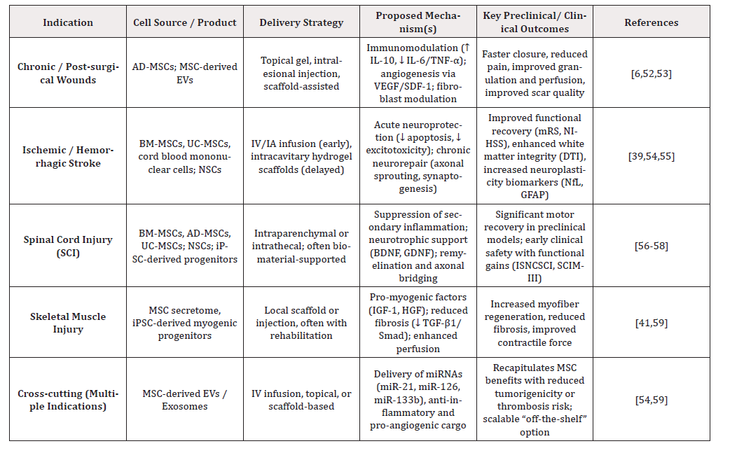
Table 1:MSC therapies by enhancing trophic signaling and mechanical cues that drive myogenesis.
Clinical Translation and Trial Design
The heterogeneity of injury contexts demands trial designs that are adaptive, mechanistically guided, and stratified by patient-specific variables. Adaptive platform trials provide an efficient framework to compare cell-based therapies with cell-free products such as exosomes, while allowing early stopping for futility or superiority [60]. Stratification should account for comorbidities, baseline inflammation, and injury chronicity, all of which modulate stem cell efficacy. Rigorous controls, including sham injections for CNS delivery and indistinguishable placebo dressings for wound trials, are essential for blinding, though ethical considerations must balance procedural risk. Combination strategies show promise. For instance, MSCs combined with platelet-rich plasma or negative-pressure wound therapy have demonstrated benefit in chronic wounds [61]; NSCs paired with biomaterial scaffolds have enhanced recovery in stroke and SCI47; and secretome-based therapies integrated with structured rehabilitation have improved outcomes in muscle injury [62]. Importantly, embedding manufacturing variables directly into trial analyses will yield actionable data to refine CQAs and optimize translational success [63]. Collectively, these design innovations will be pivotal for moving regenerative therapies from small, heterogeneous trials toward the level of rigor demanded for high-impact clinical adoption.
Biomarkers, Safety, and Regulatory Pathways
Demonstrating both efficacy and safety in stem cell-based therapies demands objective, reproducible measures of biological activity and clinical response. Circulating biomarkers, including cytokine panels and EV cargo profiles, are emerging as pharmacodynamic indicators of successful immunomodulation and angiogenesis [51]. These can be paired with advanced imaging modalities tailored to the target tissue: contrast-enhanced ultrasound or perfusion MRI to assess neovascularization in chronic wounds and skeletal muscle; DTI and functional MRI to monitor white matter integrity and cortical network remodeling in stroke and SCI [39,55]. In early- phase trials, direct cell tracking using iron labeling or reporter constructs has provided insights into biodistribution and survival, although regulatory acceptance is limited; later-phase studies increasingly prioritize validated functional biomarkers that link biological activity to patient outcomes [55].
Parallel to efficacy monitoring, rigorous attention to safety is essential. The spectrum of potential risks includes ectopic tissue formation, pro-fibrotic signaling, immune rejection, and tumorigenicity, particularly with pluripotent-derived or inadequately characterized products [64]. Intravascular administration adds a unique hazard: tissue factor expression on MSCs can trigger instant blood-mediated inflammatory reactions and thromboembolism, necessitating product-specific coagulation testing [24]. Mitigation strategies emphasize strict release criteria incorporating viability, potency, and inflammatory bias assays; staged dosing with close monitoring; and, in higher-risk populations, a shift toward cell-free derivatives such as exosomes, which carry lower risks of uncontrolled proliferation or embolic complications.
These considerations intersect with a rapidly evolving regulatory landscape. International guidelines from the ISSCR, FDA, and EMA stress the need for well-defined CQAs, xeno-free and GMP-compliant manufacturing, and long-term surveillance for delayed adverse events [65]. Ethical imperatives include transparency in informed consent, avoidance of unproven “stem cell tourism” practices, and the use of appropriate shams or matched biomaterial controls to ensure scientific rigor while minimizing risk. By aligning validated biomarkers with stringent regulatory frameworks, the field can generate high-quality evidence that not only demonstrates therapeutic promise but also satisfies the safety standards required for widespread clinical adoption (Figure 1).
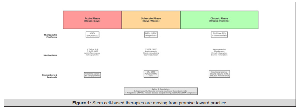
Figure 1:Stem cell-based therapies are moving from promise toward practice.
Conclusion & Future Perspectives
Stem cell-based therapies are moving from promise toward practice. They protect cells and dampen inflammation in the acute phase of injury, and they support structural repair, angiogenesis and functional recovery in the chronic phase. Evidence from wound healing, stroke, spinal cord injury and muscle regeneration shows consistent biological activity. Yet translation to routine care still lags because of variation in cell sources, manufacturing, delivery methods and outcome measurement. Progress now depends on much more than the biology of the cells. Donors and products must be screened not only for viability and phenotype but also for inflammatory bias, senescence and procoagulant activity. Standardized potency tests that mirror the intended mechanism of action are essential. Closed, xeno free production systems with clear critical quality attributes will build reliability and trust. Delivery route and combination strategy are equally important. Matching timing, dose and vehicle to the biology of each recovery phase, and combining cells or extracellular vesicles with biomaterials, growth factors, rehabilitation or neuromodulation, will likely yield benefits that no single approach can achieve. Future studies should embed these variables into trial design from the start. End points must also shift. Time to wound closure, functional independence, quality of life and reduced hospital use speak directly to health system value and to patients themselves. These pragmatic outcomes, combined with validated biomarkers of mechanism and safety, will allow regenerative therapies to show their true impact. Long term safety monitoring is indispensable. Surveillance for procoagulant events, ectopic growth, fibrosis and immune reactions must be built into every program, including cell free derivatives. Finally, large multicenter platform trials with shared biomarker cores and transparent reporting will be needed to move beyond small, heterogeneous studies. Such trials can accelerate dose finding, compare cell based and cell free products head-to-head and deliver the level of evidence required for high impact clinical adoption. By aligning mechanistic insight with careful manufacturing, adaptive trial design and patient centred outcomes, stem cell therapies can evolve from experimental promise into safe and effective regenerative treatments that change daily practice across many fields of medicine.
Validated biomarkers are essential to connect mechanism with clinical effect. In disorders of the brain and spinal cord, neurofilament light chain (NfL) and glial fibrillary acidic protein (GFAP) now stand out as sensitive blood markers of axonal injury and astroglial stress. Higher serum or CSF levels correlate with injury severity and worse outcomes after spinal cord injury, and they are increasingly used across acute neurologic conditions including stroke [66-69]. Broader panels that include S100B, neuron-specific enolase (NSE), tau and UCH-L1 help capture complementary aspects of neural damage and may enrich future trials [70]. Extracellular vesicles (EVs) add a mechanistic layer. EV-associated microRNAs such as miR-124, miR-126 and miR-133b track pathways for neuroplasticity and angiogenesis, and several reviews and experimental studies support their use as target-engagement readouts during regenerative interventions and rehabilitation after stroke or traumatic brain injury [71-75]. In vascular and endothelial contexts, miR-126 is particularly well supported as a conduit for pro-angiogenic signaling and a candidate mechanistic biomarker in EV cargo [71-74]. For chronic and diabetic wounds, fluid and swab analyses can quantify the inflammatory and protease burden that stalls healing. Elevated IL-1β relative to IL-1RA, higher CXCL8 to CXCL10 ratios, and excess matrix metalloproteinase activity (for example MMP-9 relative to TIMP-1) are repeatedly linked to non-healing trajectories and fibrosis risk; these signatures can serve as pharmacodynamic readouts when testing immunomodulatory or pro-angiogenic therapies [76-81]. Imaging should do more than show anatomy. In CNS repair, diffusion and perfusion MRI track white-matter integrity and collateral flow as structural and vascular surrogates of recovery. In wound care, hyperspectral tissue oxygenation mapping, contrast-enhanced ultrasound, and other optical methods quantify microvascular perfusion and oxygenation at the bedside, providing responsive markers for angiogenesis, granulation and infection status [71,82-85]. Embedding these readouts in trials allows early assessments of whether a therapy is altering the intended biology. These fluid, EV and imaging markers belong inside adaptive trial designs. Used together, they support early go or no-go decisions, rational dose-finding, and fair comparisons between cell-based and cell-free approaches. Safety monitoring needs the same rigor. Before intravascular use, cell and EV products should be tested for tissue factor (TF) expression and procoagulant activity (for example thrombin generation assays), given the risk of instant blood-mediated inflammatory reaction and thrombosis. Mitigation includes TF-aware release testing and anticoagulation strategies in higher- risk settings [86-93].
The next wave of regenerative therapies will move beyond proof-of-concept toward platforms that combine scalability with precision. Cell-free products-particularly extracellular vesicle- based therapeutics already demonstrate that it is possible to capture much of the trophic and immunomodulatory power of parent cells in an off-the-shelf format with improved reproducibility and a lower safety burden. At the same time, engineered cell products, from gene-edited MSCs with enhanced paracrine output to iPSC-derived progenitors with tighter lineage fidelity, are designed to overcome today’s bottlenecks of survival, integration, and donor variability. Layering precision medicine on top of these platforms will be the key. High-resolution patient phenotyping, molecular inflammatory profiles, and AI-driven stratification can guide not just who receives a therapy but also what product, at what dose, and by which route, matched to the biology of each recovery phase. This moves stem cell interventions from a “one-size-fits-all” experiment to a mechanism-matched and patient-tailored therapy.
These scientific advances must travel hand in hand with robust ethical and regulatory frameworks. Transparent adherence to ISSCR, FDA, and EMA guidance is not bureaucratic overhead but a cornerstone of public trust and protection against unregulated “stem cell tourism.” Clear critical quality attributes, xeno-free and closed manufacturing systems, and staged dose-escalation with biomarker checkpoints will shorten the distance from bench to bedside without compromising safety. Finally, the field needs large, multicenter, adaptive platform trials that can compare cell-based and cell-free products head-to-head, integrate validated biomarkers as co-primary outcomes, and incorporate long-term surveillance for delayed adverse effects such as procoagulant events, immune reactions, or ectopic growth. Such trials will allow early “go/no-go” decisions, accelerate dose finding, and establish the reproducible clinical benefit necessary for high-impact adoption.
In sum, the future of stem cell-enabled regeneration lies in integrating mechanistic insight, manufacturing rigor, biomarker-guided trial design, and patient-centered outcomes. By doing so, therapies that are still considered experimental today can evolve into safe, scalable, and effective regenerative treatments that truly change every day clinical practice across wound healing, neurological repair, and musculoskeletal regeneration.
References
- Sen CK (2025) Human Wound and Its Burden: Updated 2025 Compendium of Estimates. Adv Wound Care 14(9): 429-438.
- Tsai MH, Wu CY, Wu CH and Chen CY (2024) The Current Update of Conventional and Innovative Treatment Strategies for Central Nervous System Injury. Biomedicines 12(8): 1894.
- Waterman RS, Tomchuck SL, Henkle SL and Betancourt AM (2010) A new mesenchymal stem cell (MSC) paradigm: polarization into a pro-inflammatory MSC1 or an Immunosuppressive MSC2 phenotype. PloS One 5(4): e10088.
- Kurte M, Vega-Letter AM, Luz-Crawford P, Djouad F, Noël D, et al (2020) Time-dependent LPS exposure commands MSC immunoplasticity through TLR4 activation leading to opposite therapeutic outcome in EAE. Stem Cell Res Ther 11(1): 416.
- Pestel J, Blangero F and Eljaafari A (2023) Pathogenic Role of Adipose Tissue-Derived Mesenchymal Stem Cells in Obesity and Obesity-Related Inflammatory Diseases. Cells 12(3): 348.
- Feng H, Gong S, Liu J, Aghayants S, Liu Y, et al (2025) Adipose-derived stem cell exosomes: mechanisms and therapeutic potentials in wound healing. Biomark Res 13(1): 88.
- Hwang S, Sung DK, Kim YE, Yang M, Yoon Ahn S, et al (2023) Mesenchymal Stromal Cells Primed by Toll-like Receptors 3 and 4 Enhanced Anti-Inflammatory Effects against LPS-Induced Macrophages via Extracellular Vesicles. Int J Mol Sci 24(22): 16264.
- Voynova E, Kulebyakin K, Grigorieva O, Novoseletskaya E, Basalova N, et al (2022) Declined adipogenic potential of senescent MSCs due to shift in insulin signaling and altered exosome cargo. Front Cell Dev Biol 10: 1050489.
- Almeria C, Kreß S, Weber V, Egger D and Kasper C (2022) Heterogeneity of mesenchymal stem cell-derived extracellular vesicles is highly impacted by the tissue/cell source and culture conditions. Cell Biosci 12(1): 51.
- Li J, Wu Z, Zhao L, Liu Y, Su Y, et al (2023) The heterogeneity of mesenchymal stem cells: an important issue to be addressed in cell therapy. Stem Cell Res Ther 14(1): 381.
- Donkor ES (2018) Stroke in the 21st Century: A Snapshot of the Burden, Epidemiology, and Quality of Life. Stroke Res Treat 2018: 3238165.
- Ferdinandy P, Andreadou I, Baxter GF, Bøtker HE, Davidson SM, et al (2023) Interaction of Cardiovascular Nonmodifiable Risk Factors, Comorbidities and Comedications With Ischemia/Reperfusion Injury and Cardioprotection by Pharmacological Treatments and Ischemic Conditioning. Pharmacol Rev 75(1): 159-216.
- Hoang VT, Nguyen QT, Phan TTK, Pham TH, Dinh NTH, et al (2025) Tissue Engineering and Regenerative Medicine: Perspectives and Challenges. MedComm 6(5): e70192.
- Saceleanu VM, Toader C, Ples H, Busuioc RAC, Costin HP, et al (2023) Integrative Approaches in Acute Ischemic Stroke: From Symptom Recognition to Future Innovations. Biomedicines 11(10): 2617.
- Moll G, Ankrum JA, Kamhieh-Milz J, Bieback K, Ringdén O, et al (2019) Intravascular Mesenchymal Stromal/Stem Cell Therapy Product Diversification: Time for New Clinical Guidelines. Trends Mol Med 25(2): 149-163.
- Coppin L, Sokal E and Stéphenne X (2019) Thrombogenic Risk Induced by Intravascular Mesenchymal Stem Cell Therapy: Current Status and Future Perspectives. Cells 8(10): 1160.
- Shang Z, Wang R, Li D, Chen J, Zhang B, et al (2022) Spinal Cord Injury: A Systematic Review and Network Meta-Analysis of Therapeutic Strategies Based on 15 Types of Stem Cells in Animal Models. Front Pharmacol 13: 819861.
- Pang QM, Deng KQ, Zhang M, Wu XC, Yang RL, et al (2023) Multiple strategies enhance the efficacy of MSCs transplantation for spinal cord injury. Biomed Pharmacother Biomedecine Pharmacother 157: 114011.
- Xu Y, Wang X, Zhou X, Zeng W, Yuan J, et al (2025) Multiple strategies enhance the efficacy of MSC-Exos transplantation for spinal cord injury. Exp Neurol 383: 115038.
- Fan X, Wang JZ, Lin XM and Zhang L (2017) Stem cell transplantation for spinal cord injury: a meta-analysis of treatment effectiveness and safety. Neural Regen Res 12(5): 815-825.
- Wang R, Wang Y, Yan F, Sun J and Zhang T (2025) Assessment of mesenchymal stem cells for the treatment of spinal cord injury: a systematic review and network meta-analysis. Front Cell Neurosci 19: 1532219.
- Muthu S, Jeyaraman M, Gulati A and Arora A (2021) Current evidence on mesenchymal stem cell therapy for traumatic spinal cord injury: systematic review and meta-analysis. Cytotherapy 23(3): 186-197.
- Hoang VT, Le DS, Hoang DM, Phan TTK, Ngo LAT, et al (2024) Impact of tissue factor expression and administration routes on thrombosis development induced by mesenchymal stem/stromal cell infusions: re-evaluating the dogma. Stem Cell Res Ther 15(1): 56.
- Moll G, Ankrum JA, Kamhieh-Milz J, Bieback K, Ringdén O, et al (2019) Intravascular Mesenchymal Stromal/Stem Cell Therapy Product Diversification: Time for New Clinical Guidelines. Trends Mol Med 25(2): 149-163.
- Wang T, Huang G, Yi Z, Dai S, Zhuang W, et al (2024) Advances in extracellular vesicle-based combination therapies for spinal cord injury. Neural Regen Res 19(2): 369-374.
- Liu M, Meng Y, Ouyang S, Zhai M, Yang L, et al (2026) Neuromodulation technologies improve functional recovery after brain injury: From bench to bedside. Neural Regen Res 21(2): 506-520.
- Wang G, Yang F, Zhou W, Xiao N, Luo M, et al (2023) The initiation of oxidative stress and therapeutic strategies in wound healing. Biomed Pharmacother Biomedecine Pharmacother 157: 114004.
- Soliman AM and Barreda DR (2022) Acute Inflammation in Tissue Healing. Int J Mol Sci 24(1): 641.
- Amankwa CE, Acha LG, Dibas A, Chavala SH, Roth S, et al (2024) Neuroprotective and Anti-Inflammatory Activities of Hybrid Small-Molecule SA-10 in Ischemia/Reperfusion-Induced Retinal Neuronal Injury Models. Cells 13(5): 396.
- Yang H, He C, Bi Y, Zhu X, Deng D, et al. (2022) Synergistic effect of VEGF and SDF-1α in endothelial progenitor cells and vascular smooth muscle cells. Front Pharmacol 13: 914347.
- Younesi FS and Hinz B (2024) The Myofibroblast Fate of Therapeutic Mesenchymal Stromal Cells: Regeneration, Repair, or Despair? Int J Mol Sci 25(16): 8712.
- Yang L, Liu SC, Liu YY, Zhu FQ, Xiong MJ, et al (2024) Therapeutic role of neural stem cells in neurological diseases. Front Bioeng Biotechnol 12: 1329712.
- Farabi B, Roster K, Hirani R, Tepper K, Atak MF, et al (2024) The Efficacy of Stem Cells in Wound Healing: A Systematic Review. Int J Mol Sci 25(5): 3006.
- Kim HJ, Kim G, Lee J, Lee Y and Kim JH (2022) Secretome of Stem Cells: Roles of Extracellular Vesicles in Diseases, Stemness, Differentiation, and Reprogramming. Tissue Eng Regen Med 19(1): 19-33.
- Chen L, Xiong Y, Chopp M and Zhang Y (2024) Engineered exosomes enriched with select microRNAs amplify their therapeutic efficacy for traumatic brain injury and stroke. Front Cell Neurosci 18: 1376601.
- Bowers E and Singer K (2021) Obesity-induced inflammation: The impact of the hematopoietic stem cell niche. JCI Insight 6(3): e145295.
- Drobiova H, Sindhu S, Ahmad R, Haddad D, Al-Mulla F, et al (2023) Wharton’s jelly mesenchymal stem cells: a concise review of their secretome and prospective clinical applications. Front Cell Dev Biol 11: 1211217.
- Chen WC, Liu WF, Bai YY, Zhou YY, Zhang Y, et al (2021) Transplantation of mesenchymal stem cells for spinal cord injury: a systematic review and network meta-analysis. J Transl Med 19(1): 178.
- Detante O, Legris L, Moisan A and Rome C (2024) Cell Therapy and Functional Recovery of Stroke. Neuroscience 550: 79-88.
- Weber RZ, Rust R and Tackenberg C (2025) How neural stem cell therapy promotes brain repair after stroke. Stem Cell Rep 20(6): 102507.
- Wang YH, Wang DR, Guo YC, Liu JY and Pan J (2020) The application of bone marrow mesenchymal stem cells and biomaterials in skeletal muscle regeneration. Regen Ther 15: 285-294.
- Pastar I, Balukoff NC, Marjanovic J, Chen VY, Stone RC, et al (2023) Molecular Pathophysiology of Chronic Wounds: Current State and Future Directions. Cold Spring Harb Perspect Biol 15(4): a041243.
- Lau CS, Park SY, Ethiraj LP, Singh P, Raj G, et al (2024) Role of Adipose-Derived Mesenchymal Stem Cells in Bone Regeneration. Int J Mol Sci 25(12): 6805.
- Yu X, Liu P, Li Z and Zhang Z (2023) Function and mechanism of mesenchymal stem cells in the healing of diabetic foot wounds. Front Endocrinol 14: 1099310.
- Li X, Guan Y, Li C, Zhang T, Meng F, et al (2022) Immunomodulatory effects of mesenchymal stem cells in peripheral nerve injury. Stem Cell Res Ther 13(1): 18.
- Jiang X, Zhou L, Sun Z, Xie B, Lin H, et al (2023) MSCs overexpressing GDNF restores brain structure and neurological function in rats with intracerebral hemorrhage. Mol Biomed 4(1): 43.
- Guo W, Zhang X, Zhai J and Xue J (2022) The roles and applications of neural stem cells in spinal cord injury repair. Front Bioeng Biotechnol 10: 966866.
- Convento MB, de Oliveira AS, Boim MA and Borges FT (2024) Umbilical Cord Mesenchymal Stem Cell-Derived Extracellular Vesicles as Natural Nanocarriers in the Treatment of Nephrotoxic Injury In Vitro. Cells 13(19): 1658.
- de Freria CM, Van Niekerk E, Blesch A and Lu P (2021) Neural Stem Cells: Promoting Axonal Regeneration and Spinal Cord Connectivity. Cells 10(12): 3296.
- Yu D, Cai Z, Li D, Zhang Y, He M, et al (2021) Myogenic Differentiation of Stem Cells for Skeletal Muscle Regeneration. Stem Cells Int 2021: 8884283.
- Mei R, Wan Z, Yang C, Shen X, Wang R, et al (2024) Advances and clinical challenges of mesenchymal stem cell therapy. Front Immunol 15: 1421854.
- Nallakumarasamy A, Jeyaraman M, Maffulli N, Jeyaraman N, Suresh V, et al (2022) Mesenchymal Stromal Cell-Derived Extracellular Vesicles in Wound Healing. Life Basel Switz 12(11): 1733.
- Al-Masawa ME, Alshawsh MA, Ng CY, Hwei Ng AM, Foo JB, et al (2022) Efficacy and safety of small extracellular vesicle interventions in wound healing and skin regeneration: A systematic review and meta-analysis of animal studies. Theranostics 12(15): 6455-6508.
- Li S, Zhang J, Sun L, Yang Z, Liu X, et al (2025) Mesenchymal stem cell-derived extracellular vesicles: current advances in preparation and therapeutic applications for neurological disorders. Front Cell Dev Biol 13: 1626996.
- Hussen BM, Taheri M, Yashooa RK, Abdullah GH, Abdullah SR, et al (2024) Revolutionizing medicine: recent developments and future prospects in stem-cell therapy. Int J Surg Lond Engl 110(12): 8002-8024.
- Huang L, Fu C, Xiong F, He C and Wei Q (2021) Stem Cell Therapy for Spinal Cord Injury. Cell Transplant 30: 963689721989266.
- Xia Y, Zhu J, Yang R, Wang H, Li Y, et al (2023) Mesenchymal stem cells in the treatment of spinal cord injury: Mechanisms, current advances and future challenges. Front Immunol 14: 1141601.
- Inoue M, Yamaguchi R, He CCJ, Ikeda A, Okano H, et al (2023) Current status and prospects of regenerative medicine for spinal cord injury using human induced pluripotent stem cells: a review. Stem Cell Investig 10: 6.
- Zubair M, Abouelnazar FA, Iqbal MA, Pan J, Zheng X, et al (2025) Mesenchymal stem cell-derived exosomes as a plausible immunomodulatory therapeutic tool for inflammatory diseases. Front Cell Dev Biol. 13: 1563427.
- Ben-Eltriki M, Rafiq A, Paul A, Prabhu D, Afolabi MOS, et al (2024) Adaptive designs in clinical trials: a systematic review-part I. BMC Med Res Methodol 24(1): 229.
- Sun Y, Zhu M and Qiu L (2025) Efficacy of mesenchymal stem cells and platelet-rich plasma therapies on wound healing: A Systematic Review and meta-analysis. Regen Ther 30: 75-91.
- Nevado-Sánchez E, Rodríguez-Díaz M, Núñez-Rodríguez S, Fuente ABDL, Fuente-Anuncibay RDL, et al (2025) Effectiveness of Stem Cell Secretomes in the Regeneration and Functional Recovery of Severed Nerves in Patients with Nerve Injuries: A Systematic Review. Cells 14(7): 492.
- Dombeck C, Swezey T, Kehoe L, Kinchen K, Roe M, et al (2024) Embedding clinical trial elements into clinical practice: Experiences from trial designers and implementers. J Clin Transl Sci 8(1): 202.
- Sugai K, Nakamura M, Okano H and Nagoshi N (2025) Stem cell therapies for spinal cord injury in humans: A review of recent clinical research. Brain Spine 5: 104207.
- (2021) International Society for Stem Cell Research (ISSCR). Guidelines for Stem Cell Research and Clinical Translation.
- Park J, Jo Y, Park KM and Lee S (2024) GFAP and neurofilament light as biomarkers for neurologic disease. International Journal of Molecular Sciences 25(12): 6295.
- Stukas S, Cooper J, Gill J, Fallah N, Skinnider MA, et al (2023) Association of CSF and serum neurofilament light and glial fibrillary acidic protein, injury severity, and outcome in spinal cord injury. Neurology 100(12): e1221-e1233.
- Czeiter E, Amrein K, Gravesteijn BY, Lecky F, Menon DK, et al (2020) CENTER-TBI Participants and Investigators. (2020). Blood biomarkers on admission in acute traumatic brain injury: Relations to severity, CT findings, and outcomes in the CENTER-TBI study. EBioMedicine 56: 102785.
- Thelin EP, Nelson DW and Bellander BM (2017) A review of the clinical utility of serum S100B protein levels in the assessment of traumatic brain injury. Acta Neurochirurgica 159(2): 209-225.
- Moura LM, Luccas R, Paiva JPQd, Amaro E, Leemans A, et al (2019) Diffusion tensor imaging biomarkers to predict motor outcomes after stroke. Frontiers in Neurology 10: 445.
- Moulton E, Amor-Sahli M, Perlbarg V, Pires C, Crozier S, et al (2019) Acute diffusivity biomarkers for prediction of motor and language outcome in ischemic stroke. Stroke 50(1): 205-207.
- Barber PA, Darby DG, Desmond PM, Yang Q, Gerraty RP, et al (1998) Prediction of stroke outcome with echoplanar perfusion- and diffusion-weighted MRI. Neurology 51(2): 418-426.
- Qi X, Shao M, Sun H, Shen Y, Meng D and Huo W (2021) Exosomal miR-124 promotes neurogenesis and functional recovery after stroke. Frontiers in Molecular Biosciences 8: 685088.
- Xin H, Li Y, Buller B, Katakowski M, Zhang Y, et al (2013) Exosome-mediated transfer of miR-133b from multipotent mesenchymal stromal cells to neural cells contributes to neurite outgrowth. Stem Cells 31(12): 2737-2746.
- Bordin A, Chirivì M, Pagano F, Milan M, Iuliano M, et al (2022) Human platelet lysate-derived extracellular vesicles enhance angiogenesis through miR-126. Cell Proliferation 55(11): e13312.
- Ateeq M, Broadwin M, Sellke FW and Abid MR (2024) Extracellular vesicles’ role in angiogenesis and altering angiogenic signaling. Medical Sciences (Basel) 12(1): 4.
- Arderiu G, Espina M, Pena E, PaulT and Badimon L (2022) Endothelium-released microvesicles transport miR-126, modulating monocyte pro-angiogenic reprogramming. Frontiers in Immunology 13: 836662.
- Ding JY, Wang X, Chen BJ, Xie J and Liu YS (2023) Mesenchymal stem cell-derived extracellular vesicles in skin wound repair. Military Medical Research 10: 20.
- Rembe JD, Stallmeyer B, Qu S, Fromm T, Fromm L and Stuermer EK (2025) Immunomarker profiling of chronic wounds from minimally invasive swabs. Journal of Translational Medicine 23: 291.
- Müller M, Trocme C, Lardy B, Morel F, Halimi S, et al (2008) Matrix metalloproteinases and diabetic foot ulcers: The ratio of MMP-1 to TIMP-1 is a predictor of wound healing. Diabetic Medicine 25(4): 419-426.
- Ayuk SM, Abrahamse H and Houreld NN (2016) The role of matrix metalloproteinases in diabetic wound healing. International Journal of Endocrinology 2897656.
- Fu X, Han B, Cheng X and Hu D (2022) Inflammatory cytokines in non-healing chronic wounds: New insights and therapeutic implications. Frontiers in Pharmacology 13: 1050630.
- Saiko G, Lombardi P, Au Y, Queen D, Armstrong D, et al (2020) Hyperspectral imaging in wound care: A systematic review. International Wound Journal 17(6): 1840-1856.
- Yang Q, Sun S, Jeffcoate WJ, Clark DJ, Musgove A, et al (2018) Prediction of healing of diabetic foot ulcers using hyperspectral imaging. Journal of Imaging 4(12): 144.
- Godavarty A, Arya AK, O’Shaughnessy K and Diaz F (2023) Diabetic foot ulcer imaging: An overview and future directions. Diagnostics 13(10): 1701.
- Aziz MU, Aslan A, Nair AA, et al. (2022) Microvascular flow imaging: A state-of-the-art review. Radiology 304(1): 26-44.
- Wang Y, Tang Z, Wang S and Wang X (2024) Application of dynamic contrast-enhanced ultrasound in disease activity evaluation. Diagnostics 14(7): 672.
- Wu X, Jiang J, Gu Z, Zhang J, Chen Y, et al (2019) Mesenchymal stromal cell therapies: Immunomodulatory properties and clinical progress. Frontiers in Immunology 10: 1645.
- Schriner JB, Dutta D, Vong S, Davis S and Feldman D (2024). Low-molecular-weight heparin decreases pro-coagulant activity in clinical MSC products. Cytotherapy 26(2): 194-200.
- Wright A, Marks DC and Linden MD (2023). Procoagulant activity of umbilical cord-derived mesenchymal stromal cell extracellular vesicles. Cytotherapy 25(6): 671-685.
- Hoang VT, Pankajakshan D and Moll G (2024). Impact of tissue factor expression and administration routes on coagulation risks in MSC therapy. Stem Cell Research & Therapy 15: 117.
- Tatsumi K, Ohashi K, Matsubara Y and Uemura Y, Kohori A, et al (2013) Tissue factor triggers procoagulation in transplanted mesenchymal stem cells. Biochimica et Biophysica Acta (BBA) – General Subjects 1830(5): 2779-2789.
- Wong C, Cheung AS and Leung, K. (2024). Mesenchymal stromal cell-derived extracellular vesicles as immunomodulators and biomarkers. Frontiers in Immunology 15: 1355845.




 We use cookies to ensure you get the best experience on our website.