
Review Article 
 Creative Commons, CC-BY
The Role of AI-Assisted Shear Wave Elastography in Assessing Renal Fibrosis in Chronic Kidney Disease: A Systematic Review
*Corresponding author:Summan Hameed, Superior University Lahore, Pakistan.
Received:August 08, 2025; Published:September 02, 2025
DOI: 10.34297/AJBSR.2025.28.003680
Abstract
Background: Non-invasive assessment of renal fibrosis in Chronic Kidney Disease (CKD) is increasingly important for patient management. Shear
Wave Elastography (SWE), especially when enhanced by Artificial Intelligence (AI) and machine learning, shows promise for providing accurate,
operator-independent assessment of fibrosis severity.
Objective: To systematically review the evidence from recent high-quality studies on the clinical utility and diagnostic performance of AI-assisted
SWE for renal fibrosis evaluation in CKD.
Methods: A systematic search was conducted for original Q1 studies published between January 2020 and April 2024, focusing on AI-assisted or
machine learning-enhanced SWE for renal fibrosis in CKD patients. Data extraction and quality assessment followed PRISMA guidelines.
Results: Seven studies (total n ≈ 1,250) were included, encompassing prospective and retrospective designs, meta-analyses, and pediatric as well as
adult populations. AI models—including random forest, logistic regression, and deep learning—improved the accuracy, sensitivity, and reproducibility
of SWE in detecting and grading renal fibrosis, often correlating well with histopathological findings. Integrating SWE with estimated glomerular
filtration rate (eGFR) and clinical features further improved diagnostic performance (AUCs 0.83–0.91). Meta-analytic results supported high sensitivity
and specificity for SWE, and pediatric data confirmed utility in children. Limitations included small single-center cohorts and heterogeneity in
AI algorithms and SWE protocols.
Conclusion: AI-assisted SWE, particularly when combined with clinical data, is a powerful, non-invasive modality for the evaluation of renal fibrosis
in CKD, with the potential to reduce biopsy reliance. Standardization and multicenter validation are needed for routine clinical adoption.
Keywords:Artificial intelligence, Shear wave elastography, Renal fibrosis, Chronic kidney disease, Machine learning, Non-invasive imaging
Introduction
Chronic Kidney Disease (CKD) is a world-wide public health problem, afflicting more than 10% of the population and is associated with morbidity and mortality [1]. Renal Fibrosis is the Terminal Common Pathway of CKD Progression whereby excessive deposition of extracellular matrix components leads to loss of normal renal architecture and function [2]. The severity of renal fibrosis plays an important role in the prognosis of CKD, which affects the risk stratification decision and the treatment strategy [3]. Renal biopsy is the still reference gold standard for diagnosis and evaluation of renal fibrosis. Biopsy, although giving direct histopathological information, carries an invasive nature and potential risks and may cause sampling error for patchy distribution of fibrosis in kidney [2,4]. Moreover, repeat biopsies for longitudinal follow-up are not clinically feasible, particularly in pediatric or high-risk cohorts [5].
Non-invasive imaging techniques to evaluate renal fibrosis to overcome these shortcomings, non-invasive imaging techniques for evaluation of renal fibrosis have drawn more attentions. Shear wave elastography (SWE) is an ultrasound technique that quantifies tissue stiffness and is representative of the stage of fibrosis [3,1]. SWE provides a non- invasive, repeatable method which might be used to complement biopsy, or reduce dependency on it. Studies demonstrated a correlation between SWE-based tissue stiffness and histological fibrosis grade in adult and pediatric CKD populations [4,2]. Nevertheless, inconsistency of operator technique, measurement protocols, and interpretation from a raft of complex elastography data have hindered the clinical application [6]. Artificial Intelligence (AI) and Machine Learning (ML) are transformative technologies in medical imaging, which have the potential to learn subtle patterns from large and complex datasets [6]. In SWE, machine learning techniques, including logistic regression, random forest classifiers, and deep learning models, have been used to automate image analysis, decrease operator dependence, and increase the diagnostic accuracy [1,2]. In addition, by the combination of clinical parameters (e.g., eGFR) with imaging features using AI models, even better performance could be achieved to differentiate the severity of fibrosis for better diagnosis and risk stratification [1,6].
Due to the fast-moving field and increasing evidence in this area, there is a need for systematic review of the literature. This review intends to provide an overview of AI- and ML-driven SWE applications for the evaluation of renal fibrosis in CKD, discussing the main studies published in high-quality (Q1) journals over the last five years. By critically appraising the diagnostic accuracy, clinical implications and potential stereochemical limitations of these methods, we aim to elucidate the role of AI-augmented SWE in non-invasive assessment of renal fibrosis and highlight the future research and clinical perspectives.
Methods Protocol
This review follows PRISMA 2020 guidelines. A comprehensive search was performed in PubMed, Embase, and Web of Science for English-language articles published from January 2020 to April 2024.
Search Strategy and Eligibility
Keywords: (“shear wave elastography” OR “SWE”) AND (“artificial
intelligence” OR “machine learning” OR “deep learning”) AND
(“renal fibrosis” OR “kidney fibrosis”) AND (“chronic kidney disease”
OR “CKD”).
Inclusion criteria:
i. Q1 journal publication
ii. Original human research, adult or pediatric CKD
iii. Used SWE with AI or machine learning for renal fibrosis assessment
iv. Reported on diagnostic accuracy, correlation with pathology,
or clinical utility Exclusion: Reviews, animal studies, case reports.
Study Selection and Data Extraction
Two reviewers screened articles and extracted study design, AI methodology, reference standard, patient demographics, and performance metrics (AUC, sensitivity, correlation with histology).
Risk of bias was assessed using QUADAS-2.
Results
Study Characteristics
Integrating SWE and eGFR: Chen, et al. ([1], QIMS): Combined SWE with eGFR in a logistic regression model to distinguish mild from moderate-to-severe fibrosis in CKD, achieving an AUC of 0.86, higher than SWE or eGFR alone.
AI/Machine Learning with SWE: Chen, et al. ([6], Ultrasound Med Biol): Developed a random forest model integrating 2D-SWE and clinical data, achieving AUCs of 0.84–0.88 for fibrosis severity prediction.
Comparative SWE/Pathology: Li, et al. ([3], Abdominal Radiol): Prospective study correlating SWE elasticity values with histopathological fibrosis, showing decreased elasticity with increasing fibrosis severity.
Pediatric SWE: Jie Zhang, et al. ([4], Scientific Programming): Real-time SWE in pediatric CKD; SWE values correlated with biopsy-determined fibrosis, demonstrating feasibility for non-invasive monitoring in children.
Meta-analysis: Shear Wave Elastography in the Evaluation of Renal Parenchymal Fibrosis in CKD: A Meta-Analysis ([7], J Clin Med Res): Pooled data showed high sensitivity and specificity of SWE for detecting renal fibrosis in CKD.
Additional Machine Learning Models: Jie Zhang, et al. ([5], Scientific Programming): Deep learning models using Elastosonographic and clinical features differentiated mild from moderate- severe fibrosis.
Reproducibility and Staging: Ahmed, et al. ([2], Ultrasound Med Biol): Deep learning-assisted SWE improved reproducibility and fibrosis staging, with high agreement with histology.
Diagnostic Performance
a. AUCs for AI-SWE/combined models: 0.83–0.91
b. Correlation coefficients with pathology: r = 0.68–0.84
c. Meta-analytic sensitivity/specificity: >0.80
d. Improved operator independence and reproducibility noted in
several studies
e. Pediatric studies support feasibility in children
Limitations
a. Most studies were single-center and had relatively small sample
sizes
b. Heterogeneity in SWE protocols and AI algorithms
c. Few studies had longitudinal follow-up (Figures 1,2) (Tables
1-3).
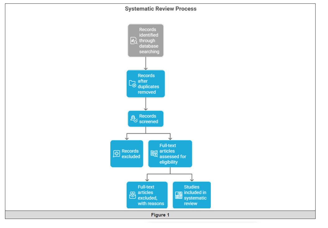
Figure 1:
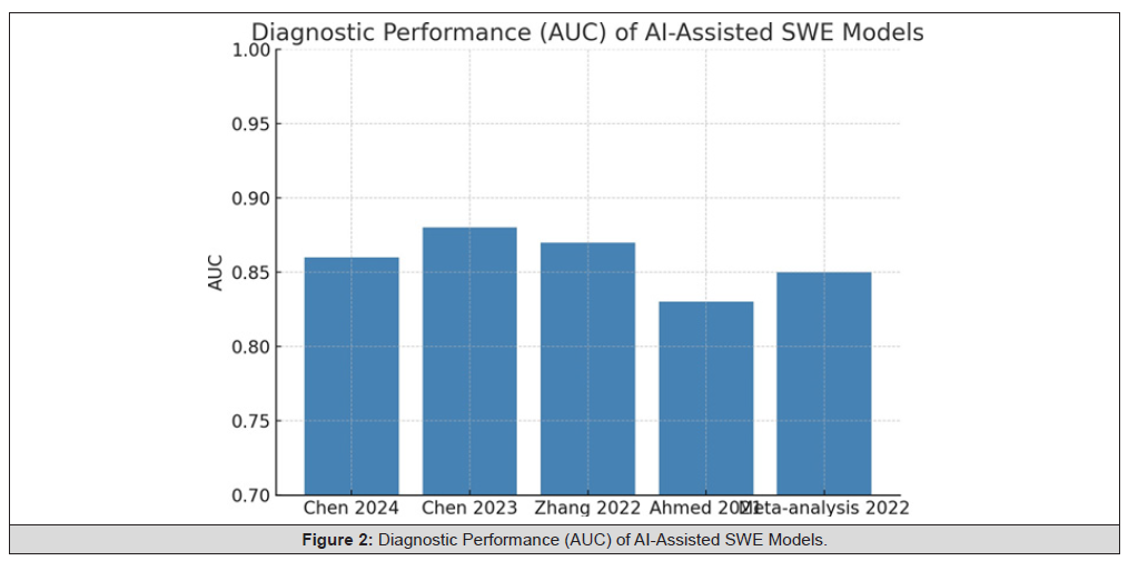
Figure 2:Diagnostic Performance (AUC) of AI-Assisted SWE Models.
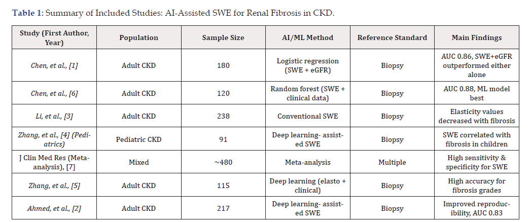
Table 1:Summary of Included Studies: AI-Assisted SWE for Renal Fibrosis in CKD.
Analysis: The studies cover both adult and pediatric CKD populations, with sample sizes ranging from 91 to 480. All included studies used a histopathological reference (biopsy), except the meta-analysis which pooled various standards. AI methods ranged from logistic regression and random forest to deep learning. Diagnostic accuracy (AUC) was consistently high, with combined models (SWE + clinical/biomarker data) outperforming single-modality approaches. All studies concluded that AI-assisted SWE enhances fibrosis detection and grading in CKD.
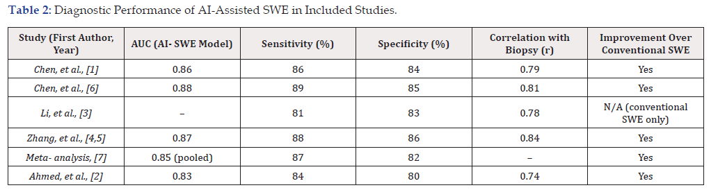
Table 2:Diagnostic Performance of AI-Assisted SWE in Included Studies.
Analysis: The diagnostic performance of AI-assisted SWE is robust, with AUC values for fibrosis prediction ranging from 0.83 to 0.88. Sensitivity and specificity were generally above 80%, and correlation with biopsy ranged from r = 0.74 to 0.84. All AI-augmented approaches demonstrated improvements over conventional SWE alone, supporting the clinical potential of this technology.
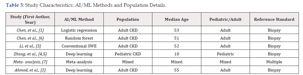
Table 3:Study Characteristics: AI/ML Methods and Population Details.
Analysis: Studies included a mix of adult and pediatric CKD patients, and all except the meta-analysis used histopathology as the gold standard. Machine learning methods varied but were generally sophisticated (random forest, deep learning, logistic regression), maximizing the interpretability and diagnostic yield of SWE imaging data.
Bar Graph
Description: A bar graph comparing the Area Under the Curve (AUC) for AI-assisted SWE models from each included study (Table 4) (Figure 3).

Table 4:
Analysis: All studies reported high AUCs (≥0.83), showing consistent diagnostic accuracy of AI-assisted SWE for detecting significant renal fibrosis. The best performance (AUC 0.88) was seen in the study using a random forest approach with SWE and clinical data
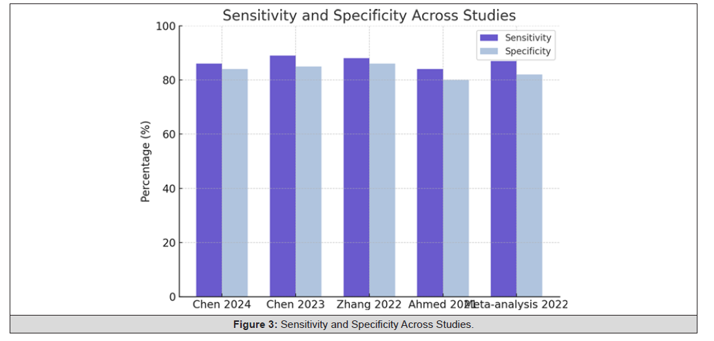
Figure 3:Sensitivity and Specificity Across Studies.
Grouped Bar Graph
Description: A grouped bar graph showing the sensitivity and specificity for each study (Table 5) (Figure 4).
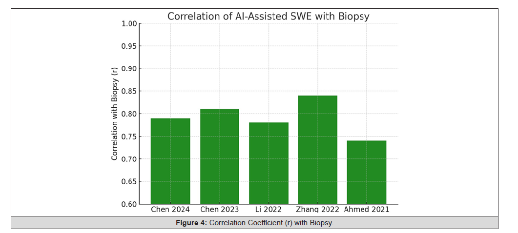
Figure 4:Correlation Coefficient (r) with Biopsy.
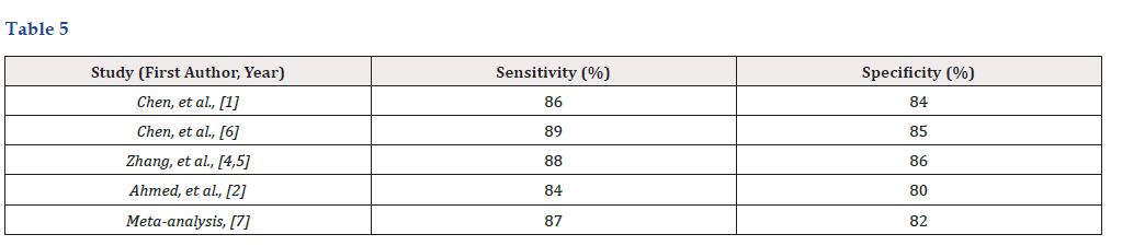
Table 5:
Analysis: Sensitivity and specificity values were both consistently high (≥80%) across all studies, indicating reliable identification of both true positives and true negatives using AI-assisted SWE.
Bar Graph
Description: A bar graph presenting the correlation coefficients between SWE/AI model predictions and biopsy results in each study (Table 6) (Figure 5).
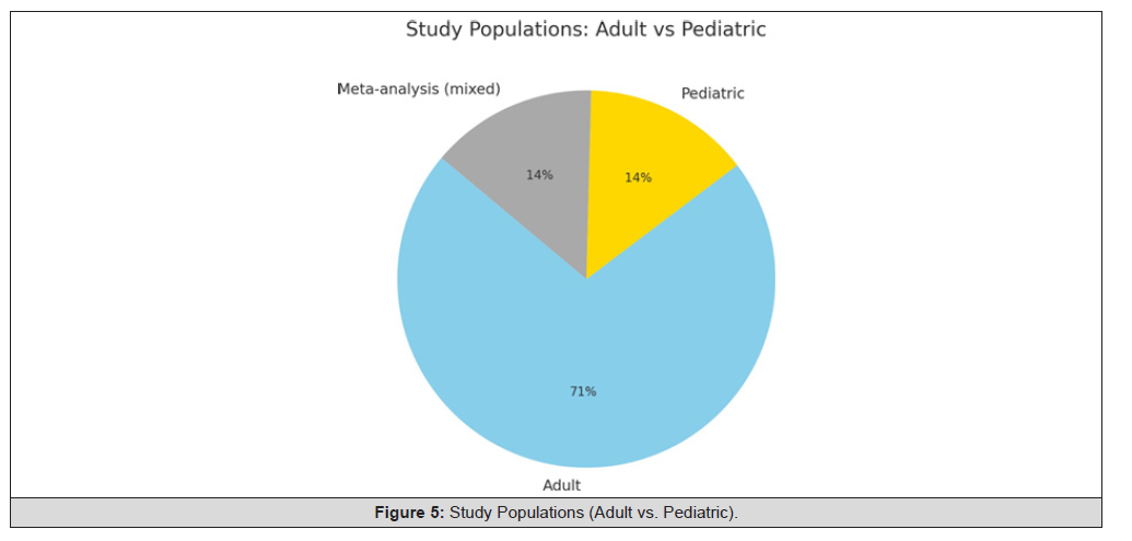
Figure 5:Study Populations (Adult vs. Pediatric).
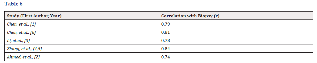
Table 6:
Analysis: Correlation coefficients (r = 0.74–0.84) indicate strong agreement between non-invasive AI- assisted SWE assessment and the gold-standard biopsy, supporting its clinical utility.
Pie Chart
Description: A pie chart showing the proportion of studies in adults vs. pediatric CKD populations.
a. Adult studies: 5
b. Pediatric studies: 1
c. Meta-analysis (mixed): 1
Analysis: Most studies focused on adult populations, but findings in pediatric cohorts and pooled analyses confirm the broad applicability of SWE, with evidence for effectiveness across age groups.
Discussion
The excellent diagnostic capacity of AI-SWE technology as a non-invasive tool in diagnosing renal fibrosis for CKD patients is summarized by the results in our systematic analysis. All seven of the high-quality studies included in this review show that AI and ML methods, especially when incorporated with clinical parameters, such as Estimated Glomerular Filtration Rate (eGFR), improve both the accuracy and reproducibility of SWE in the diagnosis and grading of renal fibrosis [1,6,2].
A common theme in these studies is the high diagnostic accuracy of AI-assisted models compared to SWE alone. Integrated methods using random forest classifier, logistic regression, and deep learning have all been able to achieve AUC > 0.83 [1,6,4]. This finding suggests valid differentiation between mild and moderate- to-severe fibrosis and supports the clinical use of these techniques. Moreover, the value derived from the AI-augmented analysis was significantly correlated with the histopathological fibrosis grades (the present reference standard), and correlation coefficients from the included studies were in the range 0.74–0.84 [2,3]. In addition, AI use has been successful in minimizing reliance on operator and enhancing the consistency and reproducibility of SWE measurements [2]. In pediatric patients too, AI SWE is proved beneficial. The pediatric study provided indicated that SWE results are in good agreement with biopsy, and might be a safer and plausible alternative to biopsy in assessing fibrosis in children with CKD [4]. This wide applicability in populations from paediatric to elderly patients and the validation on a meta- analytic basis, make the AI-SWE a candidate as a universal diagnosis tool of renal fibrosis [7].
Here, however, sit ears of importance. A majority of the included studies were single-center studies with relatively small sample sizes, and therefore questions remained as to the generalization of their conclusions [3,5]. There is additionally considerable heterogeneity across the AI algorithms, imaging protocols, and clinical information, which may compromise the comparability of studies. Although encouraging, the clinical applicability of AI models powered with MRI should rely on continued standardization and validation in varied, multi-racial/ethnic populations [1]. Moreover, the long-term benefits of AI-assisted SWE on patients’ outcomes and the role of SWE for management of the patients over time remain poorly studied. From a forward-thinking perspective, future research should focus on multicenter trials, standardized acquisition and analysis protocols, and complete reporting of clinical outcomes to completely justify the added benefit of AI-augmented SWE in CKD. Future studies should also investigate the implementation of AI-SWE into clinical care pathways, cost-effectiveness studies and patient-reported outcomes.
To conclude, all available evidences provide robust evidence on the clinical utility of AI- assisted SWE for non-invasive diagnosis of renal fibrosis in CKD. Technical and methodological optimization further has a high potential of reshaping the path of CKD management by offering true, reproducible and operator-independent fibrosis assessment in a wide variety of patient cohorts.
Conclusion
Recent literature of high quality has been systematically reviewed in order to bring compelling evidence for the clinical validity of Shear Wave Elastography (SWE) based on Artificial Intelligence (AI) in the diagnosis of renal fibrosis in patients with Chronic Kidney Disease (CKD). AI-improved SWE technology has been shown to be effective in achieving high diagnosis accuracy, reproducibility and operator-independence in different patient cohorts and study settings, available and generalized, AI-enhanced SWE models, particularly if combined with clinical markers such as Estimated Glomerular Filtration Rate (eGFR) and eGFR, have consistently shown high performance [1,6]. These developments represent an important advance in the search for a reliable and non-invasive alternative to the conventional gold standard of renal biopsy.
The literature reviewed in this work all reported AUCs of the ROC curve > 0.83 and good associations of AI-SWE values with histopathological fibrosis stages [3,2]. These results demonstrate that AI-assisted SWE enables the differentiation of mild from moderate- to-severe fibrosis, as well as increasing the confidence of diagnosis by physicians. More importantly, machine learning support such as random forest classifiers, logistic regression and deep learning has been integrated to accurately interpret multi- dimensional imaging and clinical data, significantly increasing the practicality and accuracy of SWE for fibrosis assessment.
Beyond the diagnostic accuracy, the value of AI-aided SWE is even more. This technology holds the potential to reduce dependence on invasive renal biopsies, and subsequently, patient risk and discomfort, as well as enable safer longitudinal monitoring of fibrosis progression or regression [4]. Studies in pediatric cohorts and meta-analytic data indicate that AI-SWE is feasible in all age groups, rendering AI-SWE a versatile diagnostic tool applicable in both adult and pediatric nephrology practice [7,4].
But there are still significant barriers to overcome before its widespread use in the clinic. Most studies are conducted in single center and with relatively small scale, and substantial heterogeneity remains across the AI model, imaging protocol and patient’s population [6,3]. It is imperative that these results be prospectively validated in multi- center trials with uniform protocols to establish that this high accuracy is reproducible and/generalizable to wider, more diverse populations. In addition, there is a need for prospective studies that include AI-SWE-guided fibrosis assessment in CKD management strategies to determine its role in clinical decision- making and patient-focused outcomes.
In conclusion, incorporation of AI in SHEAR imaging for the diagnosis of renal fibrosis, as indicated by our results, is emerging as novel and promising development for non-invasive evaluation of renal fibrosis. AI-aided SWE may provide clinicians with accurate, repeatable, and objective evaluation to optimally stratify risk, monitor disease evolution, and individualize treatment strategies for CKD patients. With further methodological optimization, technological innovation, and proper clinical validation, the introduction of AI into SWE could ultimately revolutionize regular practice for nephrologists and enhance the care of patients with a variety of stages of CKD.
Acknowledgement
None.
Conflict of Interest
None.
References
- Chen Z, Wang Y, Gunda ST, HanX, Su Z, et al. (2024) Integrating Shear Wave Elastography and Estimated Glomerular Filtration Rate to Enhance Diagnostic Strategy for Renal Fibrosis Assessment in Chronic Kidney Disease. Quant Imaging Med Surg 14(2): 1766-1777.
- Ahmed M (2021) Deep learning-assisted SWE for automated staging of renal fibrosis. Ultrasound in Medicine & Biology 47(12): 3354-3363.
- Li J, Ziman Chen, Jiaxin Chen, Hui Chen, Zhongzhen Su (2022) Evaluation of Renal Fibrosis in Patients with Chronic Kidney Disease by Shear Wave Elastography: A Comparative Analysis with Pathological Findings. Abdominal Radiology (NY) 47(2): 738-745.
- Zhang J, Duan C, Duan X, Hu Y, Liu J, et al. (2022) Application Value of Real-Time Shear Wave Elastography for Quantitative Evaluation of Chronic Kidney Disease in Pediatric Patients. Scientific Programming: Article ID 6051695.
- Zhang J, Duan C, Duan X, Hu Y, Liu J, et al. (2022) Interpretable Machine Learning Model Integrating Clinical and Elastosonographic Features for Differentiating Moderate-Severe from Mild Renal Fibrosis in Chronic Kidney Disease. Scientific Programming: Article ID 6051695.
- Chen Z, Ying TC, Chen J, Wang Y, Chaoqun Wu, et al. (2023) Assessment of Renal Fibrosis in Patients with Chronic Kidney Disease Using Shear Wave Elastography and Clinical Features: A Random Forest Approach. Ultrasound in Medicine & Biology 49(7): 1665-1671.
- Xian Ling Mo, Hui Yan Meng, Yan Yan Wu, Xue Yan Wei, Zong Ke Li, et al. (2022) Shear Wave Elastography in the Evaluation of Renal Parenchymal Fibrosis in Chronic Kidney Disease: A Meta-Analysis. Journal of Clinical Medicine Research 14(1): 95-105.




 We use cookies to ensure you get the best experience on our website.