Mini Review 
 Creative Commons, CC-BY
Pre-procedural Planning and Analysis of Coronary Anomalies During TAVR
*Corresponding author: Parivash Badar, Department of Cardiology, The Heart Hospital Baylor Plano, Plano TX, 4672 Thanksgiving Lane, Plano TX, USA.
Received: July 03, 2018; Published: July 12, 2019
DOI: 10.34297/AJBSR.2019.03.000740
Abstract
Variations in coronary anatomy and the presence of anomalous coronary arteries are associated with high-risk valve cases i.e. higher rates of morbidity and mortality. Therefore, it is valuable for physicians to be aware of the importance of the application of screening tools to determine the location of the coronary arteries from the aortic root. Imaging modalities such as Coronary Computed Tomography Angiography (Coronary CTA) can be utilized to evaluate and determine coronary origin, course and termination, thus allowing physicians to anticipate, prepare for and prevent complications in technical valve procedures, i.e. Transcatheter Aortic Valve Replacement (TAVR).
Keywords:Anomalous coronaries; TAVR; CTA
Introduction
The prevalence of variations in coronary anatomy and the presence of anomalous coronary arteries is known to be about 0.2– 2.3% [1-6] High rates of morbidity and mortality are associated with such high-risk cases thus, complete knowledge of the coronary anatomy is of immense clinical importance when performing technical valve procedures. Careful attention needs to be given to the topographical location of the coronary artery from the aortic root and the implanted valve for preventing coronary obstruction during TAVR.
Patient presenting with symptoms of severe aortic stenosis with complex anatomies requiring TAVR need to manage in the clinical setting and hence we discuss the importance of screening tools for the pre-procedural planning and prediction of outcomes of such complex cases.
Discussion
Water specimen collection
The presence of anomalous coronary artery is divided into: (1) right coronary artery (RCA) arising from the left coronary sinus, (2) left anterior descending coronary artery (LAD) arising from the right coronary sinus, (3) left circumflex coronary artery (LCX) arising from the right coronary sinus, (4) left main coronary artery (LMCA) arising from the right coronary sinus, and (5) anomalous location of coronary ostium outside coronary aortic sinuses. The course of a proximal anomalous coronary artery was further classified into pre-pulmonary (anterior), inter-arterial (between aorta and pulmonary artery), septal (posterior to right ventricular outflow tract within myocardium of interventricular septum) and retro-aortic [7].
It has been noted in multiple large population cohort studies, that anomalous LCA/LCX arising from the right coronary sinus is about five times less common than the anomalous origin of the RCA from the left coronary sinus [1,3]. Many cases show an increase in the rate of mortality and morbidity associated with cases of LCA and these patients are more likely to experience sudden cardiac death [6]. While some patients may remain asymptomatic until valve stenosis is severe, other patients may appear symptomatic earlier at disease onset. Symptomatic patients showing signs of dyspnea on exertion, chest pressure and syncope require surgical intervention via Trans-catheter Aortic Valve Replacement (TAVR) Hence, careful attention needs to be given to the topographical location of the coronary artery from the aortic root and the implanted valve for preventing coronary obstruction during TAVR [1].
Patients presenting with high-risk anatomy and more likely to demonstrate patterns of obstructive coronary artery disease; defined as >50 % vessel diameter reduction by qualitative assessment in at least one of LMCA, LAD, LCx or RCA [7]. The anatomical risk factors of coronary obstruction are reported to be a low coronary height (below 12 mm in height), a shallow sinus of Valsalva (below 30 mm in width), and the presence of coronary artery disease itself [8]. It was observed in many cases, that, lower-lying coronary ostium, shallow sinus of Valsalva along with irregular course taken by the coronary artery were associated anatomic factors, and despite successful treatment, acute and late mortality remained very high.
Another study analyzed multiple patients with coronary anomalies and shed light on other high-risk features: (1) luminal compression (>50 % vessel diameter reduction) between aorta and pulmonary artery, (2) proximal intramural course within aortic wall, (3) slit-like ostium (>50 % vessel diameter reduction) and, (4) acute takeoff angle (>45° between plane longitudinal to proximal CAAL and tangential plane of aortic root at level of CAAL in axial view) [7].
Conversely, coronary computed tomography angiography (CTA) is well suited to illustrate the coronary origin, course, and termination with its superb spatial and temporal resolution [6]. According to the ACC/AHA Guidelines and Appropriate Use Criteria, the use of coronary CTA has been recommended in the evaluation of anomalous coronary arteries [6,9].
CTA allows the enhancement of multi-planar images and minimal intensity projections on the scan which can enable physicians to predict possible coronary obstruction and adverse outcomes based on their CTA measurements. It is also useful to predict the possible sizing of the valve and hence avoid unnecessary surgical detours during the procedure.
Since there are only a few cases of anomalous coronary artery with TAVR procedure, not enough data is present regarding the choice of a particular valve that would be well suited for such anatomy. However, a study by Sorbets et all reported that the use of self-expandable or balloon expandable bio-prosthetic valve does not appear particularly advantageous over the other in the case of anomalous coronary artery [1,10].
The factors outlined above contribute to the outcome of patients undergoing surgical procedures with high risk anatomies and it has thus become necessary for physicians to be able to anticipate and prevent the occurrence of these complications [7,6,9].
Conclusion
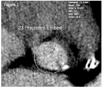
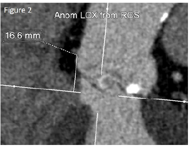
The knowledge of the coronary anatomy, particularly that of patients with coronary arteries of anomalous origin is of immense importance when treating patients with other co-morbidities and going for technical surgical valve procedures i.e. TAVR. It is important for surgeons and physicians to be aware of different methods of using imaging tools such as CTA to predict the outcomes of such cases. We hope to read about more stories of success in the future ((Figures 1 & 2)
Conflicts of interest
All authors declared that there are no conflicts of interest.
References
- Cho SW, Kim BG, Park TK, Choi SH, Gwon HC, et al. (2019) Transcatheter Aortic Valve Replacement in a Patient with Anomalous Origin of the Left Coronary Artery. J Cardiol Cases 19(4): 133-135.
- Mizote I, Conradi L, Schäfer U (2016) A Case of Anomalous Left Coronary Artery Obstruction Caused by Lotus Valve Implantation. Catheter Cardiovasc Interv 90(7): 1227-1231.
- Weich H, Ackermann C, Viljoen H, van Wyk J, Mabin T, et al. (2011) Transcatheter Aortic Valve Replacement in a Patient with an Anomalous Origin of the Right Coronary Artery. Catheter Cardiovasc Interv 78(7): 1013-1016
- Giri J, Szeto WY, Bavaria J, Herrmann HC (2012) Transcatheter Aortic Valve Replacement With Coronary Artery Protection Performed in a Patient With an Anomalous Left Main Coronary Artery. J Am Coll Cardiol 60(6): e9.
- Alexander Ralph W, George C Griffith (1956) Anomalies of the Coronary Arteries and Their Clinical Significance. Circulation 14(5): 800-805.
- Mclarry Joel, Maros Ferencik, Michael D. Shapiro (2015) Coronary Artery Anomalies: A Pictorial Review. Current Cardiovascular Imaging Reports 8(7): 23.
- Taylor AJ, Cerqueira M, Hodgson JM, Mark D, Min J, et al. (2010) ACCF/ SCCT/ACR/AHA/ASE/ASNC/NASCI/SCAI/SCMR 2010 Appropriate Use Criteria for Cardiac Computed Tomography. Circulation 122(21).
- Ribeiro HB, Webb JG, Makkar RR, Cohen MG, Kapadia SR, et al. (2013) Predictive Factors, Management, and Clinical Outcomes of Coronary Obstruction Following Transcatheter Aortic Valve Implantation. J Am Coll Cardiol 62(17): 1552-1562.
- Warnes CA, Williams RG, Bashore TM, Child JS, Connolly HM, et al. (2008) ACC/AHA 2008 Guidelines for the Management of Adults with Congenital Heart Disease. Circulation 118(23).
- Sorbets E, Choby M, Tchetche D (2012) Transcatheter Aortic Valve Implantation With Either CoreValve or SAPIEN XT Devices in Patients With a Single Coronary Artery. J Invasive Cardiol 24(7): 342-344.


 We use cookies to ensure you get the best experience on our website.