
Review Article 
 Creative Commons, CC-BY
AI -A Novel Tool for Oral Cancer Diagnosis and Prevention: Current Applications and Future Perspectives
*Corresponding author:Shaista Suhail, Department of Microbiology, BBD College of Dental Sciences, BBD University, Lucknow, India.
Received:February 17, 2026; Published:February 24, 2026
DOI: 10.34297/AJBSR.2026.30.003903
Abstract
Oral cancer represents a major global health burden, particularly in developing countries, where delayed diagnosis and limited access to specialized healthcare services contribute to poor prognosis and high mortality rates [1,2]. Despite advancements in diagnostic modalities such as clinical examination, imaging, and histopathology, early detection of oral cancer remains a significant challenge. In recent years, Artificial Intelligence (AI) has emerged as a transformative technology in healthcare, offering innovative solutions for early diagnosis, risk prediction, and prevention of oral cancer [3-6]. AI-driven systems utilize machine learning and deep learning algorithms to analyze clinical images, radiographs, histopathological slides, and molecular datasets with high accuracy and reproducibility. This review provides a comprehensive overview of the role of AI in oral cancer diagnosis and prevention. Current applications of AI in clinical image analysis, radiographic interpretation, histopathological assessment, and biomarker-based detection are critically discussed [7-12]. Additionally, the contribution of AI to preventive strategies, including risk assessment, population-level screening, and patient education, is highlighted. The integration of AI into oral healthcare has the potential to improve early detection, reduce diagnostic variability, and enhance accessibility, particularly in resource-limited settings [13]. However, challenges related to data quality, ethical concerns, algorithm transparency, and clinical validation remain barriers to widespread implementation [14-18]. This review also explores future research directions and technological advancements required for the successful translation of AI into routine oral oncology practice.
Keywords: Artificial intelligence, Oral cancer, Early detection, Prevention, Machine learning, Deep learning
Introduction
Oral cancer, predominantly Oral Squamous Cell Carcinoma (OSCC), accounts for a substantial proportion of cancer-related morbidity and mortality worldwide [1]. The burden of oral cancer is particularly high in South and Southeast Asia, including India, due to widespread tobacco use, alcohol consumption, and betel quid chewing [2,19]. Diagnosis of cancer can be divided into three categories: predicting the risk of occurrence of the disease, prediction of its recurrence and finally predicting the chances of survival of the cancer patients. Detection of cancers is most commonly done via medical imaging and histological methods. The World Health Organization (WHO), in their 2022 report, identified cancer as one of the leading causes of death, accounting for about 16% of deaths worldwide. The Cancer-Moon shot community aims to reduce the cancer death rate by half in the next 25 years and wants to improve the lives of cancer-affected people. Cancer mortality can be reduced if detected early and treated appropriately.
A major concern in oral oncology is that a significant number of cases are diagnosed at advanced stages, resulting in poor survival outcomes, aggressive treatment modalities, and reduced quality of life. Early detection of oral cancer significantly improves prognosis and treatment outcomes [20]. However, conventional diagnostic approaches such as visual inspection, palpation, and histopathological examination are often limited by subjectivity, inter-observer variability, and dependence on clinical expertise [21]. Furthermore, access to specialized diagnostic facilities remains inadequate in rural and underserved regions. These limitations emphasize the need for innovative, objective, and scalable diagnostic tools.
Artificial Intelligence (AI) has emerged as a promising adjunct in modern healthcare. AI refers to computational systems capable of simulating human intelligence, including learning, reasoning, and pattern recognition [22]. In dentistry and oral oncology, AI has demonstrated remarkable potential in assisting clinicians by enabling accurate lesion detection, risk stratification, and evidence-based decision-making [13]. The integration of AI into oral healthcare systems holds promise for transforming traditional diagnostic paradigms and improving patient outcomes.
Artificial Intelligence in Healthcare: An Overview
AI in healthcare is primarily driven by Machine Learning (ML) and Deep Learning (DL) techniques [3]. Machine learning involves algorithms that learn from structured data to identify patterns and make predictions, whereas deep learning employs artificial neural networks with multiple layers to analyze complex datasets [23]. Convolutional Neural Networks (CNNs), a subtype of deep learning, are particularly effective in image-based analysis and have revolutionized medical imaging and digital pathology [24]. AI systems are trained using large datasets comprising clinical records, imaging data, histopathological slides, and molecular profiles. These systems can process vast volumes of data rapidly and consistently, enabling detection of subtle pathological patterns that may be overlooked by human observers [5,6]. Successful AI applications have been reported across radiology, pathology, dermatology, and oncology, supporting early diagnosis and clinical decision-making [25,26]. In oral healthcare, AI models are increasingly applied to intraoral photographs, panoramic radiographs, CBCT scans, and digitized biopsy slides. The integration of multimodal data enables comprehensive diagnostic assessment, positioning AI as a powerful tool in oral cancer care
Applications of Artificial Intelligence in Oral Cancer Diagnosis
Clinical Image-Based Detection
Clinical examination is the first step in oral cancer screening; however, early lesions are often subtle and asymptomatic. AIpowered image analysis systems have demonstrated high accuracy in evaluating intraoral photographs and identifying potentially malignant oral disorders such as leukoplakia, erythroplakia, oral lichen planus, and oral submucous fibrosis [7,9]. Deep learning models trained on large image datasets can differentiate normal oral mucosa from suspicious lesions with high sensitivity and specificity [10,11]. These systems are particularly valuable in mass screening programs and tele-dentistry platforms, enabling remote assessment and early referral. AI-assisted image analysis significantly enhances screening efficiency, especially in community-based and rural healthcare settings.
Radiographic Interpretation
Radiographic imaging plays a vital role in evaluating tumor extent, bone invasion, and disease progression. Interpretation of panoramic radiographs and CBCT scans is often subjective and dependent on clinician experience. AI-based radiographic analysis systems can automatically detect pathological changes, evaluate osseous involvement, and assist in treatment planning [25,27]. Automated analysis reduces observer bias and improves diagnostic reproducibility. AI algorithms can extract quantitative features from radio-graphs, supporting early detection of malignant changes and enhancing clinical decision-making.
Histopathological Evaluation
Histopathological examination remains the gold standard for definitive diagnosis of oral cancer. However, interpretation of biopsy specimens is time-consuming and subject to inter- and intra-observer variability. AI-assisted digital pathology systems have been developed to analyze digitized histopathological slides and identify dysplasia, tumor grading, and margin status [8,12]. Machine learning and deep learning models support pathologists by highlighting areas of concern, quantifying histological features, and improving diagnostic consistency. Such systems have the potential to reduce diagnostic errors and standardize reporting in oral pathology.
Molecular and Biomarker Analysis
Recent advances have focused on salivary and molecular biomarkers for early oral cancer detection. AI- driven analytics have been applied to genomic, proteomic, and metabolomic datasets to identify complex biomarker patterns associated with malignant transformation [28-30]. Saliva-based diagnostics combined with AI offer a non-invasive, cost-effective approach for early screening and monitoring of high-risk individuals. These approaches align well with population-level preventive strategies (Table 1).
Role of Artificial Intelligence in Oral Cancer Prevention
AI plays an essential role in preventive oral oncology by enabling personalized risk assessment, population screening, and patient education. Predictive models integrate behavioral, genetic, and environmental risk factors to estimate an individual’s likelihood of developing oral cancer [13]. AI-powered mobile screening tools and telemedicine platforms support large-scale community screening programs, particularly in underserved areas [9]. These technologies reduce diagnostic delays and improve referral pathways. Furthermore, AI-driven educational platforms and chatbots provide personalized counseling and behavioral interventions aimed at reducing tobacco and alcohol use, thereby strengthening preventive efforts (Figure 1).

Table 1:Applications of AI in Oral Cancer Diagnosis.
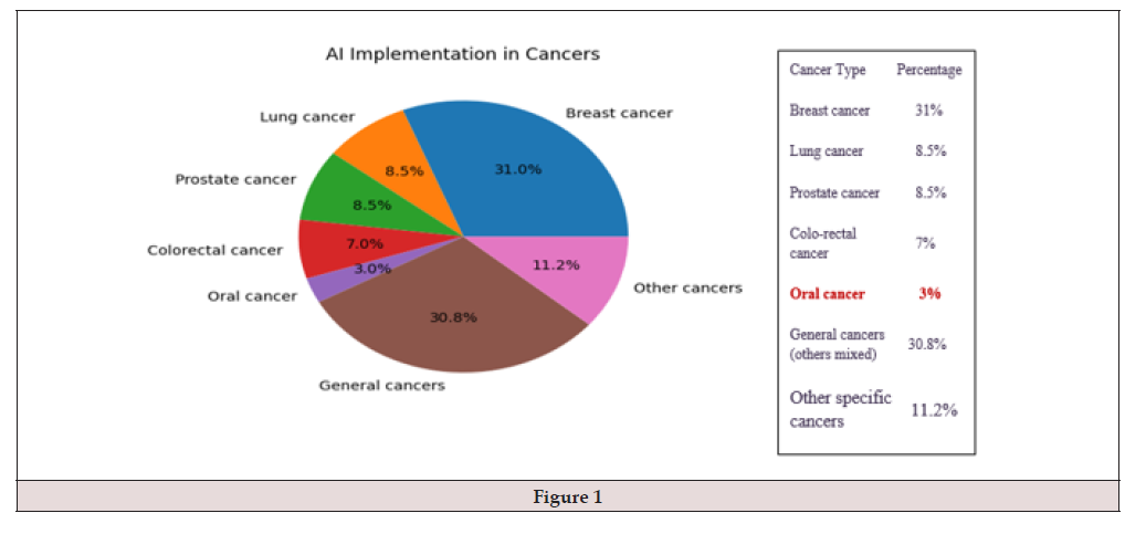
Figure 1:
Oral Cancer
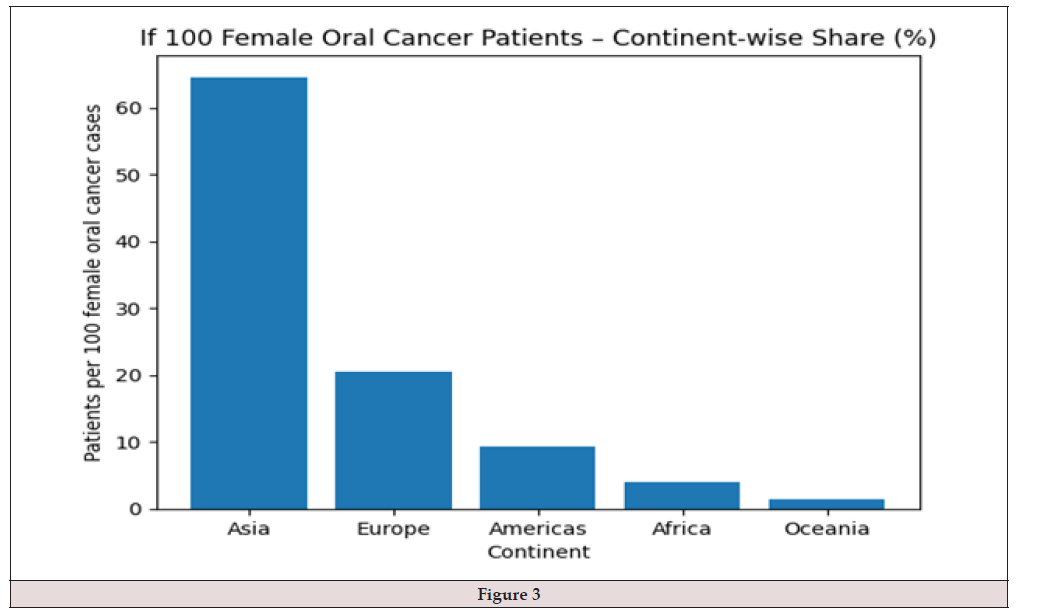
As per the statistics of WHO, 2020, oral cancer constitutes around 2–4% of all cancers globally, with a higher burden in South and Southeast Asia. The oral cavity includes the lips, buccal mucosa, tongue, floor of the mouth, hard palate, and gingiva, and is responsible for functions such as speech, mastication, and swallowing. Oral cancer mainly arises from the mucosal lining of the oral cavity, where the epithelial cells are frequently exposed to carcinogens like tobacco, alcohol, and betel nut. Oral cancer usually begins as a small lesion in the mucosa, often presenting as leukoplakia, erythroplakia, or a non-healing ulcer in the early stage, and when diagnosed early it is largely treatable. As the disease progresses, the tumor invades deeper connective tissues, muscles, and adjacent structures, making treatment more difficult in advanced stages. If left untreated, it can spread to regional lymph nodes and distant organs. Recurrence is also possible even after adequate therapy, hence regular follow-up and early diagnosis are essential. Depending on the type of cells involved, oral cancers are classified as squamous cell carcinoma (most common), verrucous carcinoma, and minor salivary gland carcinomas. Based on the extent of invasion, they are categorized as in situ, locally invasive, and metastatic oral cancers. The diagnostic techniques commonly used to detect oral cancer include clinical examination, biopsy, CT, MRI, and adjunctive imaging methods. Clinical diagnosis can be challenging due to variability in lesion appearance and similarity to benign conditions. Recently, Artificial Intelligence (AI) has emerged as a supportive tool in oral cancer detection. AI systems analyze clinical images, radiographs, and histopathological slides to assist in early detection and classification. However, challenges remain due to variations in lesion size, shape, color, and image quality, as well as differences in patient populations. Despite these challenges, AI shows promising potential in improving early diagnosis and prognosis of oral cancer (Figures 2,3).
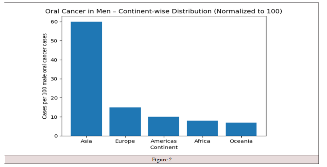
Figure 2:




 We use cookies to ensure you get the best experience on our website.