
Research Article 
 Creative Commons, CC-BY
An Audit of Fiberoptic Bronchoscopy at The Tertiary Care Unit in Central Sri Lanka
*Corresponding author:Madegedara RMD, Chair Professor of Medicine, Consultant Respiratory Physician, National Hospital Kandy, Sri Lanka. Email: dmadegedara@yahoo.com, dmadegedara@wyb.ac.lk.
Received:March 15, 2026; Published:March 26, 2026
DOI: 10.34297/AJBSR.2026.30.003953
Abstract
Background: Fiberoptic Bronchoscopy (FOB) is an essential diagnostic and therapeutic tool in respiratory medicine. While widely utilized, local data regarding its specific usage patterns and outcomes in tertiary care are limited. This audit aimed to evaluate the indications, diagnostic yield, and safety profile of FOB procedures performed at the National Hospital, Kandy.
Methodology: A retrospective descriptive study was conducted involving 621 patients who underwent fiberoptic bronchoscopy at the Respiratory and Research Unit 2 of the National Hospital Kandy between 2020 and 2024. Procedures were performed using Olympus Evis Exera III CV-190 video bronchoscopes with standard accessories (biopsy forceps, brushing, and BAL). Data regarding patient demographics, clinical indications, radiological findings, bronchoscopy observations, and procedural complications were extracted from clinical records and analyzed using SPSS software.
Results: The study population comprised 621 patients, of which 70.4% (n=437) were male. The largest age cohort was the 60–79 years group, accounting for 52.3% (n=325) of the total population. The leading indications for the procedure were suspected malignancy 33.3% (n=207), persistent undiagnosed cough 31.7% (n=197), and investigation of suspected tuberculosis 11.9% (n=75). Pre-procedural imaging revealed abnormal chest X-rays in 91.44% (n=566) of the cohort, with a statistically significant association observed between advanced age and the presence of radiological abnormalities. Upon direct visualization, endobronchial abnormalities were detected in 52.8% (n=328) of the right lung and 41.2% (n=256) of the left lung; common findings included mucosal inflammation, secretions, obstruction, and extrinsic compression. Malignancy was confirmed in 14.2% (n=88) of patients, while bronchoalveolar lavage identified tuberculosis in 9.2% (n=57) of cases. The mean duration of symptoms prior to bronchoscopy was longest for weight loss (8.63 weeks) and persistent undiagnosed cough (8.41 weeks), followed by shortness of breath (7.74 weeks) and loss of appetite (7.40 weeks), indicating delayed clinical presentation. The procedure demonstrated a high safety profile; while minor complications (primarily mild bronchospasm and minor bleeding) occurred in 11.6% (n=72) of patients, there were zero reported cases of procedure-related mortality, significant hemoptysis, pneumothorax, or respiratory failure.
Conclusions: Fiberoptic bronchoscopy remains a safe and high-yield diagnostic procedure in this setting, playing a critical role in the diagnosis of lung malignancy and pulmonary tuberculosis. The findings highlight the necessity of prioritizing this investigation for elderly patients presenting with radiological abnormalities to ensure early diagnosis and management.
Keywords: Fiberoptic bronchoscopy, Audit, Lung cancer, Tuberculosis, Safety profile
Introduction
Fiberoptic Bronchoscopy (FOB) has revolutionized the practice of respiratory medicine since its introduction in the late 1960s. It remains the gold standard for the direct visualization of the tracheobronchial tree, serving as an indispensable tool for both diagnostic and therapeutic purposes. The procedure is minimally invasive and allows for the collection of pathological and microbiological samples through techniques such as endobronchial biopsy, bronchial brushing, and Bronchoalveolar Lavage (BAL), which are critical for the management of a wide spectrum of pulmonary diseases. In the context of Sri Lanka, the dual burden of communicable and non-communicable respiratory diseases presents a unique diagnostic challenge. The incidence of bronchial carcinoma is rising, necessitating prompt tissue diagnosis for staging and management. Simultaneously, Pulmonary Tuberculosis (PTB) remains a significant public health concern, where bronchoscopy plays a vital role in diagnosing smear-negative cases. Furthermore, the evaluation of non-specific symptoms such as persistent undiagnosed cough, hemoptysis, and unexplained radiological opacities frequently relies on bronchoscopy assessment to exclude endobronchial pathology and take appropriate sampling.
Despite the widespread use of FOB in tertiary care settings, rigorous auditing of procedural outcomes is essential to maintain quality assurance and patient safety. International guidelines emphasize the importance of monitoring indications, diagnostic yields, and complication rates to ensure services align with global standards. However, there is a paucity of recently published data regarding the specific utilization patterns and safety profiles of bronchoscopy in the Central Province of Sri Lanka. Therefore, this study was conducted to audit fiberoptic bronchoscopy procedures performed at the National Hospital, Kandy. The primary objective was to retrospectively analyses the clinical indications, procedural details, bronchoscopy findings, diagnostic yield and complications. Additionally, the study aimed to ascertain the relationships between these bronchoscopy findings and patient demographics, radiological presentations, and referral indications to identify significant clinical associations in this population.
Methodology
A retrospective descriptive study (audit) was conducted at the Respiratory and Research Unit-2 of the National Hospital, Kandy, a tertiary care referral center in Sri Lanka. The study period spanned four years, from January 2020 to December 2024. The study population comprised all patients who underwent elective or emergency fiberoptic bronchoscopy at the unit during the study period. A total of 621 procedures were included in the final analysis. Patients with incomplete medical records or missing procedural data were excluded from the study.
Data were retrospectively retrieved from the unit’s endoscopy registry, Bed Head Tickets (BHTs), and pathology records using a structured data extraction form. The variables collected encompassed demographic characteristics (age and gender) and the primary clinical indications for referral, such as persistent undiagnosed cough, hemoptysis, suspected malignancy, or suspicion of tuberculosis. Radiological data were extracted regarding abnormalities observed on pre-procedural Chest X-rays (CXR) and Computed Tomography (CT) scans. The study instrument included a mandatory pre-procedural safety checklist (recording hemoglobin, platelet counts, clotting time, and written consent), a systematic anatomical assessment of the tracheobronchial tree (larynx, trachea, carina, and specific lobar segments), and documentation of the specific sampling modalities employed (bronchoalveolar lavage, bronchial washing, brushing, or biopsy). Procedural outcomes were documented, specifically visualized bronchoscopy pathologies including endobronchial masses, mucosal inflammation, anatomical distortions, or normal airways. Furthermore, all intra-operative and immediate postoperative adverse events were recorded and categorized into minor complications (e.g., transient desaturation, minor bleeding requiring no intervention) or major complications (e.g., severe hemorrhage, pneumothorax). Diagnostic yield was defined as the proportion of bronchoscopy procedures that resulted in a definitive diagnosis based on histopathology, microbiological analysis, or cytology findings obtained during fiberoptic bronchoscopy. Data was entered and analyzed using SPSS (Statistical Package for the Social Sciences) software. Descriptive statistics were utilized to summaries demographic data and frequencies of indications and outcomes. Categorical variables were expressed as percentages. The Chi-square test was employed to assess the association between patient age, radiological abnormalities, and bronchoscopy findings. A p-value of <0.05 was considered statistically significant. Ethical clearance was obtained from the hospital’s ethical committee.
Results
Study Population and Demographics
A total of 621 patients underwent fiberoptic bronchoscopy during the study period (2020–2024). The demographic analysis revealed a predominant distribution of older adults, consistent with the typical age of onset for bronchial malignancy and chronic respiratory diseases. The largest age cohort was the 60–79 years group, accounting for 52.3% of the total population. This was followed by the 40–59 years group. Patients at the extremes of age (18–39 years and >80 years) represented smaller fractions of the study population. This age distribution aligns with the increasing prevalence of suspected malignancy and chronic lung pathologies in the elderly (Table 1, Figures 1, 2).
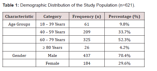
Table 1:Demographic Distribution of the Study Population (n=621).
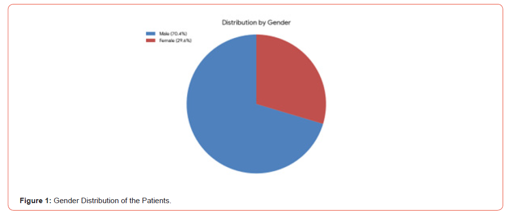
Figure 1:Gender Distribution of the Patients.
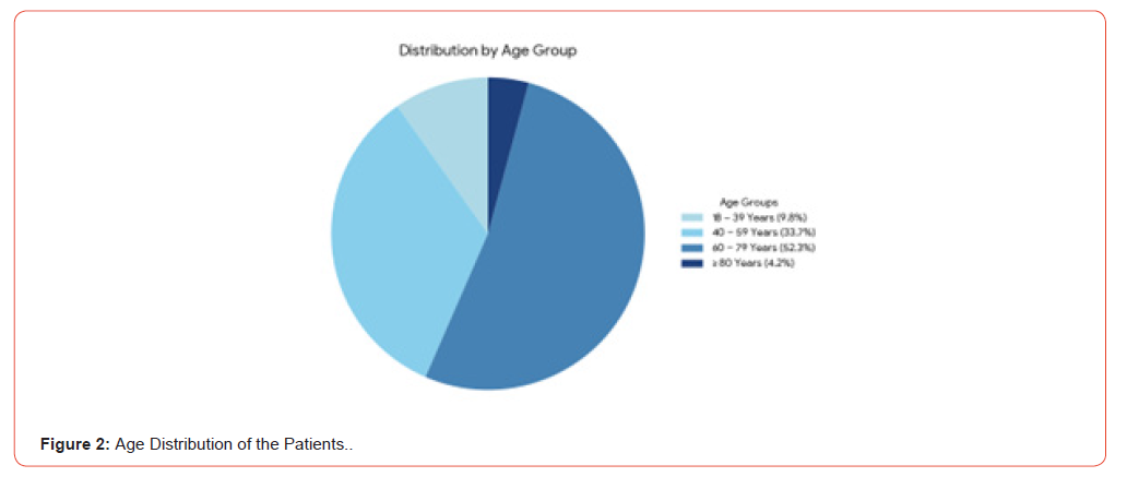
Figure 2:Age Distribution of the Patients..
Indications for Bronchoscopy
The clinical indications for fiberoptic bronchoscopy were categorised based on the primary referral reason. As summarized in Table 2, Figure 3, the leading indication was suspected malignancy, which accounted for 33.3% (n=207) of the total procedures. This reflects the unit’s role as a tertiary referral center for evaluating suspicious radiological opacities. Persistent undiagnosed cough refractory to conventional medical therapy was the second most common indication, representing 31.7% (n=197) of the cohort. The investigation of suspected Tuberculosis (TB), particularly in smearnegative cases or those requiring Bronchoalveolar Lavage (BAL) for confirmation, accounted for 11.9% (n=75). The remaining indications included investigation of hemoptysis, unexplained dyspnea, and recurrent pneumonia.

Table 2:Primary Indications for Fiberoptic Bronchoscopy (n=621).
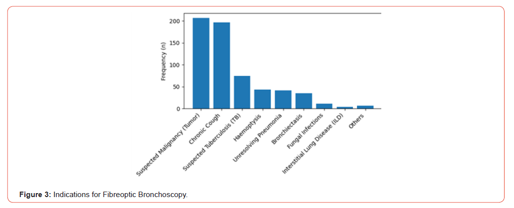
Figure 3:Indications for Fibreoptic Bronchoscopy.
Radiological Evaluation
Chest radiography was the primary screening modality, performed in 99.7% of the study population (Figure 4). Of these, 91.44% (n=566) exhibited pathological findings. Consistent with the leading referral indication of suspected malignancy, radiological evidence of a mass or tumor was observed in 30.3% (n=207) of participants. Other common findings included infiltrates and consolidations indicative of infective or inflammatory etiologies. Conversely, 8.6% (n=53) of patients had normal chest radiographs (Figure 5).
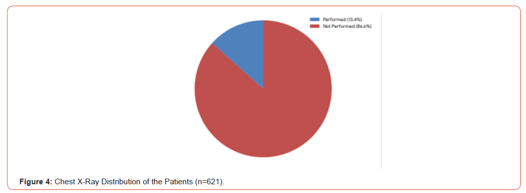
Figure 4:Chest X-Ray Distribution of the Patients (n=621).
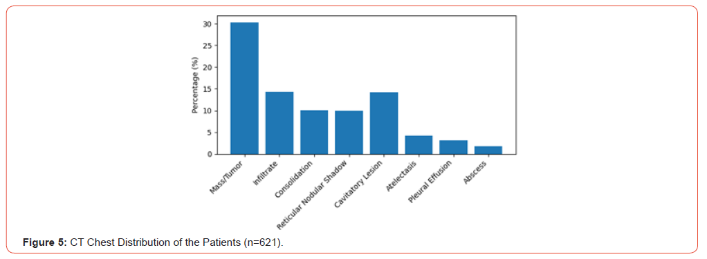
Figure 5:CT Chest Distribution of the Patients (n=621).
Computed Tomography (CT) of the chest was utilized in a subset of patients (13.4%, n=83). Among those who underwent CT imaging, 84.3% (n=70) revealed abnormalities. CT imaging demonstrated a higher yield for characterizing pathology, with 45.7% (n=32) of scans identifying pulmonary masses or tumors. Additionally, 20.0% (n=14) of CT scans revealed infiltrates, while distinct patterns such as ground-glass opacities and fibrosis were identified in patients with suspected Interstitial Lung Disease (ILD) (Figures 6,7 & Table 3).
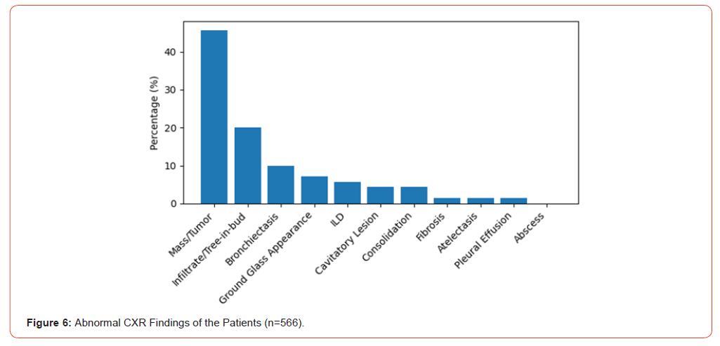
Figure 6:Abnormal CXR Findings of the Patients (n=566).
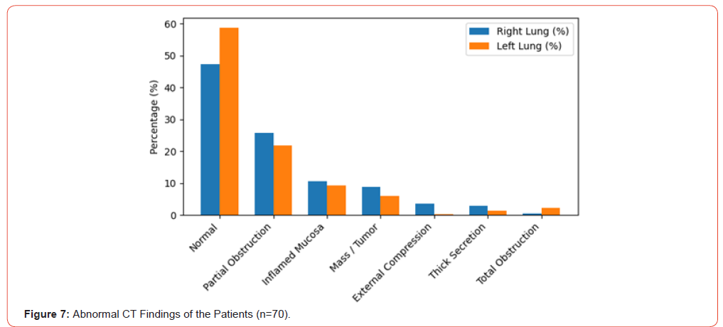
Figure 7:Abnormal CT Findings of the Patients (n=70).

Table 3:Radiological Evaluation and Findings of the Study Population.
Note: *Percentages for CXR findings are calculated based on the total study population (N=621).
**Percentages for CT findings are calculated based on those with abnormal CT scans (n=70).
Bronchoscopy Findings and Diagnostic Yield

Table 4:Distribution of Macroscopic Bronchoscopy Findings.
Macroscopic Observations
Direct bronchoscopy visualization revealed that a significant proportion of patients had anatomically normal airways (47.2% in the right lung and 58.8% in the left). However, pathological changes were frequently identified. Airway obstruction was the most common abnormality, observed in 25.8% of the right bronchial tree and 21.9% of the left. Thick secretions were noted in 18 cases (2.9%) on the right and 9 cases (1.4%) on the left. Signs of malignancy, such as masses or tumours, were identified in 8.9% of the right lung and 6.0% of the left. Additionally, external compression was identified in 3.5% of cases on the right side and 0.3% on the left (Table 4, Figure 8).
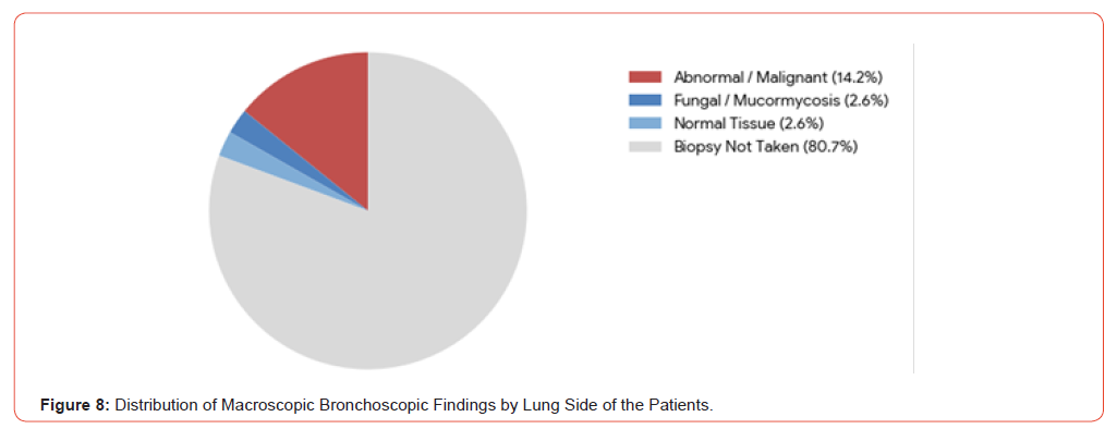
Figure 8:Distribution of Macroscopic Bronchoscopic Findings by Lung Side of the Patients.
Sampling Modalities
The selection of sampling techniques was dictated by the gross findings. Bronchoalveolar Lavage (BAL) was the predominant sampling method, utilized in 80.8% (n=503) of cases as the sole modality, particularly for suspected infections. Combined modalities were used for visible lesions, with 18.4% (n=114) of patients undergoing BAL plus Endobronchial Biopsy, and 0.6% (n=4) undergoing BAL plus Transbronchial Biopsy.
Histopathological and Microbiological Yield
The diagnostic yield varied by the type of sample obtained.
a) Biopsy Yield: Histopathological examination of
endobronchial biopsies provided the highest diagnostic yield.
Biopsies were performed in 120 patients, of which 104 (86.7%)
yielded definitive pathological diagnoses, including malignancy
(n=88) and fungal infection (n=16), while 16 samples (13.3%)
demonstrated normal histology, demonstrating a high utility
for definitive tissue diagnosis.
b) BAL Yield: BAL analysis was crucial for infectious
etiology. Bronchoalveolar lavage (BAL) was performed in 503
patients, with microbiological analysis establishing a diagnosis
in 126 cases (25.0%). BAL identified tuberculosis in 57 patients
(9.2%), pyogenic bacterial infections in 39 patients (6.3%), and
fungal infections in 30 patients (4.8%).
c) Cytology: Cytological analysis of bronchial washings
(n=621) was predominantly inflammatory (59.4%, n=369),
with atypical cells detected in 10.5% (n=65) and malignant
cells confirmed in 0.8% (n=5) of patients.
These findings highlight the complementary diagnostic roles of histopathology, microbiological analysis, and cytology in establishing definitive pathological or microbiological diagnoses through fiberoptic bronchoscopy (Table 5, Figures 9-11).
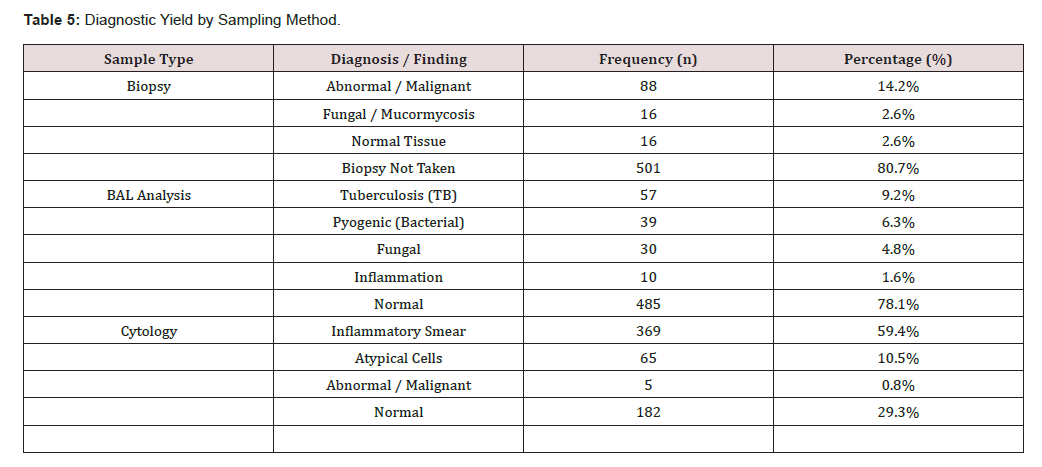
Table 5:Diagnostic Yield by Sampling Method.
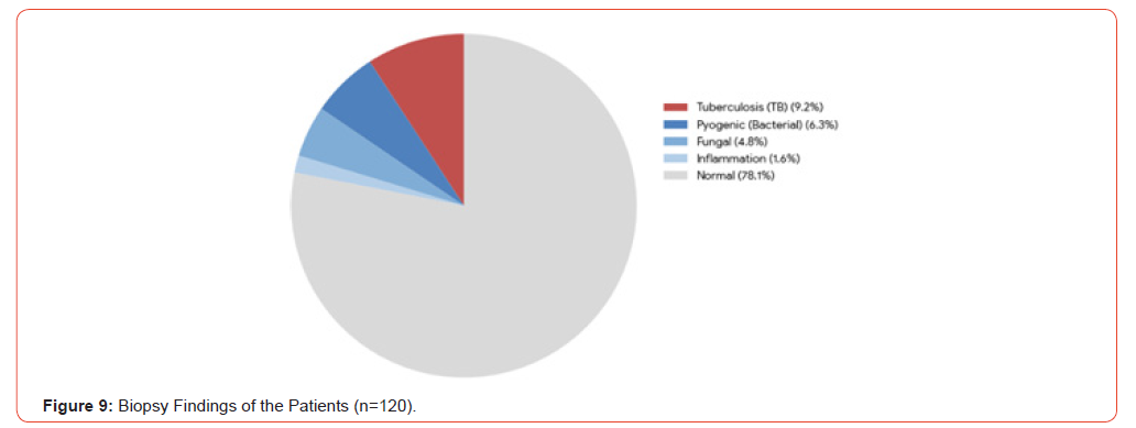
Figure 9:Biopsy Findings of the Patients (n=120).
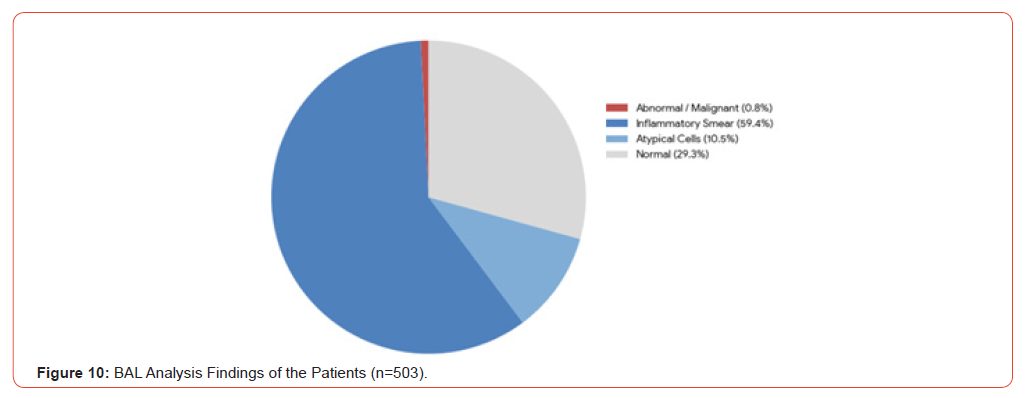
Figure 10:BAL Analysis Findings of the Patients (n=503)./p>
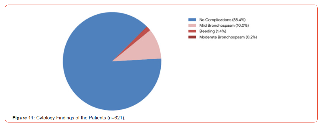
Figure 11:Cytology Findings of the Patients (n=621).
Safety Profile and Complications
The audit demonstrated a favorable safety profile for fiberoptic bronchoscopy procedures performed within the unit. While minor adverse events were noted, the incidence of severe complications was negligible. Major complications, defined as conditions requiring escalation of care or interventional management, such as tension pneumothorax, respiratory failure, hemothorax, or tracheal injury, were absent. There were zero recorded incidences of these life-threatening events. The overall adverse event rate was 11.6% (n=72), predominantly driven by mild, transient respiratory symptoms.
a) Bronchospasm: Respiratory reactivity was the
most common adverse event, but was largely minor. Mild
bronchospasm occurred in 10.0% (n=62) of patients; these
episodes were transient and self-limiting. One patient (0.2%)
experienced moderate bronchospasm requiring medical
management. Importantly, there were no cases of severe
bronchospasm recorded.
b) Bleeding: Minor hemorrhage was observed in 9 patients
(1.4%), involving small-volume bleeding from biopsy or
brushing sites. These cases resolved spontaneously or with
local cold saline/adrenaline instillation.
Consequently, 88.4% (n=549) of the study population
underwent the procedure with no complications or adverse events
(Table 6, Figure 12).
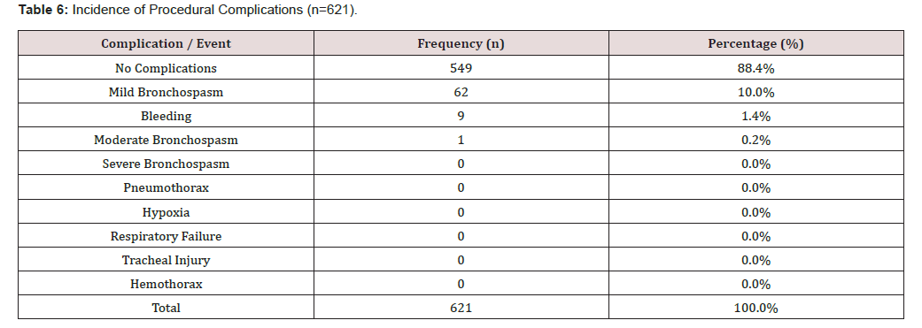
Table 6:Incidence of Procedural Complications (n=621).

Figure 12:Distribution of Procedural Complications of the Patients.
Clinical Correlations and Symptom Analysis
Statistical Associations
Further statistical analysis was conducted to evaluate the relationship between patient demographics and diagnostic outcomes. There was a highly significant positive association between advanced age and the diagnostic yield of histological sampling. Older patients were significantly more likely to have positive findings on Endobronchial Biopsy (p<0.001) and Bronchoalveolar Lavage (BAL) (p=0.018), reflecting the higher prevalence of malignancy and complex infections in the elderly cohort. Similarly, abnormal findings on CT Chest were statistically more frequent in older age groups (p=0.03). Conversely, risk assessment analysis demonstrated that there was no statistically significant association between patient age and the incidence of procedural complications (p=0.956). This finding reinforces the safety of fiberoptic bronchoscopy as a diagnostic modality for elderly patients when performed under standard monitoring protocols.
Symptom Duration Analysis
Analysis of the clinical history revealed significant delays between symptom onset and the diagnostic procedure. The mean duration of symptoms prior to bronchoscopy was highest for loss of weight (mean = 8.63) followed by patients presenting with persistent undiagnosed cough (mean = 8.41 weeks), shortness of breath (mean = 7.74 weeks) and loss of appetite (mean = 7.40 weeks). Patients presenting with fever had a shorter pre-procedural interval (mean = 5.17 weeks), likely due to the acute nature of infectious presentations prompting earlier referral (Table 7).
Discussion
Overview of Findings
This retrospective audit of 621 Fiberoptic Bronchoscopy (FOB) procedures performed at the National Hospital Kandy establishes the procedure as a safe, high-yield diagnostic tool within the Sri Lankan tertiary care setting. The demographic profile of the study population, predominantly male (70.4%) and elderly (52.3% aged 60–79 years), mirrors the global epidemiological patterns of bronchial malignancy and chronic respiratory diseases.
This male predominance aligns with regional data, such as the audit by Toori et al. in Pakistan, which reported a male population of 78% with a mean age of 48.8 years [1]. In contrast, Lyimo et al. in Tanzania observed an equal gender distribution (male: female ratio 1:1) with a median age of 58 years, likely reflecting differing referral patterns for infectious versus malignant etiologies in that setting [2]. Larger international multicenter studies, such as Faccio Longo et al. (Italy), reported a male predominance of 60.6% with a mean age of 57 ± 16 years, closely resembling our own cohort [3]. Similarly, Madhavan et al. in South India reported a male percentage of 58.4% in their general bronchoscopy population [4]. For instance, the Indian Bronchoscopy Survey involving 669 bronchoscopists across multiple centres reported a predominance of adult male patients undergoing bronchoscopy procedures, reflecting the epidemiology of respiratory diseases in the region [5]. Our findings validate the unit’s adherence to international safety standards, demonstrating a complication rate comparable to these large-scale global cohorts.
Patterns of Indications and Disease Burden
The primary indications for bronchoscopy in this cohort were suspected malignancy (33.3%) and persistent undiagnosed cough (31.7%). This distribution reflects a “mixed-burden” epidemiology characteristic of South Asia. Our malignancy referral rate aligns with findings from other regional audits, such as those reported by Toori et al. in Pakistan, where diagnostic sampling for suspected neoplasm was a leading indication [1]. However, our data contrasts with recent findings from Tanzania by Lyimo et al., where persistent cough accounted for the vast majority (82.1%) of indications, and malignancy was lower [2].
Large regional surveys further support these findings. The Indian Bronchoscopy Survey reported that bronchoscopy is commonly performed for the evaluation of suspected lung malignancy, persistent cough, and unexplained radiological abnormalities [5]. In addition, bronchoscopy procedures remain central to the diagnostic pathway for lung cancer, as emphasized in international clinical guidelines for lung cancer diagnosis and staging [6]. The higher proportion of suspected malignancy in our cohort likely reflects the National Hospital Kandy’s role as a specialized referral center for radiological abnormalities, rather than a primary filter for non-specific respiratory symptoms.
The Role of Bronchoscopy in Tuberculosis
In regions where tuberculosis remains endemic, bronchoscopy plays an important role in diagnosing smear-negative pulmonary tuberculosis. In the present study, Bronchoalveolar Lavage (BAL) confirmed tuberculosis in 9.2% of cases. Similar findings have been reported in several regional studies where BAL significantly improved diagnostic confirmation in smear-negative patients. This aligns with findings from other developing nations; for example, Toori et al. reported tuberculosis in 38.16% of abnormal bronchoscopy findings in their audit [1], while Lyimo et al. documented microbiological confirmation of tuberculosis in 4.7% of bronchoscopy procedures [2]. Bronchoscopy-guided sampling techniques such as BAL, bronchial washings, and biopsy have been widely recommended for patients with persistent radiological abnormalities when sputum studies are negative [7].
The high yield of Bronchoalveolar Lavage (BAL) in our study (9.2% for smear-negative cases) further supports its utility. This is consistent with regional data suggesting that bronchoscopy is an indispensable tool for smear-negative patients who fail to produce sputum or have non-specific radiological findings. By securing an early microbiological diagnosis, we can prevent unnecessary empirical anti-tuberculosis treatment and its associated hepatotoxicity, while also avoiding delays in diagnosing malignancies that may mimic tuberculosis radiologically [8].
Safety Profile and Risk Factors
A pivotal finding of this audit is the excellent safety profile of the unit. The overall complication rate was 11.6% (n=72), with zero procedure-related mortalities and no major complications recorded. This performance is well within the benchmarks set by the BTS Guidelines (Du Rand et al.), which cite a major complication rate of <1% for routine diagnostic bronchoscopy [9]. Furthermore, our rate of clinically significant complications compares favorably with the multicenter prospective study by Facciolo Ngo et al. (Italy), which reported a complication rate of 1.08% across 20,986 procedures4.
Studies from other regions have also confirmed the overall safety of bronchoscopy when performed by trained personnel in appropriately equipped centres10 [10]. In our cohort, when excluding minor, transient physiological reactions (such as mild bronchospasm), the incidence of complications requiring medical intervention or resulting in procedural modification was 1.6% (n=10). This slight variance is likely attributable to sample size differences rather than procedural safety, confirming that our standard of care aligns with international benchmarks. The absence of major life-threatening events in our study further mirrors the safety profile established by Faccio Longo et al.
Crucially, our analysis revealed no statistically significant association between patient age and procedural complications (p=0.956). This aligns with the prospective risk-adjusted analysis by Ernst et al., which demonstrated that age is not an independent predictor of adverse outcomes when comorbidities are managed [11]. This finding is clinically significant, confirming that advanced age (>80 years) should not be a deterrent for invasive investigation.
Sedation and Patient Comfort
Our successful completion rate suggests effective sedation and airway management protocols. While local anesthesia alone is utilized in some resource-limited settings, the impact of sedation on patient tolerance in adults remains a critical consideration in our region. Previous studies have demonstrated that conscious sedation improves patient comfort and procedural tolerance without significantly increasing complication rates.
Madhavan et al. demonstrated that bronchoscopist-directed sedation significantly improved patient comfort scores during bronchoscopy procedures [11]. Similarly, other studies have emphasized that adequate sedation improves patient satisfaction and willingness to undergo repeat procedures [12]. Our findings of high procedure completion rates and low complication rates suggest that current sedation protocols in our unit are effective and safe.
Diagnostic Yield and Limitations of Conventional Bronchoscopy
Our analysis demonstrated a highly significant positive association between advanced age and positive histological findings (p<0.001). This observation likely reflects the increased prevalence of lung malignancy among older populations. However, a limitation of conventional white-light bronchoscopy is its reduced sensitivity for small peripheral lesions compared to central masses. Advanced techniques such as Electromagnetic Navigation Bronchoscopy (ENB) have been shown by Herth et al. to significantly increase diagnostic yield in peripheral lung lesions without increasing complication rates [13]. Similarly, interventional pulmonology is increasingly evolving toward these image-guided bronchoscopy techniques [14,15]. Modern lung cancer diagnostic guidelines emphasize the importance of bronchoscopy tissue sampling for establishing histological diagnosis and staging of lung malignancies [6,16]. Endobronchial ultrasound-guided transbronchial needle aspiration has also demonstrated high diagnostic accuracy for mediastinal staging and tissue acquisition in lung cancer patients [17]. More recently, international consensus statements have highlighted the need for standardized definitions of diagnostic yield when evaluating bronchoscopy outcomes. The American Thoracic Society recommends uniform reporting criteria to improve comparability between bronchoscopy studies and facilitate evidence-based evaluation of emerging diagnostic technologies [18].
However, the study was conducted in a setting with specific infrastructural constraints regarding cross-sectional imaging. Unlike centers where Computed Tomography (CT) is routine, limited accessibility and resource constraints in our setting meant that Chest Radiography (CXR) remained the primary screening modality, CT was performed in only a subset of patients.
Symptom Duration and Delayed Presentation
Patients presented with persistent undiagnosed cough and weight loss for an average of 8.6 weeks and 8.4 weeks, respectively, prior to the procedure. This two-month diagnostic interval represents a critical delay. Unlike hemoptysis (mean 3.9 weeks), which acts as an “alarm symptom” prompting earlier referral, constitutional symptoms may be tolerated by patients or treated empirically for too long at the primary care level.
Conclusion
This audit confirms that Fiberoptic Bronchoscopy (FOB) at the National Hospital Kandy is a highly effective and safe diagnostic modality, meeting international standards for procedural safety. Bronchoscopy demonstrated high diagnostic utility, particularly through endobronchial biopsy for malignancy and bronchoalveolar lavage for infectious etiologies. When excluding minor, self-limiting physiological reactions, the rate of clinically significant complications stood at 1.6%, with no procedure-related mortalities. This low incidence of significant morbidity validates the unit’s current operational protocols and technical expertise, demonstrating that conventional bronchoscopy remains a safe and effective diagnostic tool in our setting.
The study highlights a significant burden of bronchial malignancy and tuberculosis within the referral population. Importantly, advanced age was identified as a strong predictor of positive diagnostic yield but was not associated with an increased risk of complications. This supports the continued and aggressive use of bronchoscopy for investigating radiological abnormalities in the elderly, a demographic often viewed as high-risk.
However, the diagnostic pathway is currently hindered by significant pre-procedural delays. The finding that patients experience symptoms such as persistent undiagnosed cough and weight loss for an average of two months prior to investigation suggests missed opportunities for early diagnosis. While the procedure itself is efficient, the timeline to referral remains a critical bottleneck in the management of respiratory malignancies in this setting.
Recommendations
Based on the findings of this study, the following recommendations are proposed to improve patient outcomes and service delivery:
1) TB Diagnostic Algorithm: The high yield of
Bronchoalveolar Lavage (BAL) in diagnosing smear-negative
tuberculosis (9.2%) supports its routine inclusion in the
diagnostic algorithm. BAL should be mandatory for all patients
with unexplained radiological infiltrates who remain sputum
smear-negative.
2) Earlier Referral Protocols: Given the average delay
of over 8 weeks for patients presenting with persistent
undiagnosed cough and weight loss, sensitization programs
should be conducted for primary care practitioners. Protocols
should be established to expedite referral for bronchoscopy in
any patient over 40 years of age with respiratory symptoms
persisting beyond 4 weeks, regardless of chest X-ray findings.
3) Implementation of National Protocols and Safety
Checklists: To ensure uniformity and patient safety across all
centers, a standardized National Protocol for bronchoscopy
should be developed. This should include the mandatory use of
the WHO/ BTS Safety Checklist (modified for endoscopy) prior
to every procedure to minimize errors and maintain the unit’s
high safety profile.
4) Regular Re-audits: Continuous quality improvement
is essential. Regular re-audits should be institutionalized to
monitor adherence to safety standards, track diagnostic yields,
and identify emerging trends in disease epidemiology or
procedural complications.
5) Adoption of Advanced Technology: While conventional
bronchoscopy showed high yield for central lesions, the
diagnostic limitations for peripheral nodules warrant the
introduction of Endobronchial Ultrasound (EBUS) and
Electromagnetic Navigation services. Acquiring these
technologies would significantly enhance the staging
capabilities for lung cancer in the Central Province.
6) Patient Education: Public health awareness campaigns
are necessary to educate the community about “non-alarm”
symptoms (chronic cough, unexplained weight loss) to
encourage earlier medical consultation, mirroring the rapid
response seen with hemoptysis.
Funding
This research received no specific grant from any funding agency in the public, commercial, or not-for-profit sectors.
Conflict of Interest
The authors declare no conflict of interest
References
- Toori KU, Nabi S, Hussain SW, Wadood A, Khattak S (2012) An audit of fibreoptic bronchoscopy service at KRL Hospital Islamabad. Pak J Chest Med 18(1): 1-6.
- Lyimo T, Kyaruzi VM, Byomuganyizi M, Khamisi RH (2024) Indications and outcome of flexible bronchoscopy at Muhimbili National Hospital in Dar es Salaam, Tanzania. East Afr Sci 6(1): 77-82.
- Facciolongo N, Patelli M, Gasparini S, L Lazzari Agli, M Salio, et al. (2009) Incidence of complications in bronchoscopy: multicentre prospective study of 20,986 bronchoscopies. Monaldi Arch Chest Dis 71(1): 8-14.
- Madhavan S, Chan L, Arachchi J (2022) Effect of bronchoscopist-directed sedation and other factors on patient comfort during diagnostic flexible bronchoscopy. Can Respir J 2022: 9534981.
- Madan K, Mohan A, Agarwal R, Hadda V, Khilnani GC, et al. (2018) A survey of flexible bronchoscopy practices in India: the Indian bronchoscopy survey (2017). Lung India 35(2): 98-107.
- Rivera MP, Mehta AC, Wahidi MM (2013) Establishing the diagnosis of lung cancer: diagnosis and management of lung cancer, 3rd ed. Chest 143(5 Suppl): e142S-e165S.
- Ahammed S, Sarker MSU, Ali MZ (2021) Diagnostic value of bronchoalveolar lavage examination in sputum-negative patients for acid-fast bacilli in suspected pulmonary tuberculosis. KYAMC J 12(2): 84-87.
- Korraa EA, Dwedar IA, Gomaa AA, Shata AK (2019) Evaluation of the role of bronchoscopy in the intensive care units. Egypt J Bronchol 13: 67.
- Du Rand IA, Blaikley J, Booton R, N Chaudhuri, V Gupta, et al. (2013) British Thoracic Society guideline for diagnostic flexible bronchoscopy in adults: accredited by NICE. Thorax 68(Suppl 1): i1-i44.
- Tsoka KV (2022) Diagnostic yield and complications of flexible bronchoscopy performed at a tertiary academic hospital: a 5-year retrospective study. University of the Witwatersrand Repository.
- Ernst A, Silvestri GA, Johnstone D (2008) Prospective risk-adjusted morbidity and mortality outcome analysis after therapeutic bronchoscopic procedures. Chest 134(3): 514-519.
- José RJ, Shaefi S, Navani N (2013) Sedation for flexible bronchoscopy: current and emerging evidence. Eur Respir Rev 22(128): 106-116.
- Herth FJF, Ernst A, Becker HD (2006) Electromagnetic navigation diagnostic bronchoscopy in peripheral lung lesions. Chest 130(5): A191.
- Seijo LM, Sterman DH (2001) Interventional pulmonology. N Engl J Med 344(10): 740-749.
- Wahidi MM, Herth FJF, Ernst A (2007) State of the art: interventional pulmonology. Chest 131(1): 261-274.
- Silvestri GA, Gonzalez AV, Jantz MA, Mitchell L Margolis, Michael K Gould, et al. (2013) Methods for staging non-small cell lung cancer. Chest 143(5 Suppl): e211S-e250S.
- Navani N, Lawrence DR, Kolvekar S, Sara Lock, Himender Makker, et al. (2015) Endobronchial ultrasound-guided transbronchial needle aspiration for mediastinal staging of lung cancer. Lancet Respir Med 3(4): 282-289.
- Gonzalez AV, Silvestri GA, Korevaar DA, Yaron B Gesthalter, Nisha D Almeida, et al. (2024) Assessment of Advanced Diagnostic Bronchoscopy Outcomes for Peripheral Lung Lesions: A Delphi Consensus Definition of Diagnostic Yield and Recommendations for Patient-centered Study Designs. An Official American Thoracic Society/American College of Chest Physicians Research Statement. Am J Respir Crit Care Med 209(6): 634-646.




 We use cookies to ensure you get the best experience on our website.