
Research Article 
 Creative Commons, CC-BY
Inhibitory Effects of Mir-199a-5p On Proliferation, Migration, And Differentiation Of C-Kit⁺ Cardiac Progenitor Cells
*Corresponding author:Sha Li, Department of Cardiovascular Medicine, Hebei General Hospital, Shijiazhuang Hebei-China.
Received:March 13, 2026; Published:March 30, 2026
DOI: 10.34297/AJBSR.2026.30.003959
Abstract
Background: Cardiac Progenitor Cells (CPCs) can grow, move, and change into other cell types. These abilities are important for treating Ischemic Heart Failure (IHF). MicroRNAs (miRNAs) help control heart-related processes after genes are read. This study tested how miR‑199a‑5p affects c‑kit⁺ cardiac progenitor cells.
Methods: We took c‑kit⁺ cells from mouse hearts. C57BL/6 mice were used. The cells were separated with immunomagnetic beads. Tests showed the cells were pure. The markers c‑kit, Sca‑1, and CD34 were all found in 100% of cells. CD45 was not found. Cells were placed into three groups: a control group, a negative control mimic group, and a miR‑199a‑5p mimic group. A lentivirus was used to make cells produce more miR‑199a‑5p. We then measured cell activity with the CCK‑8 test. Cell division was checked with EdU staining. A scratch test measured cell movement. We also tested for Nkx2.5, CD31, and α‑SMA using RT‑qPCR and immunofluorescence.
Results: Compared with the NC mimic group, the miR-199a-5p mimic group exhibited significantly reduced cell viability (p<0.001), EdU-positive cell rate markedly decreased (p<0.01), and 24-hour cell migration rate significantly declined (p<0.001). Furthermore, mRNA and protein expression levels of Nkx2.5, CD31, and α-SMA were all significantly downregulated (p<0.001).
Conclusions: These results show that miR‑199a‑5p slows the growth, movement, and change of c‑kit⁺ cardiac progenitor cells. This suggests miR‑199a‑5p could be a target for treating ischemic heart failure.
Keywords: C-kit⁺ cardiac progenitor cells, MiR-199a-5p, Ischemic heart failure, Cell proliferation, Cell migration, Cell differentiation
Abbreviations:IHF: Ischemic heart failure, c‑kit: Stem Cell Factor receptor, Sca‑1: Stem-cell antigen-1, CD45: Pan-leukocyte lineage marker, CD34: Hematopoietic and vascular progenitor cells marker, Nkx2.5: Cardiomyocyte marker, CD31: Platelet-endothelial cell adhesion molecule-1, α-SMA: Myofibroblast differentiation marker, HIF-1α: Hypoxia-inducible factor-1α, CPCs: Cardiac progenitor cells, OD: Optical density values.
Introduction
Ischemic Heart Failure (IHF) continues to cause many heart-related deaths across the world. After a heart attack, the heart muscle gradually changes shape and loses cells that cannot be recovered [1,2]. There are now better drugs, medical procedures, and surgical treatments available. Still, these methods do not lead to actual regrowth of heart muscle at the cellular scale [3]. Because of this, researchers are focusing more on heart regeneration as an important new direction. Among the options being studied, cardiac progenitor cells — called CPCs — show potential for helping the damaged heart muscle work properly again [4-6]. Among different types of cardiac progenitor cells, c-kit⁺ cells have received much attention. These cells can grow rapidly. They can also develop into several cell types. Studies show they help repair heart muscle [7]. Both animal studies and patient trials have reported benefits. Transplanting c-kit⁺ cells can improve heart function. It can also reduce harmful changes in hearts with poor blood supply [8,9]. Still, how these cells are controlled is not fully clear. This is especially true in the diseased heart environment of ischemic heart failure. This lack of knowledge slows their use in treatments and limits ways to improve their effects.
MicroRNAs (miRNAs:endogenous, ≈22-nucleotide, non-coding RNAs that mediate post-transcriptional gene silencing via base-pairing with complementary sequences in target mRNAs) are small molecules that help control how genes work after they are read. They play a role in many heart-related processes such as heart thickening, scarring, and damage after a heart attack [10,11]. Changes in miRNA levels have been tied to the worsening of ischemic heart failure. They also affect how stem cells and progenitor cell’s function [12,13]. One miRNA, called miR‑199a‑5p (microRNA- 199a-5p, a mature 5′-strand microRNA derived from the 3′ arm of the pre-miR-199a hairpin), is involved in heart formation, blood vessel growth, and cell death. Altered levels of miR‑199a‑5p have been found in human heart tissue that is ischemic or failing [14,15]. Previous studies show that miR‑199a‑5p can affect pathways related to low oxygen by targeting molecules like HIF‑1α [16,17]. However, how miR‑199a‑5p directly controls important activities of c‑kit⁺ cardiac progenitor cells — especially their growth, movement, and ability to change into other cell types — remains unclear and has not been fully studied.
To fill this research gap, we studied how miR-199a-5p affects c-kit⁺ cardiac progenitor cells. We increased miR-199a-5p levels in these cells using a lentiviral method. Then, we tested cell activity, growth, movement, and ability to become heart cell types. Our results show that miR-199a-5p clearly reduces these important cell functions. This suggests miR-199a-5p could be a target for treatment aimed at improving heart repair in IHF. This study helps us better understand how miRNAs control heart progenitor cells. It also offers a scientific basis for creating miRNA-based methods to improve heart muscle repair. Our findings show that miR-199a-5p acts as an inhibitor. This provides new ideas about the molecular processes that could be targeted. These targets may help make stem cell treatments for ischemic heart disease work better.
Materials and Methods
Animals and Cell Isolation
All animal experiments were approved by the Animal Ethics Committee of Hebei General Hospital. The experiments followed the relevant guidelines. We used male C57BL/6 mice. The mice were 8 weeks old. Their weight was 18–23 g. They were bought from Guangzhou Laidi Biomedical Research Institute. The mice were kept under standard lab conditions. The temperature was 22 ± 1 °C. The humidity was 55 ± 5 %. The light/dark cycle was 12 h/12 h. Food and water were available at all times. The mice adapted for two weeks. Then, they were anesthetized. We used pentobarbital sodium (50 mg/kg) given by intraperitoneal injection. Next, heart tissues were taken out under sterile conditions. Connective tissue and major vessels were removed. The heart muscle was cut into small pieces. Each piece was about 1 × 1 × 1 mm³ in size. These pieces were digested with enzymes [18]. The digestion solution contained 0.25 % trypsin and 0.1 % collagenase II. Digestion lasted 30 minutes at 37 °C with gentle shaking. We stopped the digestion by adding complete medium. Then, the cell mixture was passed through a 200‑mesh sieve. It was then centrifuged at 1000 × g for 5 minutes. After centrifugation, we collected the cell pellet. The pellet was mixed again with culture medium. The medium contained 78 % IMDM, 20 % FBS, 1 % L‑glutamine, 1 % penicillin‑streptomycin, and 55 μM β‑ Mercaptoethanol [19]. This cell suspension was placed into flasks coated with gelatin. The cells were grown at 37 °C with 5 % CO₂. We changed the medium every 2–3 days.
Immunomagnetic Sorting of c‑kit⁺ CPCs
Cells in the growth phase were collected. They were washed with PBS. The cell concentration was set to 1 × 10⁷ cells/mL. Anti c‑kit microbeads (Miltenyi Biotec) were added. We used 10 μL for every 1 × 10⁶ cells. The mixture was kept at 4 °C for 15 minutes in the dark. Then, the cells were placed in sorting buffer. This buffer was PBS with 0.5% BSA and 2 mM EDTA. The cells were passed through an MS column. The column was in a magnetic separator and was prepared first. The column was washed three times with 500 μL of sorting buffer. Cells that stuck to the magnet (c‑kit⁺ cells) were collected. These cells were spun down and placed in expansion medium. The medium contained 86% DMEM/F12, 10% FBS, 2% B27, 1% L‑glutamine, 1% penicillin‑streptomycin, and 55 μM β‑Mercaptoethanol [20]. The cells were then seeded for later tests.
Flow Cytometric Characterization
Purified cells were resuspended in PBS to reach 1 × 10⁶ cells/mL. FITC-labeled antibodies against c‑kit, Sca‑1, CD34(a trans-membrane sialomucin and canonical surface marker of hematopoietic and vascular progenitor cells), and CD45(a trans-membrane tyrosine phosphatase ubiquitously expressed on all nucleated hematopoietic cells and widely used as a pan-leukocyte lineage marker)were added. These antibodies were from Abcam. The final concentration was 10 μg/mL. The mixture was incubated at 4 °C for 30 minutes in the dark. Then, the cells were washed. Analysis was performed on a Thermo Fisher Attune NxT flow cytometer. Each sample recorded at least 10,000 events. Data analysis used FlowJo v10.0. Cells without antibody staining served as the negative control.
Lentiviral Transfection and Experimental Groups
Two lentiviral vectors were prepared. One contained a miR‑199a‑5p mimic sequence: 5′‑CCCAGUGUUCAGACUACCUGUUC‑3′. The other contained a non‑targeting control. Shanghai Sangon Biotech synthesized them using the miR‑199a‑5p reference (GenBank: NR_029505.1) [21]. c‑kit⁺ cells were seeded in 6‑well plates. Transfection began when cells covered 60–70% of the well area. Lipofectamine™ 3000 was used with an MOI of 10 [22]. The experiment included three groups: untransfected cells (CON group), cells with control lentivirus (NC mimic group), and cells with miR‑199a‑5p lentivirus (miR‑199a‑5p mimic group). Experiments were performed 72 hours after transfection.
Quantitative Real‑Time PCR (RT‑qPCR)

Table 1:
First, total RNA was extracted. We used Trizol reagent from Ta- KaRa for this [23]. Next, RNA amount and quality were measured. A NanoDrop 2000 spectrophotometer from Thermo Fisher Scientific was used. Then, cDNA was made. For this step, 1 μg of total RNA was used with a Prime Script RT kit from TaKaRa. RT‑qPCR was then performed. We used a CFX96 Touch system from Bio‑Rad. The reaction used SYBR Green Master Mix from TaKaRa. Each reaction mixture had a total volume of 20 μL. It contained: 10 μL of the master mix, 0.5 μL of forward primer, 0.5 μL of reverse primer (see Table 1 for sequences), 2 μL of the cDNA, and 7 μL of nuclease‑free water. The PCR program was set as follows: first, 95 °C for 30 seconds; then, 40 cycles of 95 °C for 5 seconds and 60 °C for 30 seconds. GAPDH was used as the reference gene. Finally, relative expression was calculated using the 2‑ΔΔCt method. Every sample was tested three separate times (Table 1).
Cell Viability Assay
Cells were seeded into 96-well plates at 10,000 cells per well. We tested them at three times: immediately (0 hours), after 24 hours, and after 48 hours. At each test time, 10 μL of CCK‑8 reagent (Beyotime) was added to each well [24]. The plates were then kept at 37 °C for 2 hours. Next, the absorbance at 450 nm was measured using a Thermo Scientific Multiskan plate reader. Each group had five replicate wells. The entire experiment was performed three separate times.
EdU Proliferation Assay
Cell division was measured with an EdU kit from Beyotime [25]. First, cells were placed in 96‑well plates. Each well received 5,000 cells. They were grown for 24 hours. Next, 50 μM EdU was added. The cells were kept at 37 °C for 2 hours. After that, cells were fixed with 4% paraformaldehyde. They were then made permeable. The Click‑iT reaction was carried out exactly as the kit manual states. Nuclei were stained with DAPI. A fluorescence microscope (Olympus IX73) was used to take pictures. ImageJ software counted the EdU‑positive cells. The proliferation rate equals (EdU‑positive cells divided by total cells) times 100%. The whole test was done three separate times.
Scratch Wound Healing Assay
Cells were grown in 6-well plates until they covered the bottom. A straight scratch was made across the well using a sterile 200‑μL pipette tip [26]. The wells were then washed with PBS to remove loose cells. After washing, serum-free medium was added. Photographs of the scratch were taken immediately (0 h) and again after 24 hours using an Olympus IX73 microscope. The distance the cells had moved into the scratch was measured using ImageJ software. The migration rate was calculated with this formula: [(scratch width at 0 h – scratch width at 24 h) / scratch width at 0 h] × 100%. Each test group was measured three times per experiment, and the entire experiment was repeated three separate times.
Immunofluorescence Staining
Cells were placed on coverslips inside 24-well plates. They were grown for 48 hours. After that, they were fixed using 4% paraformaldehyde. For staining Nkx2.5, cells were made permeable with 0.5% Triton X‑100 [27]. For staining CD31 and α‑SMA, permeabilization was not done. Next, cells were blocked with 5% BSA for 40 minutes. Primary antibodies were then applied. These were anti‑Nkx2.5 (1:2000), anti‑CD31 (1:1000), and anti‑α‑SMA (1:1000), all purchased from Abcam [28,29]. The plates were kept at 4°C overnight. The following day, plates were washed with PBS. Secondary antibodies labeled with either Cy3 or FITC were added at a 1:200 dilution. They remained for 50 minutes at room temperature.
Finally, nuclei were stained with DAPI. Images were captured using an Olympus IX73 fluorescence microscope. Fluorescence intensity was measured using ImageJ software. Three different fields were analyzed per sample. The complete experiment was performed three times independently.
Statistical Analysis
We report all values as mean ± SD. Group differences were tested with one-way ANOVA. If ANOVA showed significance, Tukey’s test followed. A p-value under 0.05 was considered significant. The software used for analysis was GraphPad Prism, version 8.0.2.
Results
Isolation and Identification of c-kit⁺ CPCs
c-kit⁺ cells were isolated from mouse cardiac tissue using immunomagnetic bead sorting targeting the cell surface antigen c-kit. Following sorting, cells were seeded in lysine-coated flasks and exhibited adherent, spindle-shaped or short rod-like morphology, showing greater uniformity compared to pre-sorted populations (Figure 1). Flow cytometry analysis verified that the isolated cells had high purity: the positive expression rates of c-kit, Sca-1, and CD34 each reaching 100%, while the hematopoietic marker CD45 was undetectable (0%) (Figure 2). These results confirm successful isolation of high-purity c-kit⁺ cardiac progenitor cells suitable for subsequent experiments.
Validation of miR-199a-5p Overexpression
RT-qPCR results showed the miR-199a-5p level in the control (CON) group. The level in the negative control mimic (NC mimic) group was similar. The difference was not significant (P > 0.05). Compared with both the CON and NC mimic groups, the miR-199a- 5p mimic group had much higher miR-199a-5p levels (P < 0.001) (Figure 3). These data confirm that the lentiviral vectors worked. They successfully raised miR-199a-5p expression in c-kit⁺ cardiac progenitor cells.
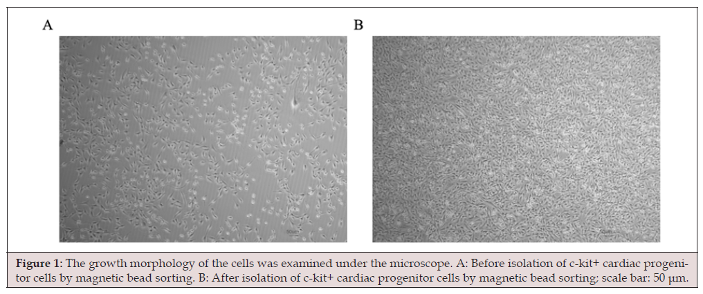
Figure 1:The growth morphology of the cells was examined under the microscope. A: Before isolation of c-kit+ cardiac progenitor cells by magnetic bead sorting. B: After isolation of c-kit+ cardiac progenitor cells by magnetic bead sorting; scale bar: 50 μm.
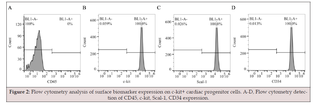
Figure 2:Flow cytometry analysis of surface biomarker expression on c-kit+ cardiac progenitor cells. A-D, Flow cytometry detection of CD45, c-kit, Scal-1, CD34 expression.
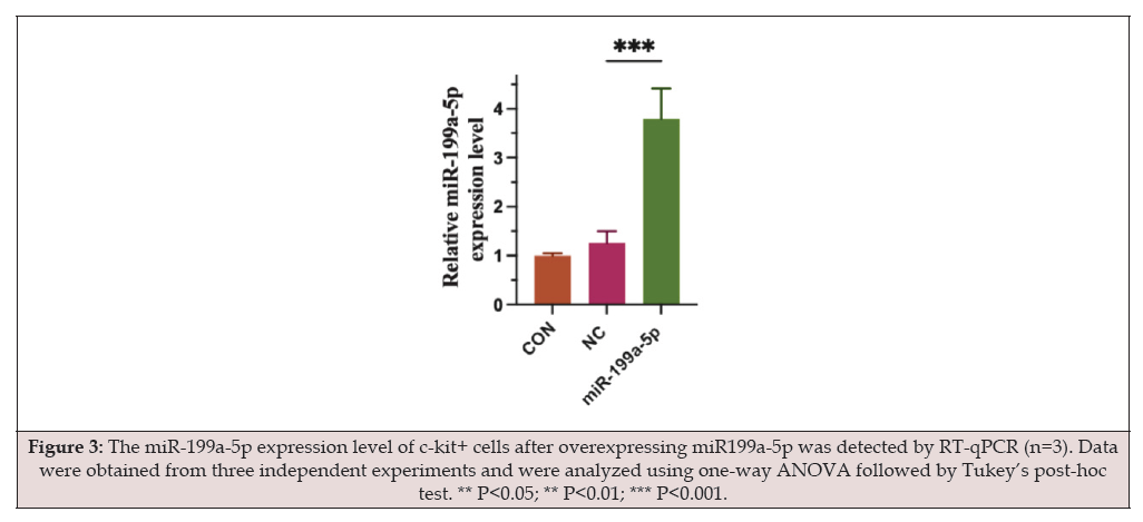
Figure 3:The miR-199a-5p expression level of c-kit+ cells after overexpressing miR199a-5p was detected by RT-qPCR (n=3). Data were obtained from three independent experiments and were analyzed using one-way ANOVA followed by Tukey’s post-hoc test. ** P<0.05; ** P<0.01; *** P<0.001.
Effect of miR-199a-5p on Cell Proliferation
Cell division was measured using EdU staining. The CON group had an EdU-positive rate of 42.36% ± 3.12%. The NC mimic group had a rate of 40.89% ± 2.87%. The miR-199a-5p mimic group had a rate of 21.57% ± 2.45%. A statistical test found that these three groups were different (F = 58.32, P < 0.001). Compared to the NC mimic group, the miR-199a-5p mimic group had a lower EdU-positive rate (P < 0.01). The CON group and the NC mimic group were not different (P > 0.05) (Figure 4). The results mean that higher miR-199a-5p levels reduce the growth of c-kit⁺ cardiac progenitor cells.
Effect of miR-199a-5p on Cell Viability
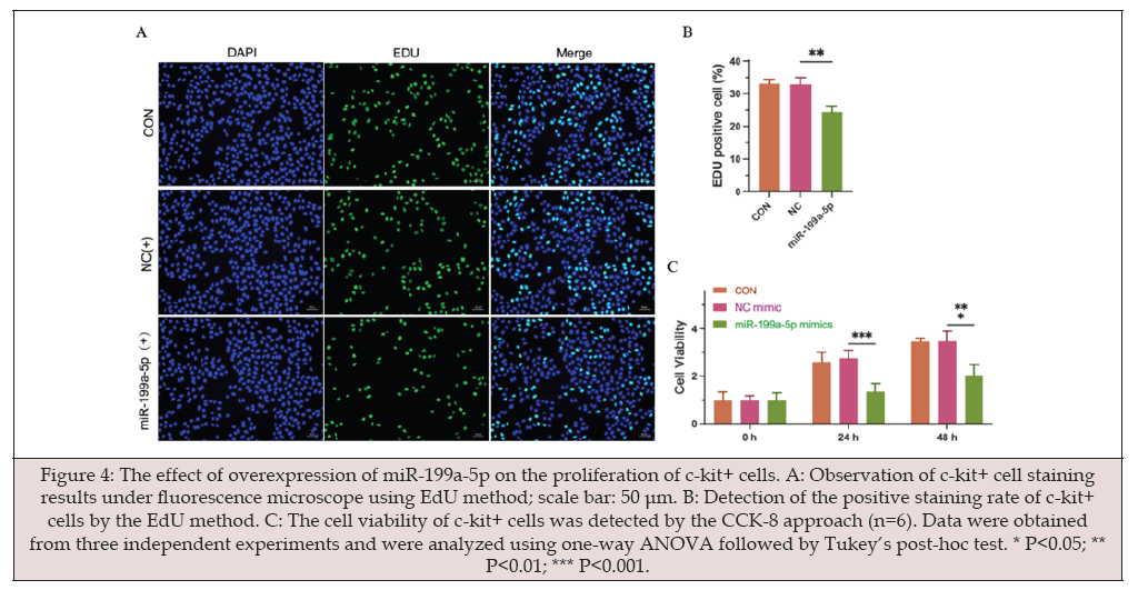
Figure 4:The effect of overexpression of miR-199a-5p on the proliferation of c-kit+ cells. A: Observation of c-kit+ cell staining results under fluorescence microscope using EdU method; scale bar: 50 μm. B: Detection of the positive staining rate of c-kit+ cells by the EdU method. C: The cell viability of c-kit+ cells was detected by the CCK-8 approach (n=6). Data were obtained from three independent experiments and were analyzed using one-way ANOVA followed by Tukey’s post-hoc test. * P<0.05; ** P<0.01; *** P<0.001.
We used the CCK-8 test to check cell viability at three times: 0, 24, and 48 hours. In every group, cell numbers grew over time. But the groups were different from each other (F = 42.67, P < 0.001). The miR-199a-5p mimic group had lower OD450 readings than the NC mimic group at 24 hours (P < 0.001). This was also true at 48 hours (P < 0.001). The CON group and the NC mimic group did not show important differences at any time point (P > 0.05) (Figure 4)
Effect of miR-199a-5p on Cell Migration
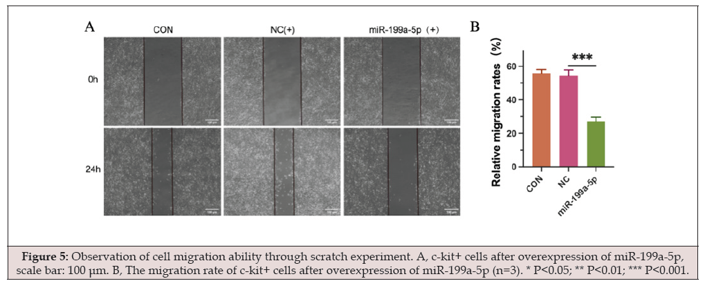
Figure 5:Observation of cell migration ability through scratch experiment. A, c-kit+ cells after overexpression of miR-199a-5p, scale bar: 100 μm. B, The migration rate of c-kit+ cells after overexpression of miR-199a-5p (n=3). * P<0.05; ** P<0.01; *** P<0.001.
We used a scratch test to check cell movement. After 24 hours, the CON group closed 68.45% ± 4.23% of the scratch. The NC mimic group closed 66.78% ± 3.98%. The miR-199a-5p mimic group closed 32.16% ± 3.57%. Statistics showed the miR-199a-5p mimic group moved less than the NC mimic group (F = 76.54, P < 0.001). The CON and NC mimic groups moved about the same amount (P > 0.05). (Figure 5) shows these results. These data indicate that more miR-199a-5p slows the movement of c‑kit⁺ cardiac progenitor cells
Effect of miR-199a-5p on Cell Differentiation
mRNA Expression of Cardiac Lineage Markers:
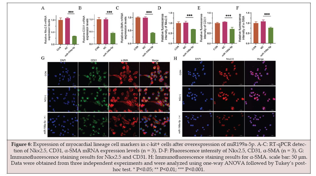
Figure 6:Expression of myocardial lineage cell markers in c-kit+ cells after overexpression of miR199a-5p. A-C: RT-qPCR detection of Nkx2.5, CD31, α-SMA mRNA expression levels (n = 3). D-F: Fluorescence intensity of Nkx2.5, CD31, α-SMA (n = 3). G: Immunofluorescence staining results for Nkx2.5 and CD31. H: Immunofluorescence staining results for α-SMA. scale bar: 50 μm. Data were obtained from three independent experiments and were analyzed using one-way ANOVA followed by Tukey’s posthoc test. * P<0.05; ** P<0.01; *** P<0.001.
RT-qPCR analysis showed that compared to the NC mimic group, the miR-199a-5p mimic group exhibited significant downregulation in the mRNA expression of the cardiac differentiation marker Nkx2.5 (F = 89.76, P < 0.001), the vascular endothelial marker CD31 (F = 92.34, P < 0.001), and the smooth muscle marker α-SMA (F = 85.43, P < 0.001). No significant differences were observed between CON and NC mimic groups (P > 0.05) (Figure 6).
Protein Expression of Cardiac Lineage Markers:
Immunofluorescence staining supported the mRNA data. We measured how much each marker was present by looking at the brightness of the stain. The miR-199a-5p mimic group showed weaker staining for Nkx2.5 (found in the nucleus), CD31 (found near the cell membrane), and α-SMA (found in the cytoplasm) compared to the NC mimic group. The statistical values were: Nkx2.5 (F = 78.65, P < 0.001); CD31 (F = 81.23, P < 0.001); α-SMA (F = 74.32, P < 0.001). The control (CON) group and the NC mimic group were not different (P > 0.05). (Figure 6) presents these results. These findings show that more miR-199a-5p reduces the ability of c‑kit⁺ cardiac progenitor cells to become heart muscle cells, blood vessel cells, or muscle wall cells.
Discussion
This work first showed how miR-199a-5p affects c-kit⁺ cardiac progenitor cells. The results indicate that miR-199a-5p reduces cell growth. It also slows cell movement. In addition, it limits the ability of these cells to become other cell types. This finding gives important experimental support. It helps explain why heart stem cells do not work well in ischemic heart failure. Before the experiments, we obtained highly pure c-kit⁺ cells. All cells expressed c-kit, Sca-1, and CD34. None expressed CD45. We then used a lentivirus to increase miR-199a-5p levels in these cells. The lentivirus system was stable. It allowed us to reliably study miR-199a-5p’s role in these cells.
Comparison with Existing Research and Theoretical Implications
Our results agree with and add to recent work on miR-199a- 5p in the heart. Earlier studies found that miR-199a-5p promotes cell death in heart muscle cells. It does this by blocking the Akt/ eNOS signaling path [30]. It also increases cell death in heart injury caused by blood flow loss and return [31]. Our study goes further. In c-kit⁺ cardiac progenitor cells, higher miR-199a-5p lowers cell activity. The CCK-8 test shows this. It also reduces cell growth. The EdU-positive cell number falls by about half. Cell movement is also slower. The scratch test healing rate drops by about half. Together, these results show that miR-199a-5p broadly weakens the heart’s repair ability.
In differentiation tests, higher miR-199a-5p clearly lowered the markers Nkx2.5, CD31, and α-SMA. This was seen at both the RNA and protein levels (P < 0.001). Most earlier studies looked at miR-199a-5p in mature heart cells [32]. Our work is different. We show for the first time that miR-199a-5p blocks heart progenitor cells from changing into different cell types. This offers a new way to think about how cell fate is controlled during heart repair.
Comparison with Existing Research and Theoretical Implications
Our findings are consistent with and extend recent research on the function of miR-199a-5p in the cardiovascular system. Previous studies have shown that miR-199a-5p facilitates ferroptosis in cardiomyocytes by suppressing the Akt/eNOS signaling pathway [30] and exerts pro-apoptotic effects in myocardial ischemia-reperfusion injury [31]. This study further demonstrates that in c-kit⁺ cardiac progenitor cells, miR-199a-5p overexpression not only reduces cell viability (significantly decreased CCK-8 OD values) but also suppresses their proliferation (approximately 50% reduction in EdU-positive rate) and migration (approximately 50% decrease in scratch healing rate). These combined effects point to a systemic inhibition of cardiac regenerative capacity by this molecule.
Notably, in terms of differentiation function, miR-199a-5p overexpression significantly downregulated the expression of the cardiomyocyte marker Nkx2.5, endothelial cell marker CD31, and smooth muscle cell marker α-SMA (both at mRNA and protein levels, P < 0.001). This finding expands the scope of previous research— most prior studies focused on the effects of miR-199a-5p on mature cardiomyocytes [32], whereas this study is the first to clarify its inhibitory role in the multilineage differentiation of cardiac progenitor cells. This provides a new perspective for understanding the regulatory networks governing cell fate determination during cardiac regeneration.
Exploration of Potential Molecular Mechanisms
In terms of possible mechanisms, our results link to the HIF-1α pathway. HIF-1α is an important factor in low-oxygen conditions. Studies show HIF-1α helps cardiac progenitor cells grow, move, change type, and form new blood vessels [33-35]. In our study, high miR-199a-5p caused problems similar to those seen when HIF-1α is reduced. It is known that miR-199a-5p can bind to HIF-1α’s 3’ untranslated region and lower its levels [14,36]. Recent research also reports that miR-199a-5p harms cardiac stem cell function by blocking HIF-1α [37]. Thus, we believe the miR-199a-5p/HIF-1α interaction may be important in controlling c-kit⁺ cell activity. This idea helps explain why these stem cells do not work well in IHF. It also supports future work on treatments that target this pathway.
Innovations and Clinical Implications
The academic contributions of this work are listed below. First, we systematically tested how miR-199a-5p acts on c-kit⁺ cardiac progenitor cells. Results show it inhibits several important cell activities. This fills a gap in experimental data in this field. Second, past research on miR-199a-5p mostly used mature heart muscle cells. Our study used heart progenitor cells. We found miR-199a-5p also affects these cells. This suggests it plays a role in heart tissue repair. Third, the results may help design new treatments for IHF. For instance, blocking miR-199a-5p might restore normal function in c-kit⁺ cells. This could improve the results of cell therapy for heart repair. Thus, miR-199a-5p could serve as a treatment target in the future.
Study Limitations and Future Directions
Our study has certain limitations. Future work should address them. First, we only tested cells in a lab dish. We did not test in live animals. Next, we should use mice with heart attacks. We can inject a miR-199a-5p blocker into the tail vein or heart muscle. Then, we can check heart function, damage size, and whether c-kit⁺ cells are recruited. This will help see if blocking miR-199a-5p works as a treatment. Second, we did not prove miR-199a-5p directly targets HIF-1α. Later studies should use a dual-luciferase reporter test to confirm binding. We can also try adding back HIF-1α. This will show if more HIF-1α can cancel out the effects of miR-199a-5p on c-kit⁺ cells. Third, we did not study how miR-199a-5p affects what c-kit⁺ cells release. These secretions are important for heart repair [10,38]. Future studies can use protein chips to measure what the cells release. This will give a full picture of how miR-199a-5p controls heart repair.
Conclusion
To summarize, we found that higher levels of miR-199a-5p slow the growth, limit the movement, and reduce the ability of c‑kit⁺ cardiac progenitor cells to develop into different heart cell types. The observed effect may occur because miR-199a-5p suppresses HIF- 1α signaling. These data add to what is known about how microRNAs influence heart tissue repair. They also offer support for new treatments that target miRNAs in ischemic heart failure. In the next stage, these results should be tested in animal models and the molecular details examined further. Doing so may help advance possible treatments based on miR-199a-5p and open up new research directions for heart regeneration.
Acknowledgements
We gratefully acknowledge the Core Facility of the Clinical Research Center, Hebei General Hospital, for providing access to flow-cytometry platforms, cell-culture suites, immunohistochemistry workstations, and real-time PCR instrumentation that were essential for the completion of this study. We also thank our colleagues in the Department of Cardiology for insightful discussions and for their sustained technical assistance throughout the entire investigation.
Author Contributions
Sha Li designed the project, performed data analysis, drafted the manuscript; Fan Wang participated in designing the experiments; Cuigai Zhang collected animal samples and performed transcriptome sequencing; Shuren Li supervised the experimental design and revised the manuscript. All authors have read and approved the final version of the manuscript.
Funding
This study was supported by the Health Commission of Hebei Provincial (Grant No. 20230296).
Data Availability
The datasets generated and analyzed during the current study are fully contained within the article and its figures. Any additional raw data that support the findings are available from the corresponding author upon reasonable request.
Declarations
Ethics approval and consent to participate
Animal procedures were conducted in the Animal Laboratory of the Clinical Research Center, Hebei General Hospital, in accordance with the guidelines of the Animal Ethics Committee of Hebei General Hospital. The protocol was reviewed and approved by the hospital’s Animal Ethics Committee, with written consent for animal use.
Consent for publication
Not applicable
Competing Interests
All the authors declare no conflict of interest
References
- Xu J, Lian W, Li L, Huang Z (2017) Generation of induced cardiac progenitor cells via somatic reprogramming. Oncotarget 8(17): 29442-29457.
- Shouman S, Zaher A, Abdelhameed A, Elshaboury S, Sakr S, et al. (2021) Cardiac Progenitor Cells. Adv Exp Med Biol 1312: 51-73.
- Fathi E, Valipour B, Vietor I, Farahzadi R (2020) An overview of the myocardial regeneration potential of cardiac c-Kit+ progenitor cells via PI3K and MAPK signaling pathways. Future Cardiol 16(3): 199-209.
- Jia G, Preussner J, Chen X, Guenther S, Yuan X, et al. (2018) Single cell RNA-seq and ATAC-seq analysis of cardiac progenitor cell transition states and lineage settlement. Nat Commun 9(1): 4877.
- Farah EN, Hu RK, Kern C, Zhang Q, Lu TY, et al. (2024) Spatially organized cellular communities form the developing human heart. Nature. 627(8005): 854-864.
- He J, Yang Y, Jiang R, Zheng Y, Yang X, et al. (2025) Integration of single-cell and spatial transcriptomics by SEU-TCA reveals the spatial origin of early cardiac progenitors. Genome Biol 26(1): 158.
- Lee JW, Lee SH, Youn YJ, Ahn MS, Kim JY, et al. (2014) A randomized, open-label, multicenter trial for the safety and efficacy of adult mesenchymal stem cells after acute myocardial infarction. J Korean Med Sci 29(1): 23-31.
- Makkar RR, Kereiakes DJ, Aguirre F, Kowalchuk G, Chakravarty T, et al. (2020) Intracoronary ALLogeneic heart STem cells to Achieve myocardial Regeneration (ALLSTAR): a randomized, placebo-controlled, double-blinded trial. Eur Heart J 41(36): 3451-3458.
- Bolli R, Mitrani RD, Hare JM, Pepine CJ, Perin EC, et al. (2021) A Phase II study of autologous mesenchymal stromal cells and c-kit positive cardiac cells, alone or in combination, in patients with ischaemic heart failure: the CCTRN CONCERT-HF trial. Eur J Heart Fail 23(4): 661-674.
- Song YS, Joo HW, Park IH, Shen GY, Lee Y, et al. (2017) Bone marrow mesenchymal stem cell-derived vascular endothelial growth factor attenuates cardiac apoptosis via regulation of cardiac miRNA-23a and miRNA-92a in a rat model of myocardial infarction. PLoS One 12(6): e0179972.
- Fernández Ruiz I (2018) Cardiac regeneration: A hydrogel-miRNA complex stimulates heart recovery. Nat Rev Cardiol 15(2): 68-69.
- Liu X, Meng H, Jiang C, Yang S, Cui F, et al. (2016) Differential microRNA Expression and Regulation in the Rat Model of Post-Infarction Heart Failure. PLoS One 11(8): e0160920.
- Lindner K, Eichelmann AK, Matuszcak C, Hussey DJ, Haier J, et al. (2018) Complex Epigenetic Regulation of Chemotherapy Resistance and Biohlogy in Esophageal Squamous Cell Carcinoma via MicroRNAs. Int J Mol Sci 19(2): 499.
- Wang K, Ding R, Ha Y, Jia Y, Liao X, et al. (2018) Hypoxia-stressed cardiomyocytes promote early cardiac differentiation of cardiac stem cells through HIF-1α/Jagged1/Notch1 signaling. Acta Pharm Sin B 8(5): 795-804.
- Liu X, Wang X, Chai B, Wu Z, Gu Z, et al. (2022) miR-199a-3p/5p regulate tumorgenesis via targeting Rheb in non-small cell lung cancer. Int J Biol Sci 18(10): 4187-4202.
- Sun Y, Zhao JT, Chi BJ, Wang KF (2020) Long noncoding RNA SNHG12 promotes vascular smooth muscle cell proliferation and migration via regulating miR-199a-5p/HIF-1α. Cell Biol Int 44(8):1714-1726.
- Guo C, Zhang M, Su W, Xu M, Zhao S (2022) miR-199a-5p Relieves Obstructive Sleep Apnea Syndrome-Related Hypertension by Targeting HIF-1α. J Immunol Res 2022:7236647.
- Chauhan SKS, Feldbrügge L, Schmelzle M, Wedemeyer H, Heinrich B (2025) Protocol for optimized mononuclear cell isolation from liver and tumor tissue using mechanical or enzymatic digestion. STAR Protoc 7(1): 104289.
- Yadav SK, Mishra PK (2019) Isolation, Characterization, and Differentiation of Cardiac Stem Cells from the Adult Mouse Heart. J Vis Exp (143):10.3791/58448.
- Beltrami AP, Barlucchi L, Torella D, Baker M, Limana F, et al. (2003) Adult cardiac stem cells are multipotent and support myocardial regeneration. Cell 114(6): 763-776.
- Kozomara A, Birgaoanu M, Griffiths Jones S (2019) miRBase: from microRNA sequences to function. Nucleic Acids Res 47(D1): D155-D62.
- Smith CL, Baek ST, Sung CY, Tallquist MD (2011) Epicardial-derived cell epithelial-to-mesenchymal transition and fate specification require PDGF receptor signaling. Circ Res 108(12): e15-26.
- Chomczynski P, Sacchi N (2006) The single-step method of RNA isolation by acid guanidinium thiocyanate-phenol-chloroform extraction: twenty-something years on. Nat Protoc 1(2): 581-585.
- Wu H, Jiang X, Fan H, Li J, Li Y, et al. (2025) Inhibition of circALPK2 enhances proliferation and therapeutic potential of human pluripotent stem cell-derived cardiomyocytes in myocardial infarction. Stem Cell Res Ther 16(1): 107.
- Wang XZ, Chen L, Sun H, Li XQ, Wang H, et al. (2023) MiR-199a-3p promotes repair of myocardial infarction by targeting NACC2. Int J Clin Exp Pathol 16(3): 57-66.
- Liang CC, Park AY, Guan JL (2007) In vitro scratch assay: a convenient and inexpensive method for analysis of cell migration in vitro. Nat Protoc 2(2): 329-333.
- Yang L, Zhu S, Li Y, Zhuang J, Chen J, et al. (2020) Overexpression of Pygo2 Increases Differentiation of Human Umbilical Cord Mesenchymal Stem Cells into Cardiomyocyte-like Cells. Curr Mol Med 20(4): 318-324.
- Deng G, Yang Y, Qing O, Linhui J, Haotao S, et al. (2025) Chrysin Attenuates Myocardial Cell Apoptosis in Mice. Cardiovasc Toxicol 25(11): 1791-1806.
- Chong JJ, Reinecke H, Iwata M, Torok Storb B, Stempien Otero A, et al. (2013) Progenitor cells identified by PDGFR-alpha expression in the developing and diseased human heart. Stem Cells Dev 22(13): 1932-1943.
- Zhang GY, Gao Y, Guo XY, Wang GH, Guo CX (2022) MiR-199a-5p promotes ferroptosis-induced cardiomyocyte death responding to oxygen-glucose deprivation/reperfusion injury via inhibiting Akt/eNOS signaling pathway. Kaohsiung J Med Sci 38(11): 1093-1102.
- Zuo Y, Wang Y, Hu H, Cui W (2016) Atorvastatin Protects Myocardium Against Ischemia-Reperfusion Injury Through Inhibiting miR-199a-5p. Cell Physiol Biochem 39(3): 1021-1030.
- Zhang H, Li S, Zhou Q, Sun Q, Shen S, et al. (2016) Qiliqiangxin Attenuates Phenylephrine-Induced Cardiac Hypertrophy through Downregulation of MiR-199a-5p. Cell Physiol Biochem 38(5): 1743-1751.
- Leri A, Rota M, Hosoda T, Goichberg P, Anversa P (2014) Cardiac stem cell niches. Stem Cell Res 13(3 Pt B):631-646.
- Hnatiuk AP, Ong SG, Olea FD, Locatelli P, Riegler J, et al. (2016) Allogeneic Mesenchymal Stromal Cells Overexpressing Mutant Human Hypoxia-Inducible Factor 1-α (HIF1-α) in an Ovine Model of Acute Myocardial Infarction. J Am Heart Assoc 5(7): e003714.
- Cheng Y, Feng Y, Xia Z, Li X, Rong J (2017) ω-Alkynyl arachidonic acid promotes anti-inflammatory macrophage M2 polarization against acute myocardial infarction via regulating the cross-talk between PKM2, HIF-1α and iNOS. Biochim Biophys Acta Mol Cell Biol Lipids 1862(12): 1595-1605.
- Yang X, Zheng Y, Tan J, Tian R, Shen P, et al. (2021) MiR-199a-5p-HIF-1α-STAT3 Positive Feedback Loop Contributes to the Progression of Non-Small Cell Lung Cancer. Front Cell Dev Biol 8: 620615.
- Li S, Zhang C, Liu Y, Meng C, Xie Y, et al. (2025) miR-199a-5p inhibited HIF-1α to suppress the proliferation, migration, and differentiation of cardiac stem cells. Eur J Histochem 69(4): 4239.
- Isanejad A, Alizadeh AM, Amani Shalamzari S, Khodayari H, Khodayari S, et al. (2016) MicroRNA-206, let-7a and microRNA-21 pathways involved in the anti-angiogenesis effects of the interval exercise training and hormone therapy in breast cancer. Life Sci 151: 30-40.




 We use cookies to ensure you get the best experience on our website.