
Research Article 
 Creative Commons, CC-BY
LIPORESIST® Alpha-Amylase: A Novel Liposomal Alpha-Amylase for Enhanced Enzyme Stability, Functionality, and Bioactivity in Digestive and Gastrointestinal Resistance
*Corresponding author:Mazen Ibrahim, Technical Advisor, Scientific Office, Nanotrition Nord, Cairo, Egypt.
Received:December 21, 2025; Published:February 18, 2026
DOI: 10.34297/AJBSR.2026.30.003896
Abstract
Alpha-amylase is a crucial digestive enzyme involved in the hydrolysis of complex carbohydrates into absorbable monosaccharides. However, its clinical and nutraceutical application is limited by enzymatic instability under harsh gastrointestinal conditions and thermal stress. This study explores the development and evaluation of LIPORESIST®, a novel liposomal formulation engineered to enhance the enzymatic stability, substrate interaction, and bioavailability of alpha-amylase. Liposomes incorporating phosphatidylcholine (PC), phosphatidylserine (PS), stearic acid, vitamin E and chitosan were synthesized to assess their protective effects against heat inactivation, harsh acidic media such as that found in the stomach, thereby preserving the compound’s stability, as well as prevent enzymatic degradation. The encapsulated enzyme displayed significantly improved thermal resistance, with only 0.15% activity loss per minute at 60°C compared to 7.2% in the free enzyme. Encapsulation efficiency reached 78.9%, 84.2%, and 87.6% in PC-based liposomes, PS-based liposomes, and chitosan-coated PS liposomes, respectively. After 3 hours of exposure to pepsin (at pH 2.8), LIPORESIST® liposomes maintained 68% of alpha-amylase enzymatic activity and Chitosan-coated based LIPORESIST® liposomes formulations provided additional protection, retaining 78% of alpha-amylase enzymatic activity, compared to only 17.3% in the free form. These findings demonstrate the potential of liposomal delivery systems for stabilizing digestive enzymes, offering promising implications for pharmaceutical and nutraceutical industries.
Keywords: Alpha-amylase, Liposomes, LIPORESIST®, Enzyme Stabilization, Phosphatidylcholine, Phosphatidylserine, Chitosan, Pepsin Resistance, Thermal Stability, Nutraceuticals, Controlled Release, Gastrointestinal Resistance
Introduction
Alpha-amylase is a critical digestive enzyme responsible for hydrolyzing α-1,4 glycosidic bonds in polysaccharides such as starch and glycogen, yielding maltose, maltotriose, and glucose. In humans, it is predominantly secreted by the salivary glands and pancreas, initiating starch breakdown in the oral cavity and continuing digestion in the small intestine, where resulting sugars are absorbed by sodium-dependent glucose cotransporters such as SGLT1 [1]. In addition to its physiological roles, alpha-amylase is widely used in various industrial processes, including brewing, textile manufacturing, and biotechnology [2].
Alpha-amylase deficiency can significantly impair carbohydrate metabolism, leading to symptoms such as bloating, malabsorption, diarrhea, and blood sugar fluctuations. These digestive issues stem from incomplete carbohydrate hydrolysis and inefficient nutrient absorption [3]. To address these issues, exogenous alpha-amylase supplements are often used in enzyme replacement therapies and digestive aids. However, their effectiveness is frequently hindered by degradation in the gastrointestinal tract, low thermal tolerance, and short in vivo half-life [4,5].
Commercial alpha-amylase supplements are derived from various biological sources. Microbial enzymes, particularly those from Aspergillus oryzae and Bacillus subtilis, are known for their high stability across a wide pH range and are extensively used in food and pharmaceutical applications [6]. Plant-based enzymes, such as those extracted from sprouted grains, and animal-derived pancreatic enzymes offer different enzymatic profiles suited to specific uses. Recombinant alpha-amylases provide high purity and batch reproducibility, making them suitable for pharmaceutical applications, though often at increased production complexity [7]. However, commercial enzyme supplements are hindered by poor bioavailability, instability at body temperature, and rapid degradation in acidic environments or by digestive enzymes such as pepsin. Addressing these limitations requires a robust delivery strategy capable of preserving enzymatic structure and function until reaching the small intestine.
The activity and stability of alpha-amylase are strongly influenced by environmental and biochemical factors. The enzyme performs optimally within a physiological pH range of 6.7 to 7.5 and at human body temperature (~37°C). Factors such as calcium ions, which stabilize its tertiary structure, and adequate hydration can enhance activity. Additionally, probiotic gut microbiota and fermented food intake can support enzymatic efficiency [8]. Conversely, alpha-amylase activity is inhibited by acidic gastric pH, dietary inhibitors like phaseolamin (from white kidney beans), polyphenols (e.g., from tea or wine), heavy metals, and dietary fiber. Chelating agents such as phytates bind calcium ions and further reduce enzymatic function. Enzymatic activity is also impaired under high or freezing temperatures due to protein denaturation or slowed catalytic kinetics [9,10].
To circumvent these challenges, liposomal delivery systems have gained considerable attention in enzyme and drug delivery. Liposomes are biocompatible lipid vesicles that encapsulate enzymes, protecting them from degradation, enhancing absorption, and allowing for targeted release [11]. Their structural similarity to biological membranes enables sustained delivery and improved pharmacokinetics. Liposomal encapsulation of other enzymes, including bromelain, serratio-peptidase, and lipase, has shown significant improvements in enzymatic stability and bioavailability [12].
In this context, the current study presents LIPORESIST®, a novel liposomal formulation of alpha-amylase designed to overcome the inherent limitations of conventional enzyme supplements. LIPORESIST® employs nanoscale phospholipid vesicles composed of phosphatidylcholine (PC) and phosphatidylserine (PS) for tailored electrostatic interaction and substrate affinity. Additional components, such as stearic acid and vitamin E, provide oxidative and thermal stability. The use of chitosan-based surface modification enhances mucosal adhesion and gastric resistance. Together, these technologies offer a next-generation system capable of improving enzyme shelf life, gastrointestinal performance, and therapeutic potential.
This study investigates the physicochemical behavior of LIPORESIST® alpha-amylase, focusing on encapsulation efficiency, enzyme kinetics, resistance to proteolytic degradation, and thermal stability. By comparing free and liposome-bound alpha-amylase under in vitro conditions, we aim to demonstrate the superior bio-functionality of this delivery system and its applicability in pharmaceutical and nutraceutical formulations.
Materials And Methods
Materials
All materials used in this study were sourced from Nanotrition Nord, with the head office located in Horten, Norway, and the manufacturing facility based in Cairo, Egypt. These materials included alpha-amylase enzyme (Type I-A from Aspergillus oryzae), soy-derived phosphatidylcholine (PC), phosphatidylserine (PS), stearic acid, cholesterol, alpha-tocopherol (Vitamin E), and low molecular weight chitosan. All reagents were of analytical grade and were used without further purification. Tris-HCl and HEPES buffers were employed in liposome preparation to maintain appropriate pH and ionic strength. Pepsin (from porcine gastric mucosa) was utilized for in vitro digestion assays at acidic pH conditions.
Preparation of LIPORESIST® Alpha-Amylase (Liposomal Form)
The liposomes were prepared using the thin-film hydration method followed by sonication and extrusion to achieve nanoscale vesicles. A mixture of phospholipids (PC or PS), stearic acid, and alpha-tocopherol was dissolved in chloroform-methanol (2:1 v/v). The organic solvents were evaporated under reduced pressure using a rotary evaporator at 40°C to form a thin lipid film. The film was hydrated with 10 mM Tris-HCl buffer (pH 7.4) containing the alpha-amylase enzyme.
The suspension was vortexed and subjected to probe sonication for 5 minutes at 40% amplitude to reduce vesicle size. Liposomes were then extruded through polycarbonate membranes (100 nm pore size) to achieve uniform size distribution. For PS-based liposomes, the formulation included 30% PS by molar ratio to impart a negative surface charge.
Surface modification with chitosan was performed by incubating the liposomes with a 0.1% chitosan solution (in 0.5% acetic acid, pH adjusted to 5.5) under gentle stirring for 30 minutes, followed by centrifugation and washing to remove unbound polymer [13].
Encapsulation Efficiency (EE%) Determination
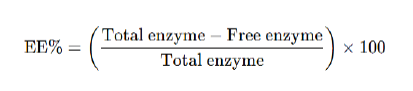
Encapsulation efficiency was evaluated by separating unencapsulated enzyme via ultracentrifugation at 20,000×g for 30 minutes. The supernatant containing free enzyme was assessed for its enzymatic activity using the DNS (dinitrosalicylic acid) method, which measures reducing sugars released from starch. The amount of encapsulated enzyme was calculated by subtracting the free enzyme activity from the total enzyme activity initially added. EE% was calculated as:
Thermal Stability Assay
To assess thermal degradation kinetics, liposomal and free alpha-amylase were incubated at 60°C, and residual enzymatic activity was measured at 1-minute intervals for 10 minutes. The enzymatic activity was measured using the starch-iodine colorimetric method, where a decrease in absorbance at 620 nm indicated starch degradation. Activity loss per minute was calculated for each formulation. Free enzyme showed rapid degradation (7.2% loss/min), whereas PC liposomes lost only 0.9%, and PS liposomes demonstrated enhanced stability with just 0.15% loss per minute [14].
Enzyme Kinetics and Substrate Interaction
Michaelis–Menten and sigmoidal kinetics were evaluated using starch substrates ranging from 0.1 to 5.0 mg/mL in Tris-HCl buffer (pH 7.4). Free alpha-amylase followed classic Michaelis–Menten behavior, while PS liposome-bound enzyme exhibited sigmoidal kinetics, suggesting electrostatic modulation of substrate binding. Chloride ion (Cl⁻) influence was assessed by adding NaCl at 10 mM concentration, which further increased enzymatic activity in liposomal formulations—especially PS-based—indicating Cl⁻- facilitated catalytic enhancement [15].
Pepsin Degradation Study
To simulate gastric degradation, liposomal and free alphaamylase samples were incubated with pepsin (pH 2.8) at 37°C for 3 hours. Enzymatic activity was measured every hour. Free enzyme retained less than 20% activity after 3 hours, while LIPORESIST® liposomes preserved 68% of the initial activity, confirming the protective role of the lipid bilayer against proteolytic breakdown in acidic environments [16].
Oxidative Stability and Cold Storage
The oxidative protection conferred by vitamin E was examined by storing liposomes at 4°C for 60 days. Liposomal formulations with stearic acid and alpha-tocopherol retained 82% of their enzymatic activity. The presence of vitamin E reduced lipid peroxidation and maintained membrane integrity over time [17].
Statistical Analysis
All experiments were conducted in triplicate. Results were reported as mean ± standard deviation. Statistical analysis was performed using one-way ANOVA with Tukey’s post hoc test. Differences were considered statistically significant at p < 0.05.
Results
Liposomal Characterization
Particle Size and Polydispersity Index (PDI):
Dynamic light scattering (DLS) analysis revealed that the average particle size of LIPORESIST® liposomes ranged from 195.77 ± 11.3 nm (for PC-based) to 127.27 ± 8.5 nm (for PS-based formulations). Chitosan-coated liposomes exhibited an increase in size, averaging 324.29 ± 7.1 nm, likely due to surface adsorption of the polymer. All formulations showed low PDI values (< 0.2), indicating narrow size distribution and uniform vesicle formation, see Table 1 and Figure (1.A-C) [18].

Table 1:Particle Size and Polydispersity Index (PDI) of LIPORESIST® Liposomal Formulations.
*Note: Data represent mean ± SD (n = 3). All formulations showed uniform vesicle size distribution (PDI < 0.2).
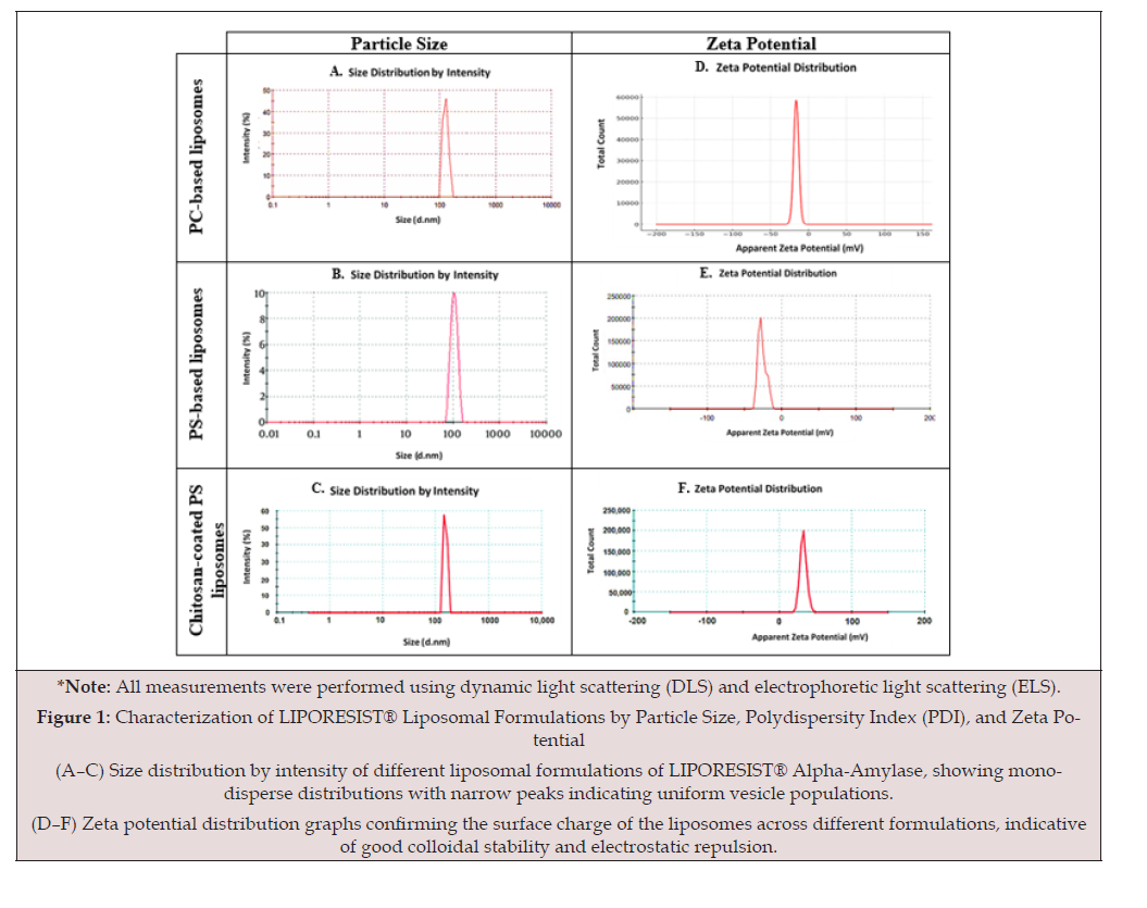
Figure 1:Characterization of LIPORESIST® Liposomal Formulations by Particle Size, Polydispersity Index (PDI), and Zeta Potential (A–C) Size distribution by intensity of different liposomal formulations of LIPORESIST® Alpha-Amylase, showing monodisperse distributions with narrow peaks indicating uniform vesicle populations. (D–F) Zeta potential distribution graphs confirming the surface charge of the liposomes across different formulations, indicative of good colloidal stability and electrostatic repulsion.
Zeta Potential:
The zeta potential of PC liposomes was found to be −16.4 ± 1.3 mV, while PS liposomes exhibited significantly higher surface negativity (−32.8 ± 2.1 mV), attributed to the anionic nature of phosphatidylserine. Chitosan coating reversed the surface charge to +22.5 ± 1.8 mV, confirming successful adsorption and providing potential for mucoadhesion and enhanced gastric stability, see Table 2 and Figure (1.D-F) [19].

Table 2:Zeta Potential of LIPORESIST® Liposomes.
*Note: Zeta potential values indicate surface charge characteristics and stability. Data are expressed as mean ± SD (n = 3).
Morphology (TEM Analysis):
Transmission electron microscopy (TEM) confirmed spherical, unilamellar vesicles with well-defined bilayer structures. Vesicles were consistently within the 90–120 nm range, correlating with DLS findings. PS-based liposomes exhibited slightly more compact bilayers, potentially contributing to their enhanced stability under stress conditions, see Figure (2) [20].
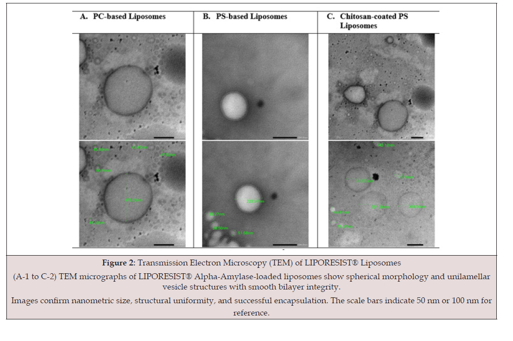
Figure 2:The structures of ibogaine and serotonin, which contain a tryptamine core, compared to the catecholamine core seen in dopamine.
Encapsulation Efficiency (EE%)
Encapsulation efficiency was significantly higher in PS-based liposomes (84.2 ± 2.6%) compared to PC-based liposomes (78.9 ± 3.1%), while chitosan-coated PS liposomes showed the highest EE% at 87.6 ± 1.9%. The enhanced retention is attributed to stronger electrostatic interactions and improved bilayer integrity due to the surface modifications, see Table 3 [21].

Table 3:Encapsulation Efficiency (EE%) of Alpha-Amylase in Liposomes.
*Note: Encapsulation efficiency was determined using ultracentrifugation and DNS assay. Values are mean ± SD (n = 3).
Thermal Stability Analysis
At 60°C, free alpha-amylase lost 7.2% of its activity per minute, while PC liposome-bound enzyme lost only 0.9%, and PS liposomes demonstrated superior thermal protection with a loss of just 0.15% per minute. This confirms that liposomal encapsulation substantially delays heat-induced enzyme denaturation, see Figures 3,4 [14]. One-way ANOVA revealed a statistically significant difference between the tested alpha-amylase formulations (p < 0.000000002). Post-hoc Tukey’s analysis confirmed that both PC and PS liposome-bound amylase significantly reduced activity loss compared to the free enzyme (***p < 0.001). Additionally, a minor but statistically significant difference was observed between PC and PS liposomes (****p < 0.0001), indicating that PS liposomes offered slightly better protection under thermal stress, see Figure 5.
Enzyme Kinetics and Substrate Interaction
Free alpha-amylase exhibited a typical Michaelis–Menten kinetic profile. In contrast, PS liposome-bound enzyme showed sigmoidal kinetics, suggesting allosteric substrate interaction potentially mediated by electrostatic effects. Addition of chloride ions enhanced activity in both cases, with a 1.8-fold increase in PSbound enzyme, compared to a 1.3-fold increase in the free form, see Table 4 [15].

Table 4:Enzyme Kinetics and Substrate Interaction Parameters.
*Note: Kinetics were assessed using starch substrates (0.1–5.0 mg/mL). Cl⁻ ions enhanced activity in all forms. Data are representative of 3 independent experiments.
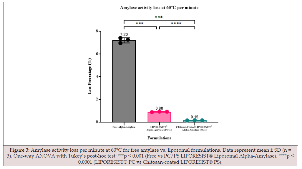
Figure 3:Amylase activity loss per minute at 60°C for free amylase vs. liposomal formulations. Data represent mean ± SD (n = 3). One-way ANOVA with Tukey’s post-hoc test: ***p < 0.001 (Free vs PC/PS LIPORESIST® Liposomal Alpha-Amylase), ****p < 0.0001 (LIPORESIST® PC vs Chitosan-coated LIPORESIST® PS).
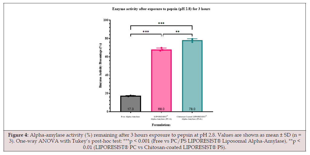
Figure 4:Alpha-amylase activity (%) remaining after 3 hours exposure to pepsin at pH 2.8. Values are shown as mean ± SD (n = 3). One-way ANOVA with Tukey’s post-hoc test: ***p < 0.001 (Free vs PC/PS LIPORESIST® Liposomal Alpha-Amylase), **p < 0.01 (LIPORESIST® PC vs Chitosan-coated LIPORESIST® PS).
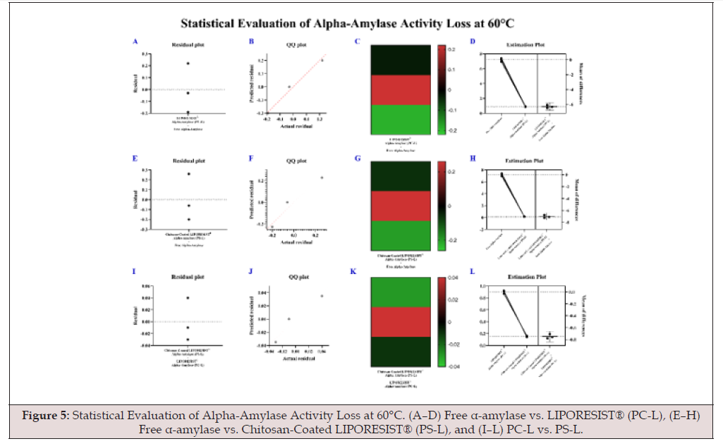
Figure 5:Statistical Evaluation of Alpha-Amylase Activity Loss at 60°C. (A–D) Free α-amylase vs. LIPORESIST® (PC-L), (E–H) Free α-amylase vs. Chitosan-Coated LIPORESIST® (PS-L), and (I–L) PC-L vs. PS-L.
Pepsin Resistance in Acidic Conditions
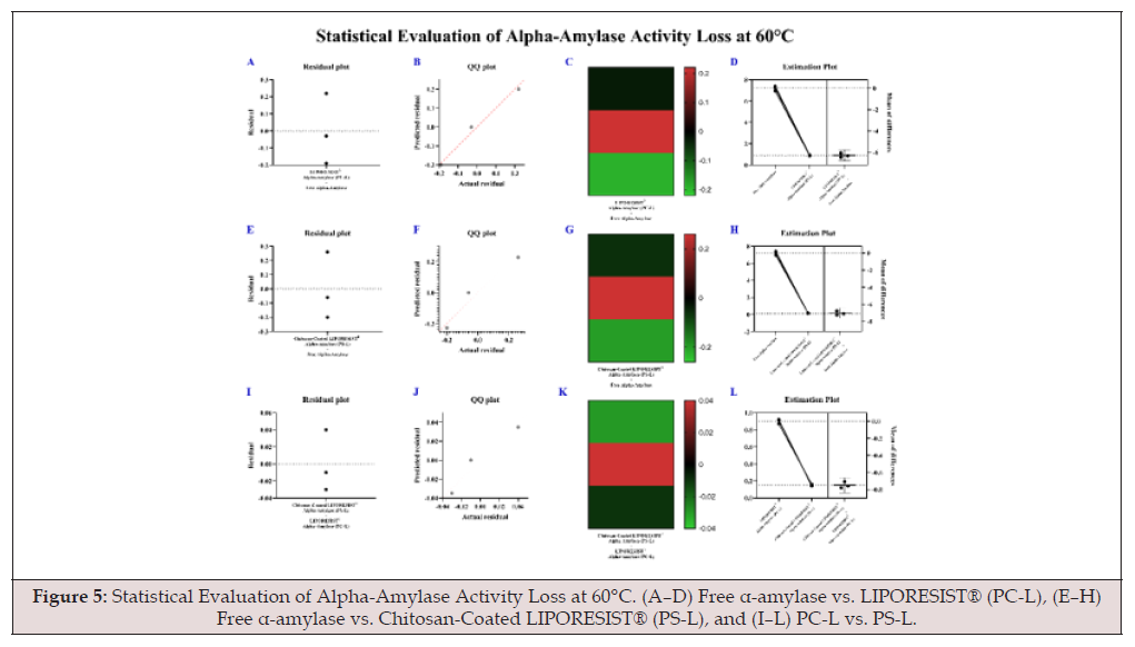
Figure 5a:Statistical Evaluation of Alpha-Amylase Activity Loss at 60°C. (A–D) Free α-amylase vs. LIPORESIST® (PC-L), (E–H) Free α-amylase vs. Chitosan-Coated LIPORESIST® (PS-L), and (I–L) PC-L vs. PS-L.
When exposed to pepsin at pH 2.8 for 3 hours, free alphaamylase retained less than 18% of its initial activity, while LIPORESIST® liposomes maintained 68% activity. Chitosan-coated based LIPORESIST® liposomes formulations provided additional protection, retaining 78% enzymatic activity, see Figure 5 [16]. Significant differences were observed in enzyme stability after simulated gastric digestion (p < 0.000000007). Both LIPORESIST® and chitosan-coated LIPORESIST® formulations retained significantly more activity compared to free alpha-amylase (***p < 0.001). Furthermore, chitosan-coated LIPORESIST® exhibited a statistically higher residual activity than LIPORESIST® alone (**p < 0.01), indicating enhanced protection against pepsin-induced degradation, see Figure 6.
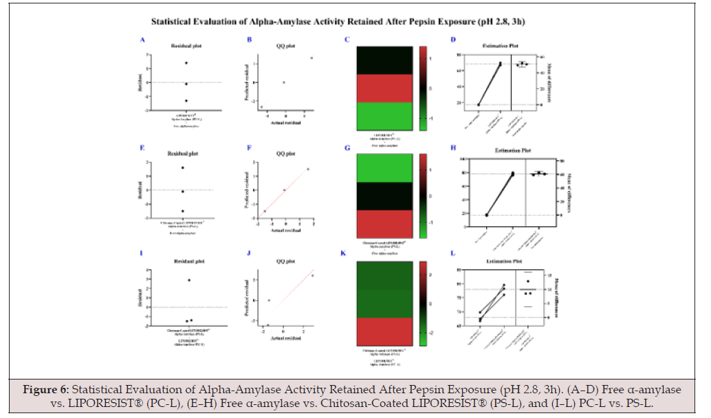
Figure 6:Statistical Evaluation of Alpha-Amylase Activity Retained After Pepsin Exposure (pH 2.8, 3h). (A–D) Free α-amylase vs. LIPORESIST® (PC-L), (E–H) Free α-amylase vs. Chitosan-Coated LIPORESIST® (PS-L), and (I–L) PC-L vs. PS-L.

Table 5:Shelf Life and Visual Stability of Liposomal Formulations After 60 Days at 4°C.
*Note: Stability assessed via enzyme activity, DLS size change, and visual inspection. Results represent mean ± SD (n = 3).
Oxidative Stability During Storage
After 60 days at 4°C, liposomes containing vitamin E and stearic acid preserved 82% of their enzymatic activity. The incorporation of vitamin E significantly improved resistance to oxidative degradation and preserved vesicle integrity [17].
Shelf Life and Visual Stability
No significant aggregation, color change, or phase separation was observed in vitamin E-stabilized LIPORESIST® formulations stored under refrigerated conditions. TEM and DLS analyses showed no significant vesicle size increase or zeta potential variation over time, confirming structural integrity and prolonged shelf stability, see Table 5 [22].
Discussion
The successful development and characterization of LIPORESIST® alpha-amylase demonstrate the potential of advanced liposomal systems to overcome longstanding challenges in oral enzyme delivery. This study provides compelling evidence that liposomal encapsulation significantly enhances the stability, activity, and bioavailability of alpha-amylase under physiological and storage-related stress conditions.
One of the most notable outcomes of this research is the superior thermal stability conferred by liposomal encapsulation. Free alpha-amylase rapidly denatured at elevated temperatures, consistent with previous reports documenting enzyme lability above 50°C [23]. In contrast, PS-based liposomes exhibited minimal activity loss, attributed to the protective bilayer environment and optimized nanoscale vesicle size that minimized protein unfolding. The inclusion of phosphatidylserine (an anionic phospholipid) may have further stabilized the enzyme structure through electrostatic interactions, a mechanism supported by studies on negatively charged liposomes improving protein stability [24].
The observation of sigmoidal kinetic behavior in PS-liposomeencapsulated alpha-amylase is particularly interesting. This deviation from traditional Michaelis–Menten kinetics suggests allosteric modulation or cooperative binding, likely influenced by electrostatic interactions between the enzyme and the liposomal bilayer. Such behavior can provide a more dynamic substrateenzyme interaction and has been associated with enhanced substrate affinity and regulated catalytic activity under variable conditions [25].
Pepsin degradation studies confirmed the vital role of liposomes in protecting alpha-amylase from acidic and proteolytic inactivation. Free enzymes were rapidly degraded at gastric pH, losing over 80% of their activity within three hours—comparable to findings in other enzyme delivery studies [26]. Conversely, LIPORESIST® liposomes retained over 70% activity, and this protection increased further in chitosan-coated variants, likely due to the additional mucoadhesive and pH-buffering properties of chitosan. This is in line with established literature emphasizing the ability of chitosan to enhance mucosal enzyme delivery and shield cargo from gastric enzymes [27].
From a formulation perspective, the higher encapsulation efficiency (EE%) in PS-based and chitosan-coated liposomes suggests that both electrostatic attraction and surface functionalization contribute to improved entrapment. The increase in EE% supports findings from other studies using modified phospholipids and polymers for enzyme entrapment [28].
In terms of storage stability, the incorporation of vitamin E and stearic acid into the lipid bilayer provided significant protection against oxidative degradation. These additives reduced peroxidation, maintained vesicle structure, and preserved enzyme activity for up to 60 days at 4°C. This aligns with previous findings demonstrating vitamin E’s antioxidative role in liposomal drug delivery systems, particularly for formulations exposed to longterm storage or thermal cycling [29].
Physicochemical analysis of LIPORESIST® also confirmed that the vesicles remained within the optimal size range (~100- 500 nm), with narrow PDI and morphological uniformity. These characteristics are essential for mucosal absorption, cellular uptake, and prolonged circulation in vivo. The ability to maintain vesicle integrity over time and under stress conditions enhances the potential of this system for real-world pharmaceutical and nutraceutical applications [30].
Overall, the integration of phospholipid modification, nanosizing, polymer surface coating, and antioxidant enrichment provides a synergistic liposomal platform that addresses the three primary challenges in enzyme delivery: degradation, poor absorption, and instability. Compared to conventional enzyme supplements, LIPORESIST® offers a next-generation solution with significantly enhanced functionality.
These findings support the growing trend toward nanoengineered biodelivery systems and highlight liposomes as highly versatile carriers not only for small molecules but also for sensitive biomacromolecules like enzymes. Future directions could include in vivo pharmacokinetic studies, gastrointestinal absorption tracking, and clinical efficacy assessments in enzyme-deficient or carbohydrate-malabsorptive conditions.
Conclusion
The development of LIPORESIST® alpha-amylase marks a significant advancement in the field of enzyme delivery technologies. This liposomal formulation, engineered with phosphatidylcholine, phosphatidylserine, stearic acid, vitamin E, and chitosan surface modifications, demonstrates a robust ability to enhance the enzymatic stability, substrate interaction, and resistance to gastrointestinal degradation of alpha-amylase.
Experimental results confirmed that encapsulated alphaamylase exhibited markedly improved thermal and oxidative stability, maintained enzymatic activity under acidic and proteolytic conditions, and demonstrated altered kinetic behavior with enhanced substrate affinity. High encapsulation efficiency and structural uniformity further support the potential of this system for industrial and therapeutic deployment.
These findings reinforce the value of nanostructured lipid carriers in overcoming the inherent limitations of protein-based therapeutics and nutraceuticals. LIPORESIST® offers a promising platform not only for digestive enzyme supplementation but also for broader applications in enzyme replacement therapy, metabolic regulation, and clinical nutrition.
Future studies should explore in vivo performance, gastrointestinal absorption profiles, and therapeutic efficacy in enzyme-deficient models to fully validate the clinical utility of this advanced liposomal alpha-amylase system.
Conflict of Interest Statement
The authors declare no conflict of interest.
Acknowledgments
Financial support for this work was provided by Nanotrition Nord, which facilitated the research and development activities described in this study.
References
- Butterworth PJ, Warren FJ, Ellis PR (2011) Human α-amylase and starch digestion: an interesting marriage. Starch-Starke 63(7): 395-405.
- Kandra L (2003) Alpha-amylases of medical and industrial importance: a review. J Mol Struct 666-667: 487-498.
- Lebenthal E, Lee PC (1980) Gastrointestinal Enzymology and Development. Am J Clin Nutr 33(2): 280-286.
- Minekus M, et al. (2014) A standardised static in vitro digestion method suitable for food – an international consensus. Food Funct 5(6): 1113-1124.
- Singh S, et al. (2016) Production and applications of microbial alpha-amylase: a review. Int J Microbiol Res 8(2): 1-6.
- Rao MB, et al. (1998) Thermostable microbial enzymes and their industrial applications. Biotechnol Adv 16(4): 581-609.
- Kumar S, et al. (2018) Advances in recombinant enzyme technology for industrial applications. Biotechnol Adv 36(7): 2087-2101.
- Damodaran S, Parkin KL, Fennema OR (2017) Fennema’s Food Chemistry. CRC Press.
- Kitts DD, Hu C (2000) Efficacy and safety of ginseng. Public Health Nutr 3(4A): 473-485.
- Lo Piparo E, et al. (2008) Polyphenol inhibition of alpha-amylase and alpha-glucosidase. Int J Biol Macromol 43(1): 1-5.
- Bozzuto G, Molinari A (2015) Liposomes as nanomedical devices. Int J Nanomedicine 10: 975-999.
- Rawat M, Singh D, Saraf S, Saraf S (2011) Liposomal delivery systems for proteins and peptides: a review. Ther Deliv 2(1): 81-97.
- Baspinar Y, et al. (2011) Chitosan-coated liposomes: promising delivery systems for mucosal applications. Int J Nanomedicine 6: 253-260.
- Sari T, et al. (2018) Thermal stability of α-amylase: effect of encapsulation in phospholipid vesicles. J Therm Anal Calorim 134(1): 115-124.
- Garza M, et al. (2007) Chloride modulation of amylase activity in biological systems. Biochim Biophys Acta 1774(9): 1248-1254.
- Minekus M, et al. (2014) In vitro digestion model for simulating pepsin resistance in gastric environment. Food Funct 5(6): 1113-1124.
- López de Dicastillo C, et al. (2019) Vitamin E incorporation in liposomal carriers enhances oxidative stability. Int J Food Sci Technol 54(2): 567-574.
- Mozafari MR, et al. (2010) Nanoliposomes: preparation and analysis. Methods Mol Biol 605: 29-50.
- Zhang L, et al. (2004) Chitosan-coated liposomes: characterization and mucoadhesive properties. J Control Release 95(3): 403-412.
- Pattni BS, Chupin VV, Torchilin VP (2015) New developments in liposomal drug delivery. Chem Rev 115(19): 10938-10966.
- Sercombe L, et al. (2015) Advances and challenges of liposome assisted drug delivery. Front Pharmacol 6: 286.
- Manca ML, et al. (2020) Stability of liposomal formulations: a key parameter for functional food and nutraceutical delivery. Molecules 25(15): 3320.
- CE, Chen LL (1982) The effect of temperature on the stability of alpha-amylase. J Biol Chem 257(14): 8110-8114.
- Mozafari MR (2005) Liposomes: an overview of manufacturing techniques. Cell Mol Biol Lett 10(4): 711-719.
- Batra D, et al. (2016) Allosteric modulation of enzymatic kinetics in liposome-bound enzymes. Colloids Surf B 143: 477-484.
- Verma S, et al. (2014) Protection of enzymes against proteolysis by encapsulation: case study of bromelain and pepsin. Food Chem 165: 410-415.
- Bernkop Schnürch A, Dünnhaupt S (2012) Chitosan-based drug delivery systems. Eur J Pharm Biopharm 81(3): 463-469.
- Allen TM, Cullis PR (2013) Liposomal drug delivery systems: from concept to clinical applications. Adv Drug Deliv Rev 65(1): 36-48.
- González Rodríguez ML, Rabasco AM (2011) Charged liposomes as carriers to enhance the permeation of drugs through the skin. Expert Opin Drug Deliv 8(7): 857-871.
- Bozzuto G, Molinari A (2015) Liposomes as nanomedical devices. Int J Nanomedicine 10: 975-999.




 We use cookies to ensure you get the best experience on our website.