
Research Article 
 Creative Commons, CC-BY
Cardiac Fulcrum Functions
*Corresponding author:Jorge Carlos Trainini, Residente Perón Hospital. National University of Avellaneda, Argentina.
Received:July 13, 2026; Published:July 22, 2026
DOI: 10.34297/AJBSR.2026.31.004082
Abstract
Objective: The biomechanical support function of the cardiac fulcrum is not its only function. In our research, we have analyzed
other aspects that contribute to the physiology of the heart. These include its role in stabilizing the cardiac helix and its function,
together with the AV node, as an electromechanical unit. It also participates in the continuous self-organizing process that programs
the heart’s 100,000 daily heartbeats.
Material and Methods: The methods used in this research to explain consisted of: 1) The use of 94 hearts from a morgue,
slaughterhouse and breeding farms: a) 17 humans; b) 57 bovines; c) 10 porcine; d) 10 anurans (Rhinella arenarum). 2) Histological
and histochemical analysis of the anatomical samples. 3) Immunostaining technique for neurofilaments. 4) The endo- and epicardial
electrical activation of the left ventricle was studied in 5 humans using three-dimensional electroanatomical mapping. The following
studies were performed: 1. Anatomical, histological, and histochemical; 2. Endocardial and epicardial electrical activation of the left
ventricle was studied in five humans using three-dimensional electroanatomical mapping; 3. Imaging studies of the cardiac fulcrum
with magnetic resonance imaging, computed tomography, radiology and ultrasound.
Results: The research reveals that the cardiac fulcrum is the heart´s support point. Furthermore, the key reference points that
define the organizational pattern of cardiac function. These are: the anatomical contiguity between the AV node and the cardiac
fulcrum; the continuous presence of the filaments that structure the AV node with the fulcrum-where the myocardium originates-
clearly forming an electromechanical unit; and the pathway of activation through the myocardium that enables helical torsion,
achieved through the anatomical, anisotropic, and functional spatial arrangement between the descending and ascending segments
at the septal level, as confirmed by echocardiographic and electrophysiological studies
Conclusion: During this investigation, in human and other species’ hearts, histological analysis revealed that the fulcrum (the
beginning and end of the continuous myocardium) is adjacent to the AV node, defining a space rich in plexuses with interconnected
neurofilaments between both structures. The structural relationship between myocardial stimulation and its mechanical product
was analyzed, based on the assumption that this electromechanical unit is undergoing a continuous process of self-organization in
which time plays a fundamental role as a fourth dimension added to the three spatial dimensions. The mechanical consequence of
the initiation of stimulation in the anatomical and functional unit between the AV node and the cardiac fulcrum, and the continuity of
myocardial activation up to the anisotropic zone at the contiguity between the descending and ascending segments, is what generates
myocardial torsion through the sequential opposition of movements. Each heartbeat is then a result of a reading of fluctuations
in an external and internal environment, which enters through the autonomic system to the AV node in sync with the Fulcrum that
generates rhythmic feedback with coupling and decoupling similar to a self-organizing process.
Keywords:Cardiac Fulcrum, AV node, Self-Organizing System
Introduction
The study of the anatomy and histology of the myocardium provides evidence that myocardial fibers constitute a continuous muscle that describes a double helix to form both ventricles [1-3] and that, to fulfill its muscular function, it needs a point of support that we have investigated, found, and named the cardiac fulcrum. Thus, the myocardium has the following characteristics derived from the anatomical and histological analysis performed:
a) It is composed of a single, continuous, coiled muscle that forms
a helix with two spirals.
b) The myocardium is attached at its origin and termination, as
is any muscle, to a support we have described and called the
cardiac fulcrum, to act as a lever. The muscle fibers surrounding
the atrioventricular rings have no insertion into them.
c) The helical spatial arrangement forces the muscle to overlap
segments in its spatial configuration.
d) This anatomical situation is closely related to sequential
myocardial movements and the stimulation that travels
through its segments.
e) The transverse interconnections between the muscle tracts do
not invalidate the concept of a continuous myocardium. This
helical configuration is understood as a result of evolutionary
development aimed at achieving structural strength in strict
relation to its functional potential.
f) The fulcrum is located next to the AV node, which, with its
specialized fibers, surrounds and invades it.
The function of the myocardium requires it to have a point of support at both its origin and its termination. If the myocardium did not have this helical anatomical configuration, but rather an insertion at its ends located at the base of the heart and did not remain free at the apex, that is, a pendulum in the thorax for cardiac recoil after systole; and furthermore, if it did not have a stimulation that allows its torsion and detorsion, it could not fulfill its extraordinary muscular power. Echocardiography with speckletracking techniques has demonstrated shortening and lengthening movements during the systolic and suction phases, respectively. To calculate twist, the echocardiographers’ algorithm performs an algebraic subtraction (adding the positive apex rotation to the negative base rotation). In normal subjects, it is around +19 ± 9 degrees, with apex rotation always predominating [4-8].
The biomechanical support function of the cardiac fulcrum is not its only function. In our research, we have verified other capabilities that contribute to the physiology of the heart. Thus, we can mention that it acts as a stabilizer of the cardiac helix and that, together with the AV node, it constitutes an electromechanical unit, also participating in the self-organizing process for programming the 100,000 cycles per day that the heart has.
Material and Methods
In this study, 94 hearts were used, obtained from the morgue, slaughterhouses and breeding grounds:
a. 57 two-year-old bovine hearts weighing between 1300 and
1900 g (average 1650 g);
b. 17 human hearts (three at 8, 16, and 23 weeks of gestation,
respectively; four infants at 30 days, 36 days, 10 weeks, and 27
weeks of age; one 4-year-old child; one 10-year-old child; and
eight adults with an average weight of 300 g);
c. 10 porcine hearts (400 g);
d. 10 anuran hearts (Rhinella arenarum).
The following studies were performed:
1) Anatomical, histological, and histochemical. The hearts were
fixed in 10% buffered formalin. Histology was performed using
hematoxylin and eosin staining, Masson’s trichrome staining,
and four-micron sections. 10% formalin was used as buffer.
Immunostaining (S100 neurofilaments) was also performed
[9].
2) Endocardial and epicardial electrical activation of the left
ventricle was studied in five humans using three-dimensional
electroanatomical mapping.
3) Imaging studies of the cardiac fulcrum with magnetic
resonance imaging, computed tomography, radiology and
ultrasound.
The single, continuous, helical myocardium was unfolded by anatomical dissection according to techniques published in our previous work [10,11]. A fundamental concept is at the beginning of the unfolding process, since any attempt to disregard the axes along which the myocardium folds helically during dissection will result in a rupture of the cardiac mass. The junction between the origin and termination of the cardiac muscle at the cardiac fulcrum constitutes a meeting point between the right and ascending segments, the origin and termination of the myocardium. Thus, both ends are located in the same place, with the origin of the myocardial fibers in a plane anterior to their termination. Samples were taken from the AV node and the His bundle in Koch’s triangle (Figure 1, 2)
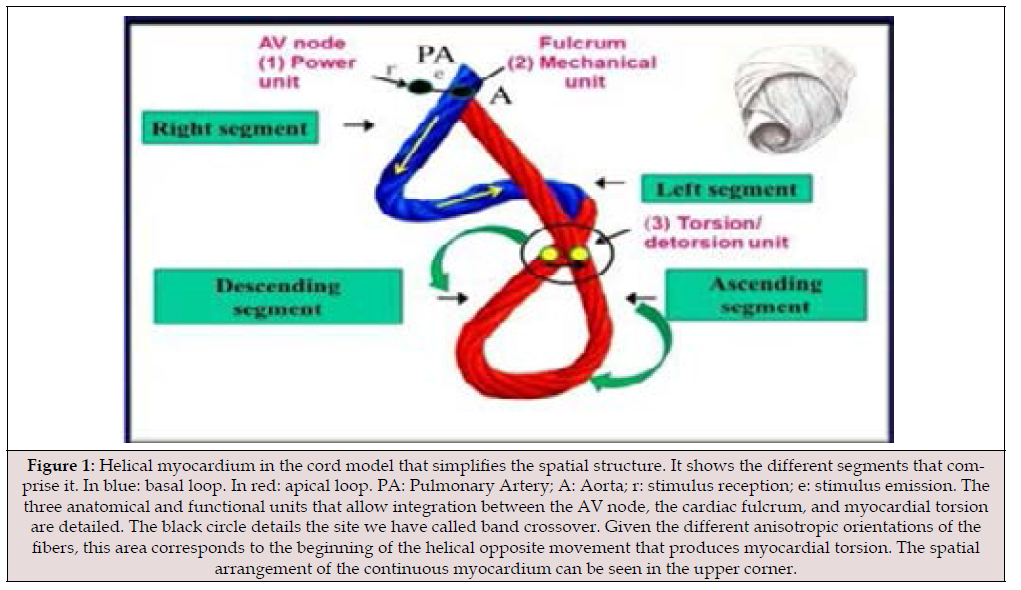
Figure 1:Helical myocardium in the cord model that simplifies the spatial structure. It shows the different segments that comprise it. In blue: basal loop. In red: apical loop. PA: Pulmonary Artery; A: Aorta; r: stimulus reception; e: stimulus emission. The three anatomical and functional units that allow integration between the AV node, the cardiac fulcrum, and myocardial torsion are detailed. The black circle details the site we have called band crossover. Given the different anisotropic orientations of the fibers, this area corresponds to the beginning of the helical opposite movement that produces myocardial torsion. The spatial arrangement of the continuous myocardium can be seen in the upper corner.
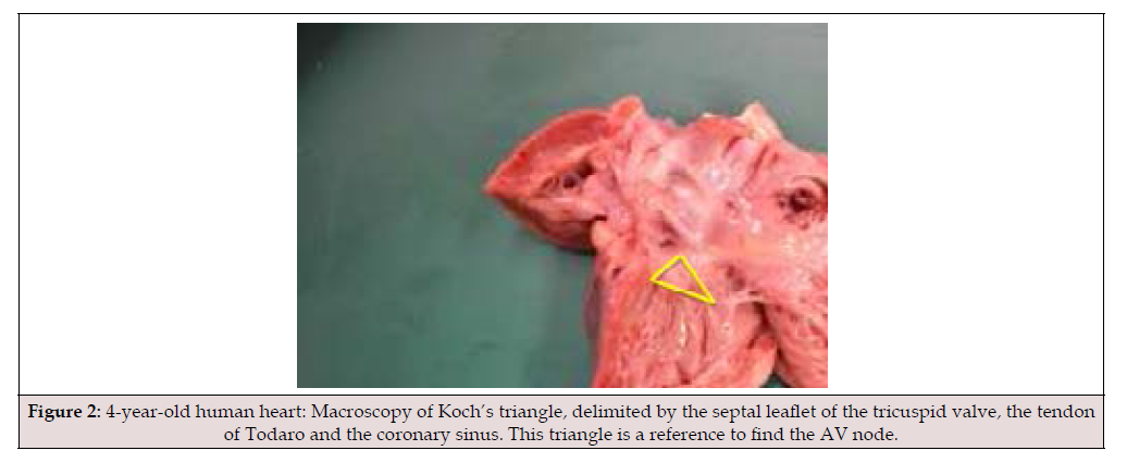
Figure 2:4-year-old human heart: Macroscopy of Koch’s triangle, delimited by the septal leaflet of the tricuspid valve, the tendon of Todaro and the coronary sinus. This triangle is a reference to find the AV node.
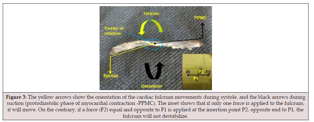
Figure 3:The yellow arrows show the orientation of the cardiac fulcrum movements during systole, and the black arrows during suction (protodiastolic phase of myocardial contraction -PPMC). The inset shows that if only one force is applied to the fulcrum, it will move. On the contrary, if a force (F2) equal and opposite to F1 is applied at the insertion point P2, opposite end to P1, the fulcrum will not destabilize.
Results
Kinetics of the Cardiac Fulcrum
The contraction and recoil of myocardial muscle fibres, whose ends must rest on some “fixed point” to have mechanical effect, would not be effective without the existence of the cardiac fulcrum. The fulcrum is located at the centre of gravity of the lever, which ensures its functionality in the cardiac system, allowing the forces to be equally distributed. For this mechanism to be subjected to tractions in a magnitude of one hundred thousand times a day, it must meet certain conditions:
a. Stability
b. Resistance
c. Heterogeneity
d. Anisotropy
e. Elasticity
f. Plasticity
These faculties allow it to reach a certain level of stress when subjected to loads and then recover its shape when the loads are removed [12].
As can be seen in (Figure 3), if only a force (F1) is applied to the fulcrum at a point (P1) on the left insertion line, the fulcrum would move in that direction, since it is a semi-floating body. But if a force (F2) equal and opposite to (F1) is applied at the insertion point (P2), opposite to (P1), on the right insertion line, the fulcrum would not move. As both forces are produced by the same muscle fiber of the myocardium, which originates at (P1) and ends at (P2), both forces are equal (similar to those of an elastic band). The same can be applied to the totality of myocardial muscle fibers inserted in the myocardium along the lines of insertion (Figure 3). Therefore, to maintain the equilibrium of a system of forces applied on both sides, these forces must be equal and opposite [13].
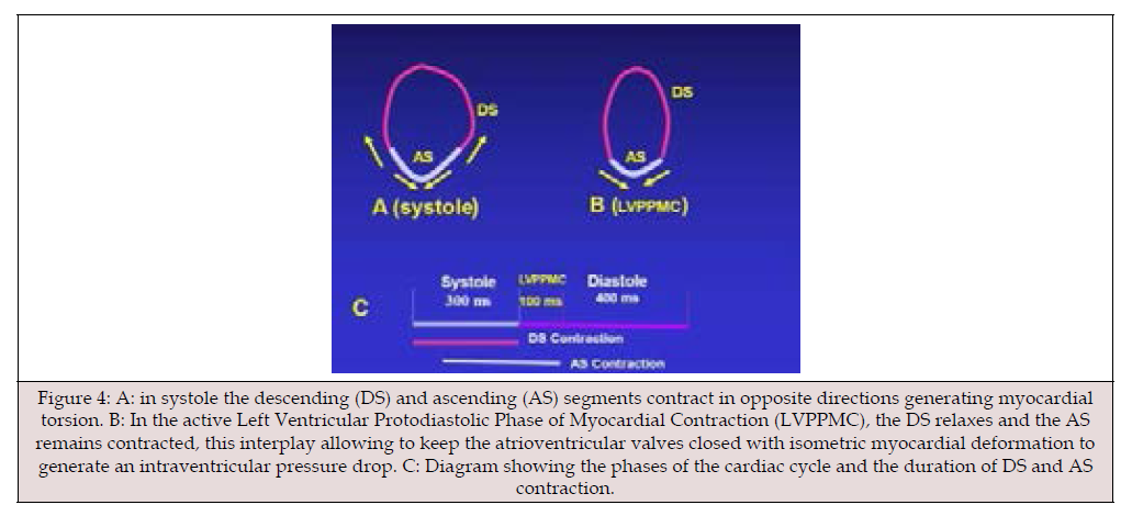
Figure 4:A: in systole the descending (DS) and ascending (AS) segments contract in opposite directions generating myocardial torsion. B: In the active Left Ventricular Protodiastolic Phase of Myocardial Contraction (LVPPMC), the DS relaxes and the AS remains contracted, this interplay allowing to keep the atrioventricular valves closed with isometric myocardial deformation to generate an intraventricular pressure drop. C: Diagram showing the phases of the cardiac cycle and the duration of DS and AS contraction.
The fulcrum acts like a seesaw during cardiac movements. During systole, it approaches the apex, primarily at its right end, where the continuous myocardium originates, while the opposite end, where the fibers of the ascending segment insert, rises. During the beginning of diastole, in its first 80-100 ms, there is muscle activation with energy expenditure. This phase, classically called the isovolumic diastolic phase, in light of these findings in our research, we have named it the Protodiastolic Phase of Myocardial Contraction (PPMC). At this point in the cardiac cycle, the fulcrum undergoes movements opposite to those previously described, thus performing the movements of a seesaw. In turn, the fulcrum’s torque is molded by twisting and untwisting movements, alternately during systolic ejection and PPMC. During diastole, it returns to its initial resting position. To achieve this hysteresis, reformulation requires external (electrical) stimulation; hence, it is called extrinsic hysteresis, as opposed to hysteresis achieved through endogenous factors (intrinsic hysteresis, e.g., rubber). In the sequential movements of the myocardium, each segment, through its action, favors the other (Figure 3).
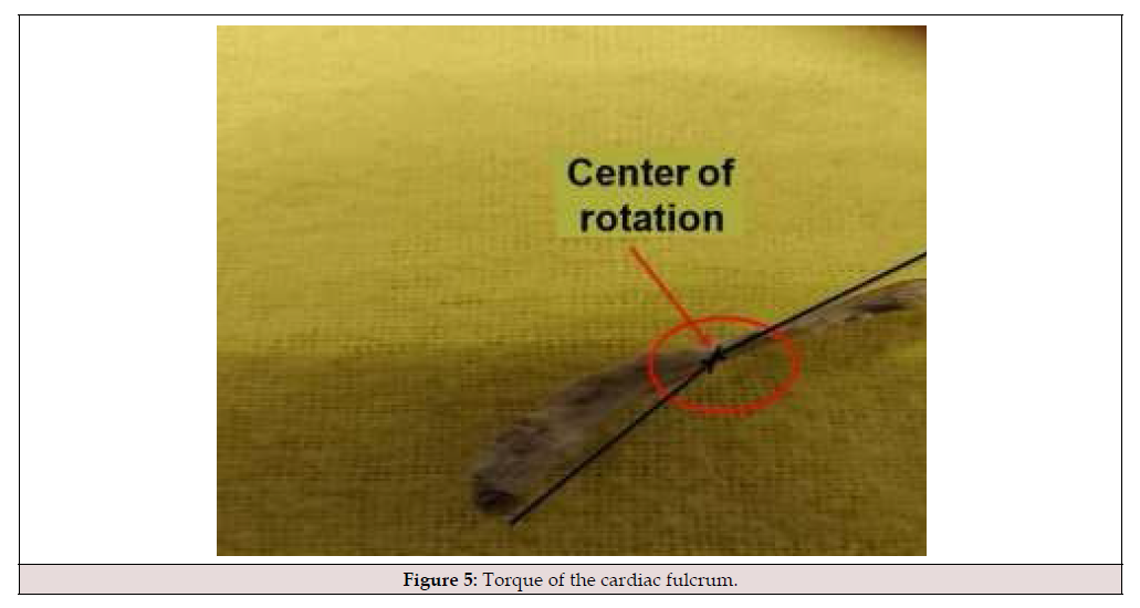
Figure 5:Torque of the cardiac fulcrum.
As the fulcrum is subjected to the forces of the myocardial segments attached to its structure, it obviously registers modifications in the space that even lead it to model its morphology. This concept is explained by the fact that the myocardial movements are sequential and superimposed on the myocardial segments, determining tensions that act as a lever arm with epicenter at the point of support, i.e. the fulcrum [12]. During systole, the different segments contract sequentially and synchronously according to the stimulation pattern we have investigated. This begins in the right segment, tethered to the right and anterior sector of the cardiac fulcrum, with continuation in the left, descending and ascending segments. The fundamental peculiarity of this activation is that although at its origin it is unidirectional, upon reaching the junction of the descending and ascending bands, simultaneity is produced -by transverse activation- in both, generating a helical movement essential for the myocardium to eject the ventricular content at a speed of 200 cm/s (Figure 1) [14-17]. This systolic activation produces longitudinal shortening of the myocardium with circular narrowing and the muscular torsion that characterizes its helical conformation, which implies that the fulcrum undergoes a downward displacement, accompanied by a torque from its right end toward the opposite end due to torsion (Figure 4).
When the ejection period ends, the ascending segment remains in an active process of contraction in its terminal part, that is, in its attachment to the cardiac fulcrum, occurring mainly in its anteroinferior portion and posterior surface. This phase occupies about 80-100 ms and is intermediate between systole and diastole. We have called it the Protodiastolic Phase of Myocardial Contraction (PPMC) and it is caused by the process of generating negative intracavitary ventricular pressure, which, upon opening of the atrioventricular valves, will precipitate the entry of blood into the ventricles by a suction mechanism. During this phase the myocardium lengthens, narrows and untwists [18,19]. Under these stresses the fulcrum undergoes an upward displacement and generates a torque opposite to that during systole. Obviously, this continuous torque in coupling models the fulcrum with a torsion that is well observed in a profile view (Figure 3). The torque is a demonstration of the opposing forces that stress the fulcrum; thus, the ascent-descent of the continuous myocardium together with torsion-detorsion models its morphology.
The section of the fulcrum is not axisymmetric in relation to its axis, therefore the torsion-detorsion it undergoes in each cardiac cycle determines deformations at each point of its structure. Thus, the torque (twisting force) (Figure 5) applied at each of its ends during the cardiac cycle causes the free ends of the bar to rotate at an angle Φ, which is called the twisting angle or twist. In this deformation the maximum shear strain is produced in the middle of the fulcrum surfaces and around the axis at which it rotates. When this system of forces is applied, a rotation effect is produced, without achieving a manifest shift. In the fulcrum we would be in the presence of two couplings (a system of parallel forces of equal intensity but opposite directions, with resultant equal to 0) meeting at point 0. In synthesis, the continuous myocardium meeting at its origin and end with the fulcrum develops a moment of force (torque) which determines its rotation in opposite directions that are equalized at point 0 (center of rotation). When these two forces counteract each other, the sum is zero.
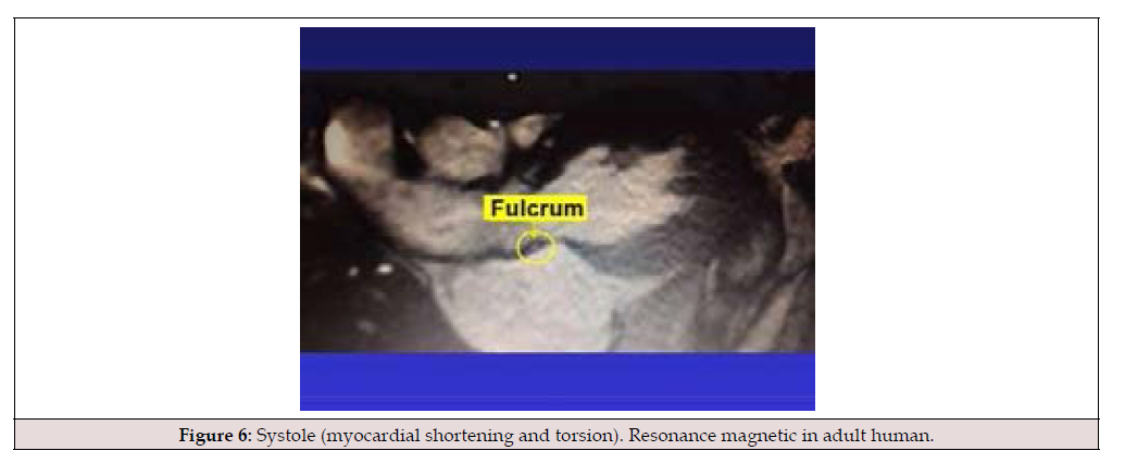
Figure 6:Systole (myocardial shortening and torsion). Resonance magnetic in adult human.
It was also possible to document the fulcrum during movement. We have observed fulcrum displacements in adult humans during the phases of the cardiac cycle in resonance magnetic, which implies possibilities of knowledge of cardiac mechanics and also of pathological situations. In systole (Figure 6) the cardiac fulcrum descends horizontally due to the concomitant myocardial shortening and torsion (clockwise rotation), which allows the image to be outlined. During PPMC (Figure 7), when the terminal end of the ascending segment contracts, the heart undergoes elongation accompanied by detorsion (counterclockwise rotation). In this phase of the cardiac cycle the fulcrum is observed with a certain pause in the movement according to PPMC, without volumetric changes in the ventricular chambers and with geometric deformation of the myocardium. In diastole the fulcrum is shown in its full magnitude with its characteristic morphology (Figure 8).
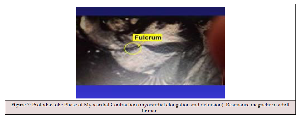
Figure 7:Protodiastolic Phase of Myocardial Contraction (myocardial elongation and detorsion). Resonance magnetic in adult human.
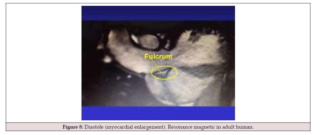
Figure 8:Diastole (myocardial enlargement). Resonance magnetic in adult human.
The fulcrum is not only a support for the myocardium. The heart, with its movements, generates tensions that are absorbed by the fulcrum, avoiding their transfer to the aorta. In this way, it prevents the aorta from traction and rotation, which would produce resistance to blood ejection. From the anatomy the fulcrum can be confirmed to present on its upper edge a ledge that adapts to the aortic annulus determining a proof of the organization pattern of the helical heart (Figure 9).
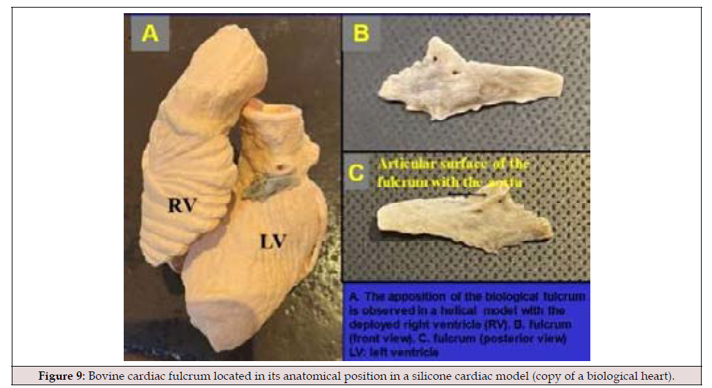
Figure 9:Bovine cardiac fulcrum located in its anatomical position in a silicone cardiac model (copy of a biological heart).
Relationship of the Cardiac Fulcrum to the AV Node
Histological analysis revealed that the heart’s support structure, which we have named the cardiac fulcrum [1,2,20,21], is adjacent to the AV node, forming a cellular cluster rich in neurofilament plexuses (Figures 10-14). This proximity between the two structures was found in all specimens studied. The key finding of this research is that neurofilaments are also located within the cardiac fulcrum, in contact with the myocardial muscle fibers that insert into its AV node occupies the apex of Koch’s triangle (Figure 4). Its anterior border is defined by the septal leaflet of the tricuspid valve, and its posterior border by the fibrous tendon of Todaro. The AV node also has a compact portion and a marginal portion (transitional cells) located between the right atrium and the node, forming its outer layer. The compact portion is continuous with the center of the cardiac fulcrum.
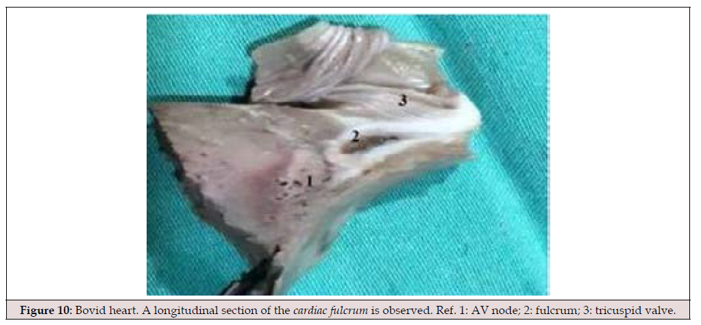
Figure 10:Bovid heart. A longitudinal section of the cardiac fulcrum is observed. Ref. 1: AV node; 2: fulcrum; 3: tricuspid valve.
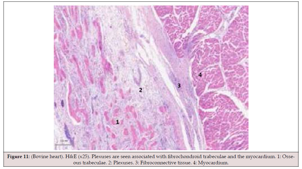
Figure 11:(Bovine heart). H&E (x25). Plexuses are seen associated with fibrochondroid trabeculae and the myocardium. 1: Osseous trabeculae. 2: Plexuses. 3: Fibroconnective tissue. 4: Myocardium.
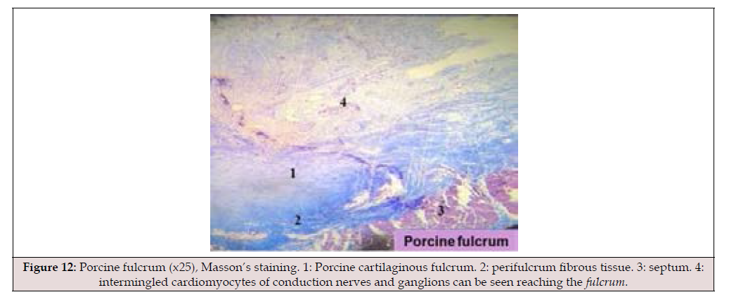
Figure 12:Porcine fulcrum (x25), Masson’s staining. 1: Porcine cartilaginous fulcrum. 2: perifulcrum fibrous tissue. 3: septum. 4: intermingled cardiomyocytes of conduction nerves and ganglions can be seen reaching the fulcrum.
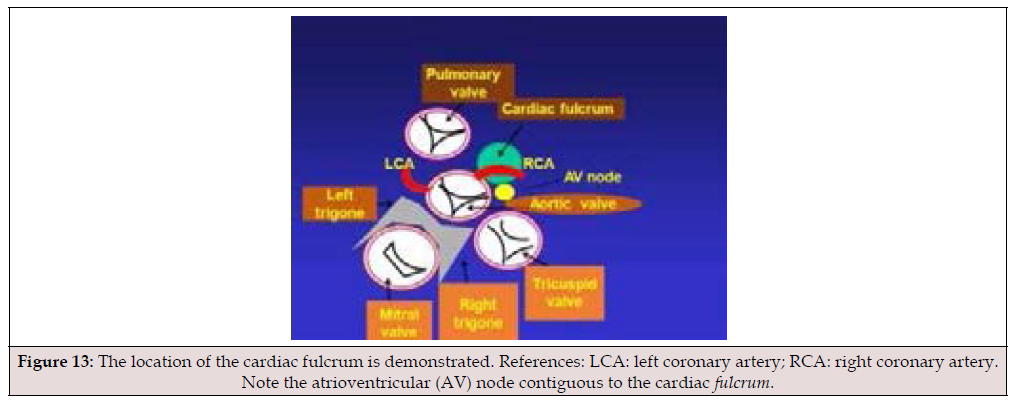
Figure 13:The location of the cardiac fulcrum is demonstrated. References: LCA: left coronary artery; RCA: right coronary artery. Note the atrioventricular (AV) node contiguous to the cardiac fulcrum.
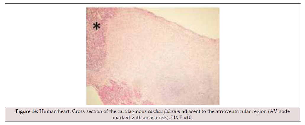
Figure 14:Human heart. Cross-section of the cartilaginous cardiac fulcrum adjacent to the atrioventricular region (AV node marked with an asterisk). H&E x10.
The cardiac conduction system is embryologically nourished by myocardial precursor cells originating from the splanchnic mesocardium, but the innervation of cardiomyocytes via ganglion cells and fibers also comes from the neural crest. This profound interrelationship between both embryological layers demonstrates an anatomical and functional electromechanical unity. Furthermore, from a histological perspective, the cellular transition from the right atrial wall to the AV node is gradual, making its precise onset uncertain, as stated by Rushmer [22-24]. The same occurs at its point of continuity with the bundle of His. Histochemically, the AV node can be divided into two distinct zones: a compact zone (fast conduction) and an inferior nodal zone (slow conduction). The location of the AV node, adjacent to the cardiac fulcrum, between it and the septal leaflet of the tricuspid valve, is strategic, as it allows for the modulation of stimulation impulses in the continuous myocardium that originates from the fulcrum along its helical course. In this way, both structures constitute the electromechanical unit of the myocardium (Figures 15-17).
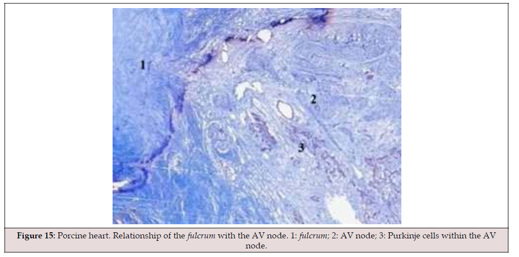
Figure 15:Porcine heart. Relationship of the fulcrum with the AV node. 1: fulcrum; 2: AV node; 3: Purkinje cells within the AV node.
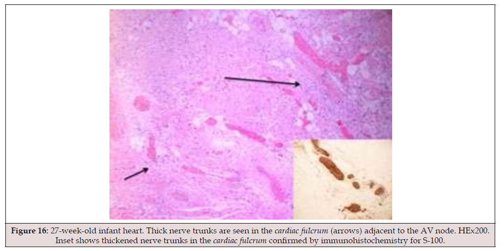
Figure 16:27-week-old infant heart. Thick nerve trunks are seen in the cardiac fulcrum (arrows) adjacent to the AV node. HEx200. Inset shows thickened nerve trunks in the cardiac fulcrum confirmed by immunohistochemistry for S-100.
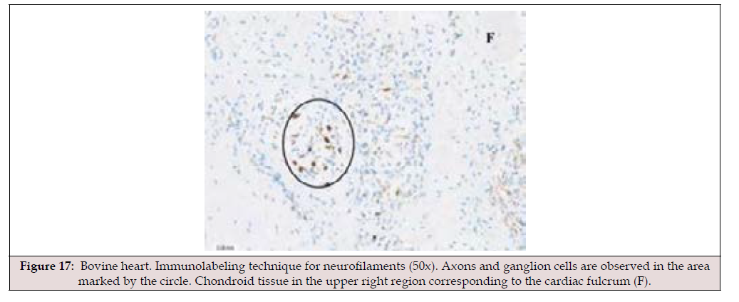
Figure 17:Bovine heart. Immunolabeling technique for neurofilaments (50x). Axons and ganglion cells are observed in the area marked by the circle. Chondroid tissue in the upper right region corresponding to the cardiac fulcrum (F).
Research of cardiac electrical activation with 3D mapping
Material and Methods: The endo and epicardial electrical activation sequence of the Left Ventricle (LV) have been studied by means of three-dimensional electroanatomic mapping (MET) with a Carto navigation and mapping system (Biosense Webster, California, USA) that allows a three-dimensional anatomical representation, with activation maps and electrical propagation. Isochronic and activation sequence maps were generated, correlating them with the surface ECG. Endo and epicardial ventricular activation maps were also produced, obtaining detailed high-density recordings with apical, lateral and basal views. The study was conducted at Presidente Perón Hospital (Buenos Aires, Argentina) including patients who provided their informed consent. The research was previously approved by the Institutional Ethics Committee. All patients were in sinus rhythm, with a normal QRS and did not have evident structural heart disease by Doppler echocardiography and in gamma camera stress and rest studies (Table 1).

Table 1:Patient characteristics.
The MET mapping was performed during the course of radiofrequency ablation for arrhythmias owing to probable abnormal occult epicardial pathways. Mapping was carried out at the onset of studies, followed by ablation maneuvers. No complications developed. The presence of abnormal pathways did not interfere with mapping, as during the whole procedure baseline sinus rhythm was preserved with normal QRS complexes, both in duration and morphology, without antegrade preexcitation. As the muscular structure of the Left Ventricle (LV) is made up of an endocardial layer (descending segment) and an epicardial layer (left and ascending segments), two approaches were used to carry out mapping. The endocardial access was performed through a conventional atrial transseptal puncture. The epicardial access was obtained by means of a percutaneous approach in the pericardial cavity [25] with an ablation catheter. Endo and epicardial mappings were performed consecutively and immediately and were later superimposed, synchronizing them by electrocardiographic timing. Thus, a simultaneous mapping of both surfaces was obtained. In addition, the propagation times of electrical activation through the myocardium were measured in milliseconds (ms).
Until now, the theory of the continuous myocardium lacked an essential investigation, due to the absence of documentation on the electrophysiological mechanism that would support the mechanical activation sequence of the helical anatomical model. The advent of MET managed to overcome this limitation, since it not only allows independent recording of the various ventricular zones, but also those of the endocardium and epicardium, either exclusively or integrated. We mapped the activation of the LV on its endocavitary and epicardial surfaces according to the methodology described. Mapping was performed simultaneously with the surface ECG. This provided a unified temporal frame of reference, allowing on the one hand to correlate both records, and on the other hand to obtain a synchronized view of the simultaneous activation observed in various electro anatomical incidents (Video 1).
Percutaneous access technique The Carto system was used for MET to obtain voltage, activation and propagation maps. Epicardial mapping was done in the pericardial cavity through the left paraxiphoid space (Figure 18) [25]. A decapolar catheter in the coronary sinus and a quadripolar catheter in the bundle of His were placed as fluoroscopic reference. Once the epicardial recording of both ventricles was obtained, left ventricular intracavitary mapping was performed. The LV was accessed by transeptal puncture through the right femoral vein using standard technique. In addition, the propagation times of electrical activation through the myocardium were measured in milliseconds (ms).
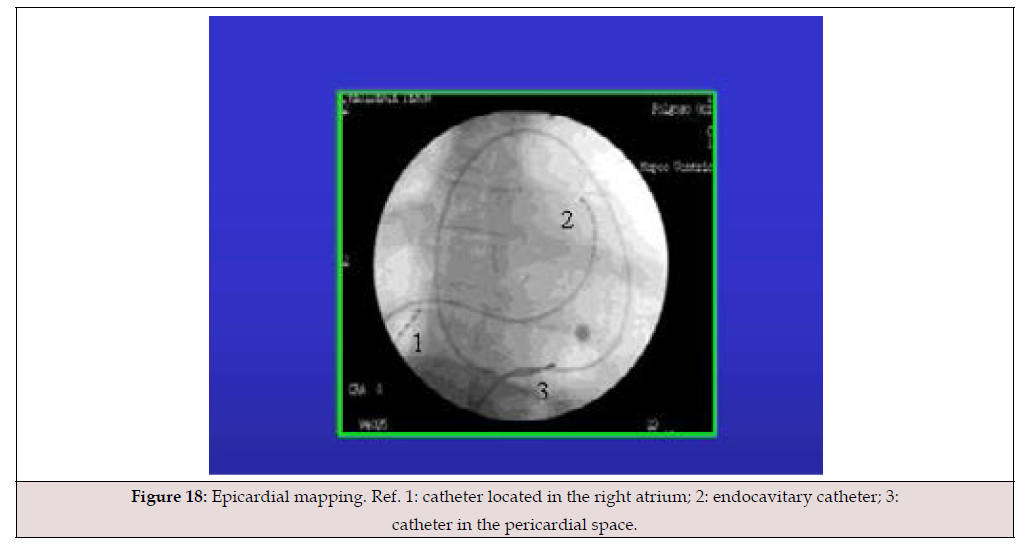
Figure 18:Epicardial mapping. Ref. 1: catheter located in the right atrium; 2: endocavitary catheter; 3: catheter in the pericardial space.
Results: Mapping allowed a detailed activation recording (Figure 19). Although the activation sequence varied in its details, in each case it was similar in its general aspects, forming an endoepicardial depolarization matrix. As MET corresponded to the LV, the activation wave previously generated in the right ventricle was not obtained. The MET lasted approximately 20 minutes. There were no complications associated with the procedure or any of the accesses. (Figures 20-22) show the propagation of endocardial an epicardial electrical activation. In all the figures the right projection is observed in the left panel and the simultaneous left anterior oblique projection in the right panel. At each time, the activated zones are detailed in red. The lateral inset represents the activation of the descending and ascending muscle bands that make up the ventricular continuous structure of the myocardium in the cord model, a simplification of the three-dimensional myocardial spatial structure. In all figures, the area depolarized at that time is represented in red and those that were previously activated and are in refractory period are represented in blue. Below the cord model, the average electrical propagation time along the myocardium can be seen measured in milliseconds (ms) at the analyzed site (Table 2,3).

Table 2:Activation times of the different cardiac structures (in ms) (See explanation in the text of the cited Figures). References. ms: milliseconds; X: Mean; SD: Standard deviation.

Table 3:Radial propagation time (in ms). References. ms: milliseconds Pat: patient; X: Mean; SD: Standard deviation.
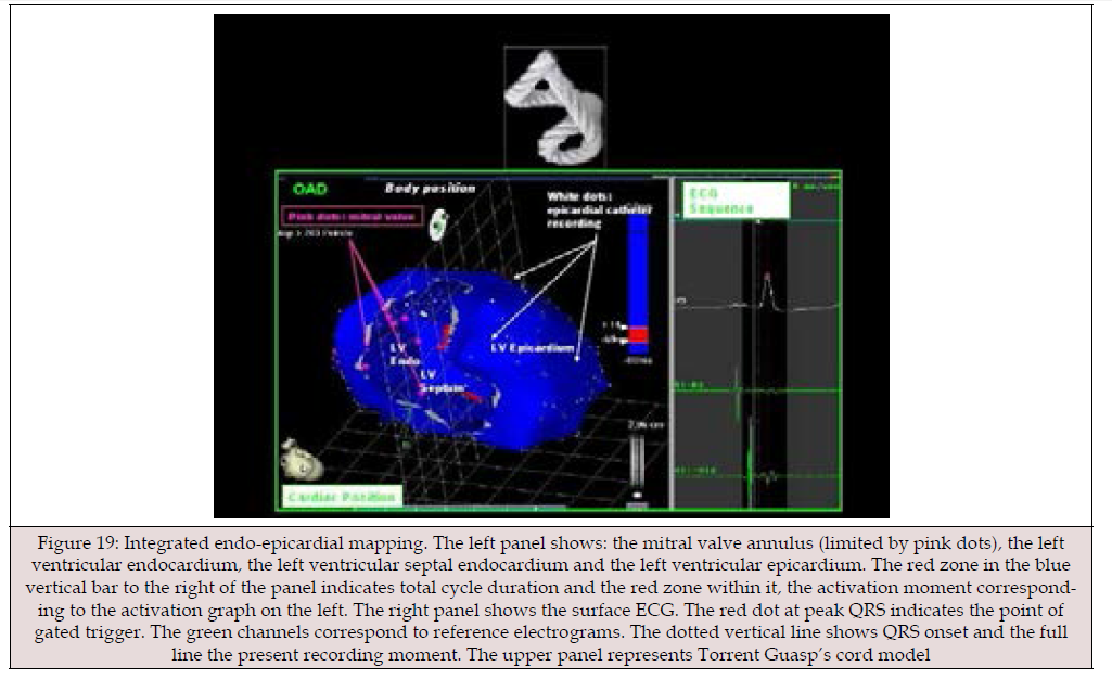
Figure 19:Integrated endo-epicardial mapping. The left panel shows: the mitral valve annulus (limited by pink dots), the left ventricular endocardium, the left ventricular septal endocardium and the left ventricular epicardium. The red zone in the blue vertical bar to the right of the panel indicates total cycle duration and the red zone within it, the activation moment corresponding to the activation graph on the left. The right panel shows the surface ECG. The red dot at peak QRS indicates the point of gated trigger. The green channels correspond to reference electrograms. The dotted vertical line shows QRS onset and the full line the present recording moment. The upper panel represents Torrent Guasp’s cord model
Left ventricular activation occurs 12.4 ms±1.816 ms after the onset in the interventricular septum (Figure 20 A). This contraction is determined by the bundle of His and its conduction fibers that give origin to the Purkinje network. Based on the anatomy that these fibers occupy, the endocardium is the first area of the LV to receive electromechanical activation. At that moment it also propagates to an epicardial area - ascending band- evidencing a transverse activation at a point we call “band crossover” which is produced 25.8±1.483 ms after septal stimulation (Figure 20 B) (Table 3) and at 38.2±2.135 ms from the onset of cardiac activation. This leads to subendocardial shortening and subepicardial lengthening, resulting in opposite rotations between the ventricular base (counterclockwise) and apex (clockwise). In echocardiographic studies verifying stimulation-function at this time of the cardiac cycle, greater systolic torsion was found in the mid-interventricular septum than in the anterior walls. Synchronously, following the anatomical arrangement of the descending band, the activation propagates longitudinally towards the ventricular apex, reaching it at 58±2.0 ms (Figures 21A, B) (Table 2).
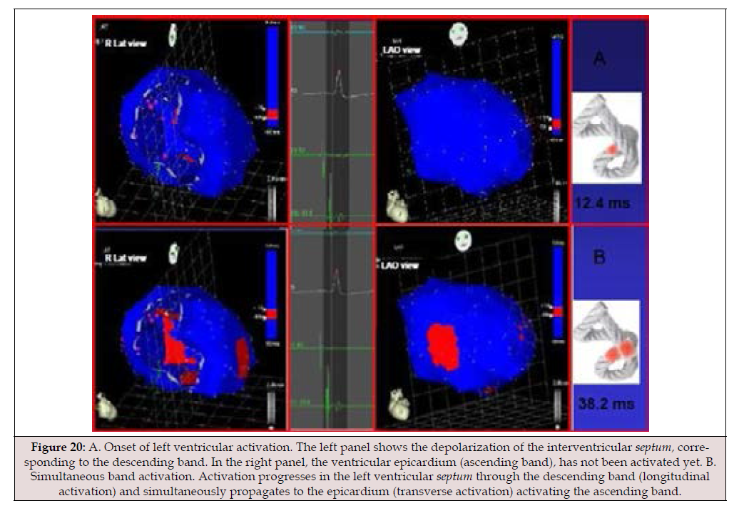
Figure 20:A. Onset of left ventricular activation. The left panel shows the depolarization of the interventricular septum, corresponding to the descending band. In the right panel, the ventricular epicardium (ascending band), has not been activated yet. B. Simultaneous band activation. Activation progresses in the left ventricular septum through the descending band (longitudinal activation) and simultaneously propagates to the epicardium (transverse activation) activating the ascending band.
From “band crossover” onwards the activation loses its
unidirectional character and becomes slightly more complex. Three
simultaneous wave fronts are generated:
a) The distal activation of the descending band towards the apical
loop
b) The depolarization of the ascending band from band crossover
towards the apex
c) The activation of this band from the crossover points towards
its final portion in its insertion in the cardiac fulcrum,
myocardial support.
Figures 21 B, 22 A & B show the continuation and completion of this process. Intracavitary activation ends long before QRS termination (Figure 22 A). The rest of the QRS corresponds to the late activation of the distal portion of the ascending band, which justifies the persistence of its contraction during the isovolumic diastolic phase. This contraction constitutes the basis of the ventricular suction mechanism (Figure 22 B), and for this reason, it will be now termed Left Ventricular Protodiastolic Phase of Myocardial Contraction (LVPPMC), as in it there is contraction and no relaxation. A synthesis of the stimulation found in this study is shown with the cord model in (Figure 23) (upper panel)
In our research, the right ventricle initiates systolic activity 12.4 ms before the left ventricle. The pulmonic valve opens as soon as its intraventricular pressure rises to 8-10 mm Hg. The inflow region of the right ventricle contracts very early. This right ventricular ejection phase precedes left ventricular systole. Only at an average of 38.2 ms in our research, after the beginning of the cardiac cycle with right ventricular contraction, are the ascending and descending segments stimulated, which would imply left ventricular ejection at that moment, as the aortic valve opens. These 40 ms differences in the earlier opening of the pulmonic valve compared to the aortic valve is logical, since with both ventricles ejecting and filling simultaneously, circulation would be practically interrupted, lacking the necessary continuity and falling to levels even lower than the gradients exhibited by normal circulatory return.
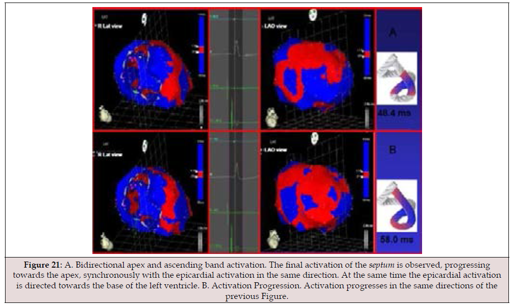
Figure 21:A. Bidirectional apex and ascending band activation. The final activation of the septum is observed, progressing towards the apex, synchronously with the epicardial activation in the same direction. At the same time the epicardial activation is directed towards the base of the left ventricle. B. Activation Progression. Activation progresses in the same directions of the previous Figure.
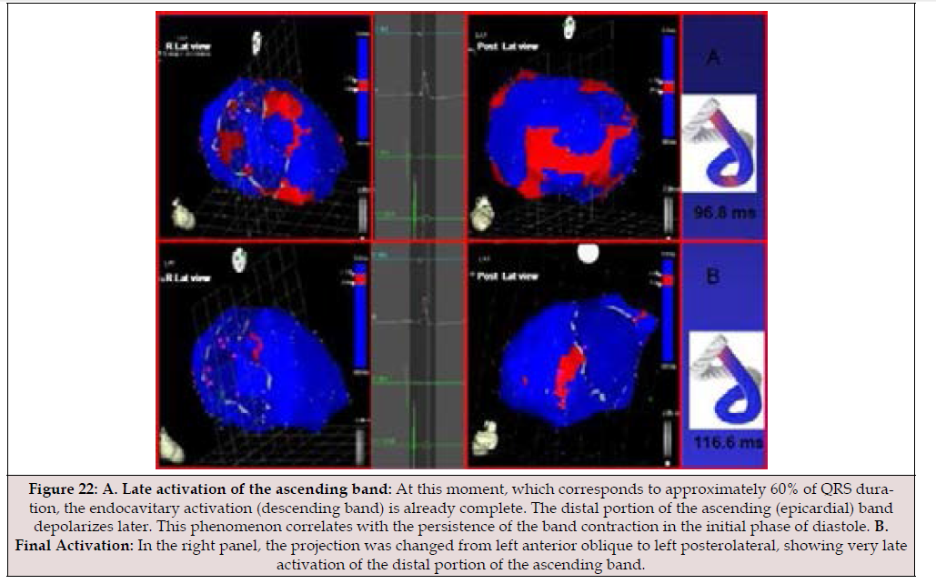
Figure 22:A. Late activation of the ascending band: At this moment, which corresponds to approximately 60% of QRS duration, the endocavitary activation (descending band) is already complete. The distal portion of the ascending (epicardial) band depolarizes later. This phenomenon correlates with the persistence of the band contraction in the initial phase of diastole. B. Final Activation: In the right panel, the projection was changed from left anterior oblique to left posterolateral, showing very late activation of the distal portion of the ascending band.
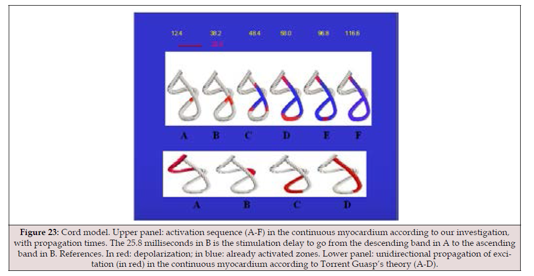
Figure 23:Cord model. Upper panel: activation sequence (A-F) in the continuous myocardium according to our investigation, with propagation times. The 25.8 milliseconds in B is the stimulation delay to go from the descending band in A to the ascending band in B. References. In red: depolarization; in blue: already activated zones. Lower panel: unidirectional propagation of excitation (in red) in the continuous myocardium according to Torrent Guasp’s theory (A-D).
The two synchronized active functions of ejection and suction between both ventricles correspond to circulatory continuity. This raises the question: how is the early opening of the pulmonic valve relative to the aortic valve justified? The ventricles take advantage of the asynchronous impulse of approximately 40 ms, or 5s of the 800 ms duration of the cardiac cycle, so that one of them, the right ventricle, assists the other, the left, during the loading phase, through the complementarity between ejection and suction. This earlier opening of the pulmonic valve relative to the aortic valve, observed in our human studies, has also been seen in fish. Thus, in teleost’s (Cretaceous period), which have four cardiac chambers in a line (sinus venosus, atrium, ventricle, and bulbus arteriosus), a similar 40 ms delay in myocardial deflection has been found after the discharge of the electrical spike, between the proximal and distal ventricles at the level of the bulbus, analogous to the right and left ventricles of mammals (Figure 24) [26]. This correlation implies an evolutionary biological matrix that transcends species. In this estimated interval of at least 38.2 ms on average in our research, between the opening of the pulmonic and aortic valves, the final phase of left ventricular filling (diastolic period 3) occurs in the left ventricle.
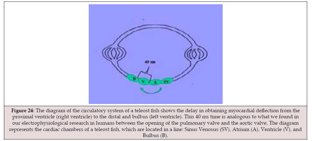
Figure 24:The diagram of the circulatory system of a teleost fish shows the delay in obtaining myocardial deflection from the proximal ventricle (right ventricle) to the distal and bulbus (left ventricle). This 40 ms time is analogous to what we found in our electrophysiological research in humans between the opening of the pulmonary valve and the aortic valve. The diagram represents the cardiac chambers of a teleost fish, which are located in a line: Sinus Venosus (SV), Atrium (A), Ventricle (V), and Bulbus (B).
The interpretation of the anatomical relationships between the Cardiac Fulcrum and the AV Node implies the complementarity of the anatomy with the physiology of the continuous helical myocardium, since their contiguity is found at the site where stimulation begins and ends, the production of which determines the mechanical action of torsion and detorsion in the systolic and suction phases of the ventricles. The cardiac fulcrum, the support and insertion point of the myocardium that acts as a lever during its movements, is located adjacent to the AV node of Aschoff- Tawara (described by Sunao Tawara in 1906). Thus, the two form an electromechanical unit located at the beginning and end of the continuous, helical myocardium. This anatomical and functional arrangement of the myocardium is supported by a rich plexus of specialized filaments located within the cardiac fulcrum that interact with the cardiomyocytes responsible for mechanical work [27].
The Quantum Biology of the Cardiac Cycle: Scientific progress is usually achieved by unifying concepts that were previously separate, generally because they arise from different disciplines. For example, the unification of thermodynamics and mechanics through statistical mechanics. This alchemy also occurs between optics and electromagnetism, thanks to Maxwell’s theory of magnetic fields, and through chemistry and atomic physics, which are unified in quantum mechanics [28]. If there is no convergence between different disciplines, the increase in entropy tends to hinder scientific development. The information transmitted thus becomes the cornerstone of the evolution of the sysmets. We must consider that interpretations that differ from the cultural reality we experience imply an intellectual debate, which, if conducted honestly, leads to progress in knowledge.
In the last century, quantum physics has blurred the boundary between observer and observed, making the observation of the world far less objective. Another advance has been the understanding, through the observation of living beings, that there is an opposition between the mechanistic interpretation and the properties that determine their behavior. In this regard, information theory introduced the concept of a program to describe the information contained within an organism. The implexion (from the Greek implexus), that is, the entanglement between the anatomy and physiology of the heart and quantum physics, is possible without violating the principles governing biological epismetology in research.
In view of the structure found in the continuous myocardium with its helical conformation (Figure 2), the following questions arose, some quite unsettling: Does the relationship between the cardiac fulcrum and the AV node constitute an electromechanical unit open to stimuli from outside the organism? Why does the myocardium originate and terminate at the same point, the cardiac fulcrum, after a double-helix path? Are we witnessing, with this myocardial anatomy, a self-organizing process with the accumulation of negentropic internal information? Does this electromechanical unit behave as an attractor of the cardiac stimulus based on the circular nature of the myocardium, unique in myology? Does the myocardium have a fourth dimension, time? Does myocardial activation combined with time constitute an inseparable activation-time equation? In order to remain within the bounds of scientific reasoning, we have sought answers to the questions posed, focusing on the concept that the study of the heart’s properties would inevitably lead to a better understanding of its function. In this research, we analyzed the anatomical and histological relationship between the cardiac fulcrum and the atrioventricular node in human, bovine, porcine, and anuran hearts, as well as the possible functionality between these two structures and the actions derived from this electromechanical unit-a situation we will investigate in detail given its importance.
The fulcrum [1,2], located at the atrioventricular junction at the insertion of the interventricular septum, below the aorta and pulmonary artery, is adjacent to the AV node of Aschoff-Tawara, which lies to its right (Figures 10-13). The AV node is located at the atrioventricular junction, at the base of the muscular septum, below the origin of the great vessels, adjacent to the cardiac fulcrum, situated between it and the insertion of the septal leaflet of the tricuspid valve. It constitutes a cluster of cells (specialized myocytes) that R.F. Rushmer defines as a spherical or bulbous end composed of bundles of fibers [22-24] for the purpose of transmitting electrical impulses to the myocardial mass. Continuing along its length, it transforms subtly into the bundle of His, which is short in length, sometimes even non-existent. In humans it measures approximately 5 mm in length, 3 mm in width and 1 mm in thickness and is irrigated by a branch of the coronary arteries, generally coming from the right in 90% of cases and from the circumflex in the remaining minority [20,21].
In short, we believe this approach can help us advance our understanding of cardiac physiology and its therapeutic possibilities, given that living sysmets must be understood as open sysmets subject to a constant flow of energy, matter, and information (Figure 25-28). From a genetic perspective, the structure, through its one-dimensional form, determines twodimensional cellular production, which then transitions to threedimensionality in organs, acquiring a four-dimensional behavior. This extra dimension can be interpreted as time (Einstein’s spacetime), which will be duly explained, as there is a possible horizon defined by time in each cardiac cycle. The evolution of a dynamic system, such as cardiac activity, implies giving significance to the time of its cycle. In its dynamics, it returns to its initial state. It must be understood that there is a reconstruction within the selforganizing phenomenon in the cardiac system.
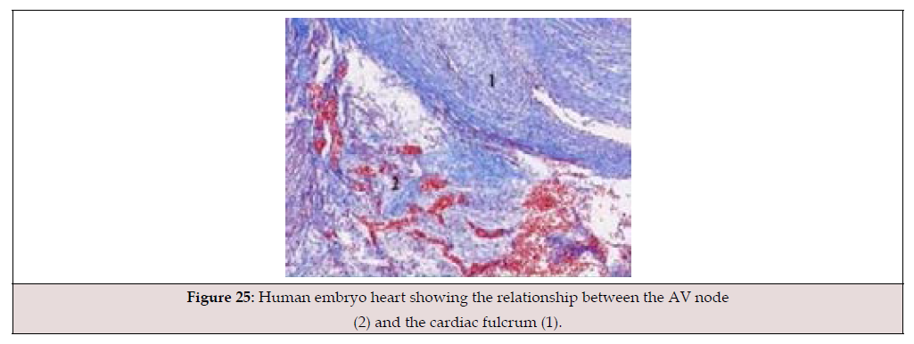
Figure 25:Human embryo heart showing the relationship between the AV node (2) and the cardiac fulcrum (1).
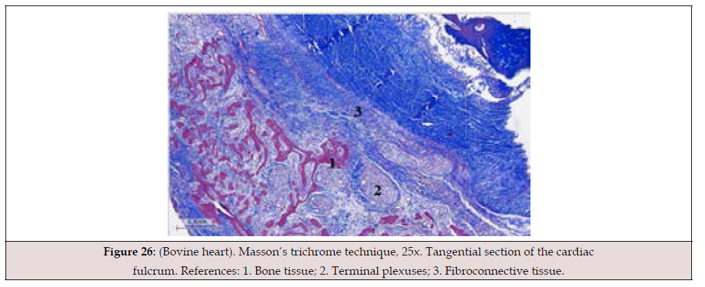
Figure 26:(Bovine heart). Masson’s trichrome technique, 25x. Tangential section of the cardiac fulcrum. References: 1. Bone tissue; 2. Terminal plexuses; 3. Fibroconnective tissue.
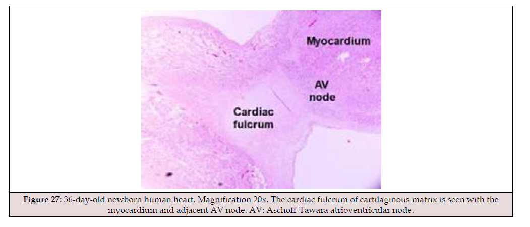
Figure 27:36-day-old newborn human heart. Magnification 20x. The cardiac fulcrum of cartilaginous matrix is seen with the myocardium and adjacent AV node. AV: Aschoff-Tawara atrioventricular node.
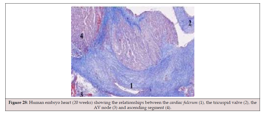
Figure 28:Human embryo heart (20 weeks) showing the relationships between the cardiac fulcrum (1), the tricuspid valve (2), the AV node (3) and ascending segment (4).
Just as the structure of the human body is supported by the insertion of its muscles, upon which forces act to coordinate movement and enable work, the same is true of the heart. It is logical to assume that its helical shape and the remarkable physiological characteristics of the myocardium necessitate a point of support to fulfill its function as a pump, both expelling (torsion) and suctioning (detorsion). The heart is roughly the size of a human fist and weighs an average of 270 grams. It pumps a volume of blood ranging from 4 to 6 liters per minute at a speed of 200 cm/s; it consumes only 10 watts, operates continuously for 80 years without maintenance and almost silently, producing 100,000 beats per day. Its task is equivalent to extracting 1 ton of water daily from a depth of 1 m with a mechanical efficiency (work/energy ratio) of 50%, a feat not achieved by man-made machines, which reach only 30%. Its effectiveness allows it to expel 70% of the left ventricular contents with only a 12% shortening of its contractile unit, the sarcomere [1,2]. Without myocardial support at the cardiac fulcrum, this shortening would not be possible.
Unfolding the heart, layer by layer of muscle, allows us to visualize its true internal architecture, and among these findings, we glimpse the cardiac fulcrum, which emerges from its hidden position to the surface (Figures 29,30). The heart must be unfolded from its helical conformation to be understood. The heart is organized through a series of successive integrations. The crucial question is: how does the heart function with this double-helix arrangement of its muscle layers?
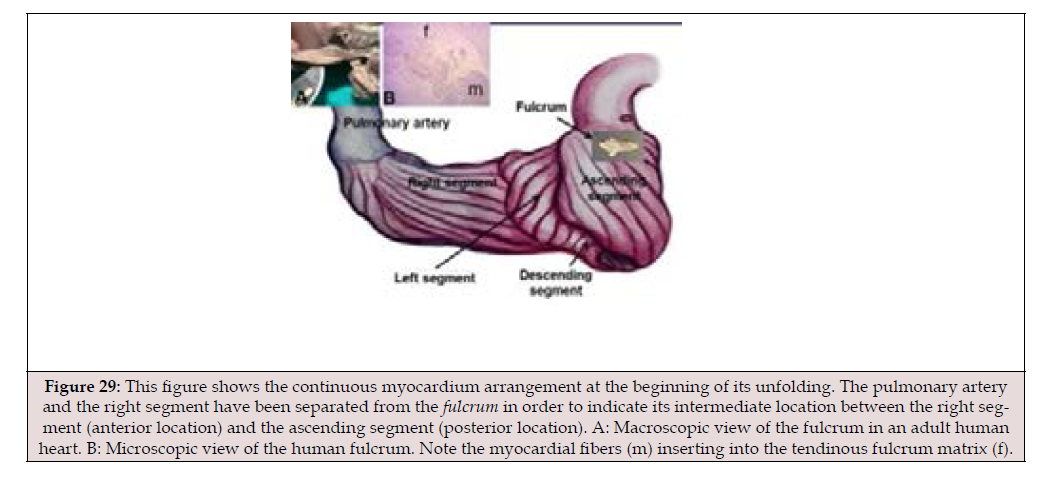
Figure 29:This figure shows the continuous myocardium arrangement at the beginning of its unfolding. The pulmonary artery and the right segment have been separated from the fulcrum in order to indicate its intermediate location between the right segment (anterior location) and the ascending segment (posterior location). A: Macroscopic view of the fulcrum in an adult human heart. B: Microscopic view of the human fulcrum. Note the myocardial fibers (m) inserting into the tendinous fulcrum matrix (f).
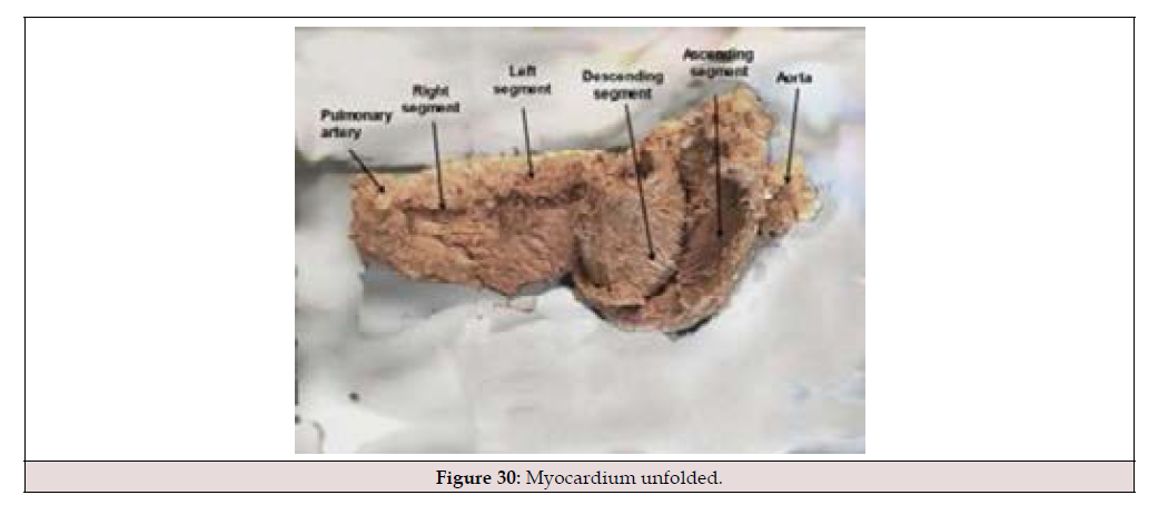
Figure 30:Myocardium unfolded.
Electrocardiographic Correlation: The onset of the QRS complex correlates with the early endocardial depolarization of the anteroseptal wall of the left ventricle. Epicardial depolarization begins approximately 38.2 ms later, according to our research, after the endocardial segment. Corresponding to approximately the final 40% of the QRS complex, coinciding with the positive peak of dP/dt, there is persistent contraction of the ascending segment of the apical loop, as demonstrated in the endoepicardial electrophysiological mapping recordings we performed after aortic valve closure, during the first 80-100 ms of diastole. We have termed this phase the Protodiastolic Phase of Myocardial Contraction (PPMC) due to its energetic activity. It corresponds to the phase classically known as the isovolumetric diastolic phase, which is erroneously considered inactive. Of course, it should be noted that there are small differences between the start and end times of the QRS complex and of the intracavitary and epicardial potentials similar to those recorded in electrophysiological studies.
Since Harvey and later Einthoven, both the mechanical contraction and electrical activation of the heart have been considered almost linear and homogeneous processes. According to the classical concept, normal ventricular activation should begin in the septum, continue at the apex, and end in the basal region. In this way, contraction would occur en bloc during systole, and relaxation would be almost homogeneous during diastole. Systole is synonymous with cardiac contraction, and diastole with relaxation. These have been the classical foundations. At this stage of our understanding, they must be considered more complex, as they were conceptualized without taking into account the helical structure of the continuous myocardium and its sequential dynamics.
There are some aspects of our research that allow for theorizing:
a. While there is no problem in defining the beginning of systole as the moment the mitral valve closes and the beginning of diastole as the moment the aortic valve closes, this in no way implies that the contraction and relaxation processes begin at those precise moments. On the contrary, it is obvious that the ventricular contraction that generates the pressure increase required for mitral valve closure occurs before that point (i.e., during diastole), and the relaxation that reduces intraventricular pressure and allows aortic valve closure begins before that point, i.e., during systole. It is also logical to infer that not all myocardial fibers contract or relax simultaneously, given the constitution of the continuous myocardium in a helix with a sequential movement, but rather that in certain period (impossible to quantify in each individual fiber in its duration) fibers coexist in various stages of contraction and relaxation, both during systole and during diastole.
b. The final state of contraction achieved (and therefore the geometric configuration of the ventricle in general and its cavity in particular) must depend to some extent, perhaps a very significant one, on the contraction or relaxation state of the adjacent fibers and structures. It would seem logical to assume that the first fibers to contract, surrounded by relaxed fibers, can reach their maximum degree of potential contraction; conversely, if the adjacent fibers are already contracted and “pulling” in the opposite direction, their effective degree of contraction would be less.
c. Electromechanical coupling. Although electrical depolarization triggers mechanical contraction, from that moment on these two processes are distinct and autonomous. Membrane activation can occur without mechanical contraction; evidence of this is neuronal electrical activation, which leads to the release of neurotransmitters but not to contractile activity.
We assume that if, for example, a selective inhibitor of actinmyosin activation existed, we would have normal depolarization without muscle contraction. Furthermore, the temporal relationship between electrical activation and mechanical contraction is not linear. It is well known that there is a latency between electrical and mechanical activation. Thus, the time between electrical activation and contraction (at least maximal contraction) likely varies depending on multiple factors: the type of fiber, its metabolic state, its location within the myocardium, the intracellular calcium level, the contraction or relaxation of neighboring fibers, and so on.
Let’s consider typical situations: the QRS complex normally lasts around 100 ms. Systole, plus the PPMC, lasts 400 ms. Therefore, the effect of the QRS complex on total ventricular contraction “lasts” for at least 300 ms after its termination. Fibers activated at the end of the QRS complex contract or maintain their contraction at the beginning of diastole, during the first 100 ms of PPMC. It would not be necessary, therefore, for depolarization potentials to exist within the T wave to justify muscle contractions during diastole (which, as we have already mentioned, is not synonymous with the relaxation of all cardiac fibers).
Given this situation of electromechanical decoupling, there is a crucial point to develop, which is the interrelation between energy (stimulus) and matter (myocardium) with its continuous selforganization process in the production of the sequence of cardiac movements. This is what we have investigated and termed the electromechanical unit.
We reiterate that, during this investigation, in both human and bovine hearts, histological study revealed that the fulcrum (beginning and end of the continuous myocardium) is adjacent to the AV node, defining a synergistic space rich in plexuses with neurofilaments. A fundamental and crucial finding is that the neurofilaments also occupy the cardiac fulcrum in their innervation of the myocardial fibers, constituting an electromechanical unit (Figure 31) [29-31]. We performed a mapping of left ventricular activation on its endocavitary and epicardial surfaces according to the described methodology. The mapping was performed simultaneously with the surface ECG. This provided a unified temporal frame of reference, allowing us to correlate both recordings and obtain a synchronized view of the simultaneous activation observed in various electroanatomical views.
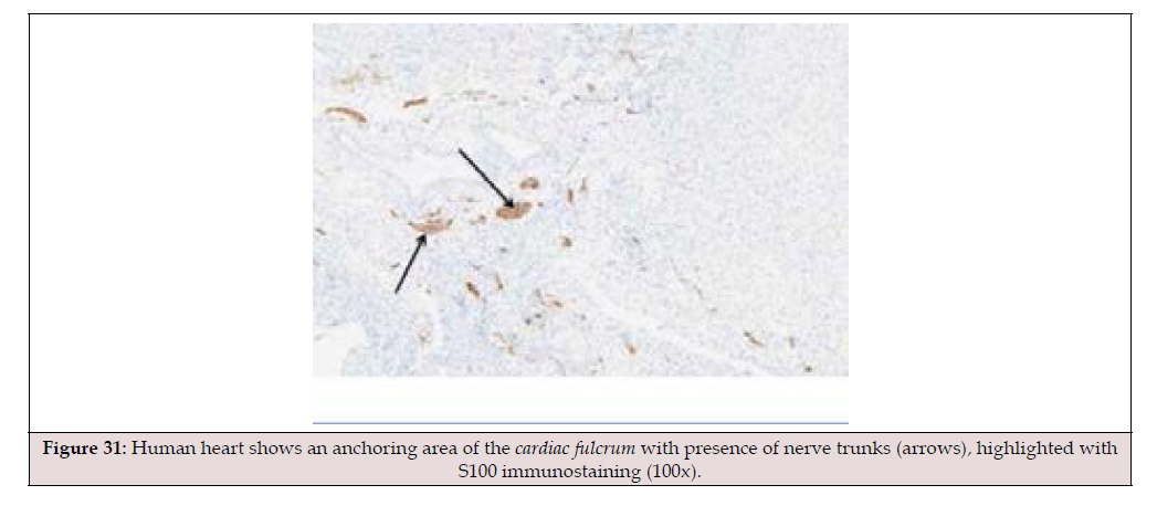
Figure 31:Human heart shows an anchoring area of the cardiac fulcrum with presence of nerve trunks (arrows), highlighted with S100 immunostaining (100x).
This research found a relationship between myocardial stimulation and its mechanical product. The mechanical consequence in the cardiac structure is the initiation of stimulation in the electromechanical anatomical-functional unit between the AV node and the cardiac fulcrum, and its continuation in myocardial activation up to the zone of simultaneous activation with opposing movements between the descending and ascending segments. This generates ventricular myocardial torsion through opposing rotation between the base and the apex, with simultaneous shortening of both ventricles.
At this point in the discussion, the essential question remains: if the QRS complex normally lasts around 100 ms, why does the mechanical contraction (systole + PPMC) last an additional 300 ms? Therefore, the effect of the QRS complex on the total ventricular contraction “persists” for at least those 300 ms after its termination. Given this situation, we must analyze the AV node’s behavior and its relationship to the fulcrum, the site where the continuous myocardium begins and ends after its helical configuration. It can be conjectured that the cardiac fulcrum-Atrioventricular Node (AVN) complex acts as the electro-mechanical self-organizing unit of the cardiac system. The fulcrum is not merely an inert anchor point. Having a central section that acts as a lever and a pole in contact with the AVN, it involves a signal in the heart’s mechanics (the contraction and torsion of the muscular band) that the fulcrum transforms into a pulsatile tilting movement. This suggests that the fulcrum allows the AVN to “feel” the mechanical tension.
Discussion
Interpretation of Myocardial Activation in Relation to the AV Node, Cardiac Fulcrum, and the Torsion Mechanism
We were able to begin this path of analyzing the selforganization of the helical myocardium with the knowledge of the morphological, histological, and functional relationship between the AV node and the cardiac fulcrum, the latter being the site where the myocardium originates and terminates after its helical course [1,2]. For a proper understanding of the heart as a participant in the quantum concepts of current science, it seems appropriate to first explain the interpretation of myocardial activation in relation to the cardiac fulcrum and the torsion mechanism (Figure 2). In this sense, the words of León Rosenfeld are relevant: “a theory is understood through its limits” [32].
The heart has historically been studied in terms of its individual components, exhibiting a global and homogeneous contraction, simultaneous throughout its muscular structure; but not with a clear understanding that the movements in its different phases occur sequentially and overlappingly, contributing to its complex function in an infinitesimal time of less than one second per cycle and 100,000 cycles per day. The heart has been studied only partially, undoubtedly because attributing the complexity of its function to the duration of its cycle implies the difficulty of observing the reality that unfolds within it. Due to the rapidity of cardiac movement, Girolamo Fracastoro (Verona, 1478-1553) had expressed the impossibility of understanding its mechanism. It is understandable to consider that the integrity proposed by the general systems theory will remain relegated as long as adherence to purely analytical models continue, as the whole is reduced to the sole study of its parts.
Our previous publications concerning the cardiac fulcrum as a support for the myocardium, its relationship with the Aschoff- Tawara node, and the sequential activation of the heart in a clear organizational arrangement to achieve torsional movement [1,2], led us to analyze these structures in terms of their potential and functional aspects [11]. Thus, we arrived at the concept that the heart acts through three fundamental units: activation, electromechanical unit, and myocardial torsion, which are interrelated in their anatomy and organization to act in accordance with a highly efficient system (Figure 2). In summary, the research results reveal the key reference points that define the organizational pattern of cardiac function. These are: the anatomical contiguity between the AV node and the cardiac fulcrum; the continuous presence of the filaments that structure the AV node with the fulcrum-where the myocardium originates-clearly forming an electromechanical unit; and the pathway of activation through the myocardium that enables helical torsion, achieved through the anatomical, anisotropic, and functional spatial arrangement between the descending and ascending segments at the septal level, as confirmed by echocardiographic and electrophysiological studies. At this point, we must analyze how these elements, including structures and circuits of energy and mechanical transfer, interact.
The analysis is deeply correlated with myocardial movements and the stimulation that travels through its segments, according to the electrophysiological studies we have carried out [1,2]. The interpretation of the anatomical relationships between the cardiac fulcrum and the AV node implies the complementarity of the anatomy with the physiology of the continuous helical myocardium, since their contiguity is found at the site where stimulation begins and ends, with the production of the mechanical action of torsion and detorsion in the systolic and suction phases of the ventricles as the different segments of the myocardium are activated.
Myocardial Torsion
Although various aspects of the propagation of the electrical stimulus through the ventricles have been known for a long time, the advent of three-dimensional navigation systems and electroanatomical mapping has allowed a much more detailed study of it in the human heart in totally physiological clinical situations.
As previously mentioned, left ventricular activation begins in the descending endocardial segment, which depolarizes longitudinally and transversely with a 12.4 ms delay relative to the baseline loop (right and left segments). At the point of contact with the ascending segment, activation propagates from the endocardium to the epicardium (transverse propagation), that is, from the descending to the ascending segment, with a 25.4 ms delay, thus totaling 38.2 ms from the start of cardiac stimulation. From this point, the ascending segment depolarizes in both directions: toward the apex and toward the base, while the descending segment completes its activation toward the apex (Figures 21, 22). Thus, as we have analyzed, two primary phenomena occur:
• The apical loop, upon depolarization from the crossing
of the segments, with two simultaneouswavefronts (from the
descending and ascending segments), generates a synchronized
contraction of both.
• Activation of the ascending segment occurs from
the crossing in two opposing directions: toward the apex and
toward the base. The resulting mechanical contraction will also
have an opposing direction, giving rise to clockwise (apex) and
counterclockwise (base) rotations.
In a cardiac cross-section (Figure 32) below the atrioventricular valves, we can observe that the descending segment is located internally, surrounded by the ascending segment on the free wall of the left ventricle [33]. The ascending and descending segments move in opposite directions, both during systole and diastole, to achieve ventricular ejection and suction, generating friction between their surfaces. This topic has been addressed in other publications, where we found abundant amounts of hyaluronic acid, a lubricating substance, in both the myocardium and the Thebesian vessels in studies conducted on anurans, cattle, and humans (Figures 32-35) [34,35]. The histology in the inset of (Figure 32) clearly shows the different orientation of the fibers of the descending segment in relation to the ascending segment, which explains their opposing movements. The arrangement of myocardial fibers in the epicardium and endocardium, with a 180° change in angle, results in the epicardial fibers being oriented in the opposite direction to the endocardial fibers. Given the different anisotropic orientations of the fibers, this area corresponds to the beginning of the opposing movement that produces myocardial torsion. The structure of the septum, with the contiguity between the descending and ascending segments, allows for continuous activation between them with opposing movements and the consequent myocardial torsion (Figure 36). The interventricular septum plays a predominant role in myocardial function, given its essential position in biventricular interdependence [36].
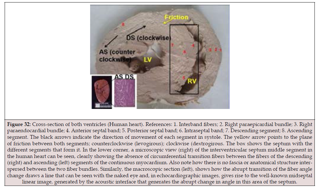
Figure 32:Cross-section of both ventricles (Human heart). References: 1. Interband fibers; 2. Right paraepicardial bundle; 3. Right paraendocardial bundle; 4. Anterior septal band; 5. Posterior septal band; 6. Intraseptal band; 7. Descending segment; 8. Ascending segment. The black arrows indicate the direction of movement of each segment in systole. The yellow arrow points to the plane of friction between both segments; counterclockwise (levogirous); clockwise (dextrogirous. The box shows the septum with the different segments that form it. In the lower corner, a microscopic view (right) of the interventricular septum middle segment in the human heart can be seen, clearly showing the absence of circumferential transition fibers between the fibers of the descending (right) and ascending (left) segments of the continuous myocardium. Also note how there is no fascia or anatomical structure interspersed between the two fiber bundles. Similarly, the macroscopic section (left), shows how the abrupt transition of the fiber angle change draws a line that can be seen with the naked eye and, in echocardiographic img, gives rise to the well-known midseptal linear image, generated by the acoustic interface that generates the abrupt change in angle in this area of the septum.
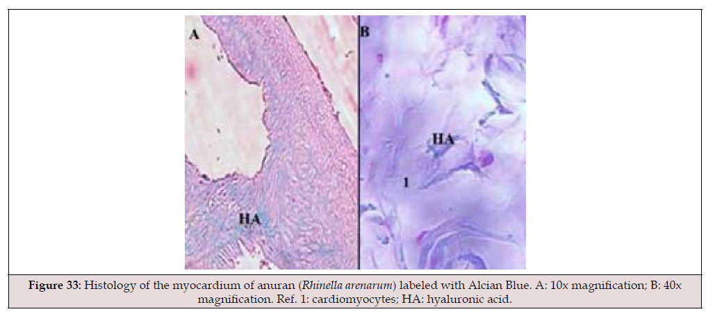
Figure 33:Histology of the myocardium of anuran (Rhinella arenarum) labeled with Alcian Blue. A: 10x magnification; B: 40x magnification. Ref. 1: cardiomyocytes; HA: hyaluronic acid..
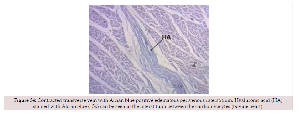
Figure 34:Contracted transverse vein with Alcian blue positive edematous perivenous interstitium. Hyaluronic acid (HA) stained with Alcian blue (15x) can be seen in the interstitium between the cardiomyocytes (bovine heart).
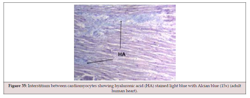
Figure 35:Interstitium between cardiomyocytes showing hyaluronic acid (HA) stained light blue with Alcian blue (15x) (adult human heart).
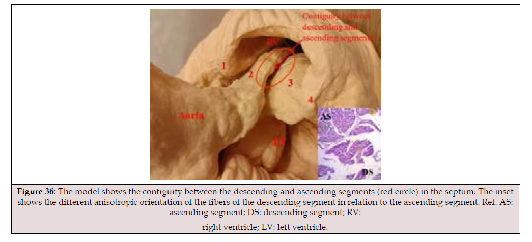
Figure 36:The model shows the contiguity between the descending and ascending segments (red circle) in the septum. The inset shows the different anisotropic orientation of the fibers of the descending segment in relation to the ascending segment. Ref. AS: ascending segment; DS: descending segment; RV: right ventricle; LV: left ventricle.
The research on the stimulation sequence allowed for the determination of electrophysiological propagation in the continuous myocardium and also led to deductions regarding ventricular torsion and the suction effect in lef ventricle Protodiastolic Phase of Myocardial Contraction [37]. The orientation of the fibers in the continuous myocardium and their activation involve a concatenation of muscle movements in cardiac mechanics. These movements occur in four phases: constriction, shortening-torsion, lengthening-detorsion, and widening, which allows it to perform its functions of systole, suction, and diastole. The fundamental movements in which the different segments of the continuous myocardium participate during systole and suction are shown in (Table 4).

Table 4:Segment function. Reference. PPMC: Protodiastolic Phase Miocardial Contraction.
This sequential activation correlates with well-established fundamental phenomena, such as the opposing clockwise and counterclockwise torsion of the apex and base of the left ventricle, responsible for its mechanical efficiency (Video 2). In order to attempt to explain the mechanism of this muscular torsion, this research aimed to analyze the sequence of ventricular electrical activation using simultaneous endo-epicardial band MET of the segments from their origin at the anatomical and functional contiguity between the AV node and the cardiac fulcrum.
During the narrowing phase, there is a sequential contraction of the right (free wall of the right ventricle) and left (edge of the mitral orifice) segments, which together form the basal loop. According to Armour (1970) [38], this contraction forms an external covering within which the apical loop will contract. In reality, the crescentshaped free wall of the right ventricle is located laterally to the rest of the ventricular mass (septum and left ventricle), since the left segment forms part of the posterior epicardial wall of the left ventricle in its superior portion, encircling the mitral annulus, while the remainder is covered externally by the ascending segment. In this layer (basal loop), stimulation progresses from the subepicardium to the subendocardium. This stimulation then extends to the descending segment, and at an average of 25.8 ms in our study, the ascending segment is activated. The termination of stimulation in the myocardium occurs at the terminal portion of the ascending segment, near its insertion into the cardiac fulcrum, during the first 80-100 ms of diastole, in the period traditionally known as the diastolic isovolumetric phase, which we have more accurately termed PPMC [1,2].
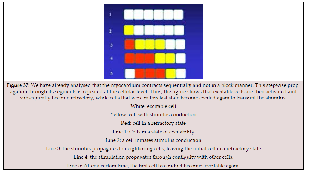
Figure 37:We have already analysed that the myocardium contracts sequentially and not in a block manner. This stepwise propagation
through its segments is repeated at the cellular level. Thus, the figure shows that excitable cells are then activated and
subsequently become refractory, while cells that were in this last state become excited again to transmit the stimulus.
White: excitable cell
Yellow: cell with stimulus conduction
Red: cell in a refractory state
Line 1: Cells in a state of excitability
Line 2: a cell initiates stimulus conduction
Line 3: the stimulus propagates to neighboring cells, leaving the initial cell in a refractory state
Line 4: the stimulation propagates through contiguity with other cells.
Line 5: After a certain time, the first cell to conduct becomes excitable again.
Extensive knowledge of the helical myocardium explains that cardiac movements are sequential [39]. Similarly, cellular stimulation is illustrated in the diagram in (Figure 37). In the myocardium, actin and myosin are fundamental structural and contractile proteins in muscles, organized into sarcomeres to produce movement. Myosin (thick filament) uses energy from ATP to bind to and slide along actin (thin filament), causing muscle contraction and shortening the sarcomere. Tropomyosin and troponin are key regulatory proteins in the thin filaments of skeletal and cardiac muscle. Together they control contraction: tropomyosin blocks myosin from binding to actin at rest, while troponin, by binding to calcium, displaces tropomyosin to allow muscle contraction. The MET study explains the torsional phase of the heart, defined as the opposing rotational movement of the base and apex. Activation, at the point where the descending and ascending segments cross, propagates from the endocardium to the epicardium (transverse propagation), that is, from the descending to the ascending segment.
While there is a progression of electrical conduction along the continuous myocardium, this isolated activation does not explain the generation of a force capable of ejecting ventricular contents at a speed of 200 cm/s with low energy expenditure. This research found that transverse propagation from the descending to the ascending segment plays a fundamental role in ventricular torsion, allowing opposing forces on its longitudinal axis to generate the intraventricular pressure necessary for the abrupt expulsion of blood. This would produce a torsion mechanism similar to “wringing out a towel,” as described by Giovanni Borelli and Richard Lower [40].
Cardiac Self-Organization
At this stage of the research, we understood that the threedimensional spatial helix of the heart, in relation to a fourth dimension-the time of its cycle-implied that the relationship of the cardiac fulcrum with stimulation and the consequent myocardial torsion played an organizing role, as energy returns to that site after completing an 800 ms cycle that is repeated 100,000 times daily. During this journey through the myocardium, stimulation not only provides energy that is transformed into work and heat, but also accumulates information (negentropy). In this respect, entropy, not energy, is what allows us to evaluate the cost of functional existence. This recurrent journey to the electromechanical unit allows it to accumulate information to reconfigure the next cycle, given the interrelationship between the cardiac fulcrum and the AV node. This situation allows it to optimize data and deliver to the electromechanical unit the information and energy required for the efficiency of the next pulse [41-43]. En este aspecto hay que correlacionar el carácter de atractor del fulcro cardíaco en la continuidad de la contracción miocárdica, o sea luego del cierre de la válvula aórtica durante la PPMC, con la posibilidad de acumular información para el siguiente ciclo.
The sensory organs in mammals filter an enormous amount of information from the outside world, which implies an organizational reality provided by the nervous system. This represents a mass of information that serves daily life. It also constitutes a factor of selective pressure [44] on biological behavior and also on human thought.
A fundamental point in this analysis is the interconnection between the AV node and the cardiac fulcrum. The latter acts as an attractor of the stimulus, which travels along the same myocardial circuit, originating and terminating in the myocardial fibers that insert into the fulcrum, the electromechanical unit. This concept is based on Henri Poincaré’s (1854-1912) recurrence theorem, which states that every dynamic system will eventually return to its initial state. This characteristic is found in stable systems, as it is invalid in unstable ones. The attractor is a point in the helical myocardium with minimal entropy production. It is worth asking whether this periodicity transmits information that originates and returns to the heart in variables such as amplitude, voltage, and frequency. The pulse width defines the energy transmitted to the muscle fiber. The wider the pulse, the more energy. This is called Pulse Width Modulation.
Cardiac conduction can be represented as a self-generating dynamic system, since cardiac automaticity is the heart’s intrinsic capacity to produce rhythmic electrical impulses, but it also operates in a regime open to external influences and undergoes a process of self-organization. Information theory is useful here, as it is characterized by quantifying the information content of a sequence of biomolecules.
AV Node
The AV node receives the energy destined to stimulate the myocardial muscle fibers, as well as the control signals derived from this energy. The AV node receives a constant level of energy, which is used to excite the muscle fibers via neurotransmitters. To generate the output impulses from the AV node that reach the muscle fibers, control pulses are required, which respond to the Central Autonomic Nervous System (Figure 38).
Each subsystem owns its own process and can modify it, positively impacting it and thus reducing the disorder inherent in any activity or process. Shannon’s information theory [28] is applicable here (Figure 38):
S = k (Pa Log2 (1/Pa) + Pb Log2 (1/Pb) + Pc Log2 (1/Pc) + Pd Log2 (1/Pd))
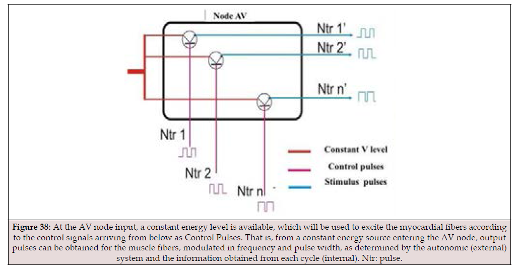
Figure 38:At the AV node input, a constant energy level is available, which will be used to excite the myocardial fibers according to the control signals arriving from below as Control Pulses. That is, from a constant energy source entering the AV node, output pulses can be obtained for the muscle fibers, modulated in frequency and pulse width, as determined by the autonomic (external) system and the information obtained from each cycle (internal). Ntr: pulse.
Where Pa, Pb, Pc, and Pd are the probabilities of occurrence of events with decreasing qualities, with “a” being the optimal quality and “d” the inferior one. If the standard allows for the existence of all four probabilities, the resulting S1 will be greater than the S2 that would result from eliminating the worst qualities, leaving only “a”.
In the case of the heart’s self-organization, the local subsystem (AV node) appears to be both cause and effect, a situation ensured by the cyclical feedback that allows it to reorganize and improve its management according to the body’s needs. This process incorporates information and energy (negentropy). In other words, from a constant energy source entering the AV node, output pulses can be generated to the muscle fibers, modulated in frequency and pulse width, as determined by the autonomic nervous system (external) and the information obtained from each cardiac cycle (internal).
In myocardial stimulation, there is a sequential property of the myocardium, which is repetitive. This creates a link between this property of excitation, frequency, and energy, resulting in the continuous events of cardiac cycle stimulation. In complex organisms, there is the possibility of information selection [45]. This concept implies what would be a considerable challenge: verifying the energy (or voltage) levels that reach the AV node. It also involves verifying the control pulse trains and, consequently, the stimulus pulse trains.
The electromechanical unit would function by controlling the current flow between its terminals (emitter, base, and collector) in a manner analogous to a transistor (an invention of physicists John Bardeen, Walter Brattain, and William Shockley at Bell Laboratories in December 1947). In this way, the electromechanical unit would have a programming plan for the next cardiac cycle, receiving information from external sources (the nervous system) as well as from its own myocardial function (voltage, amplitude, and frequency) in a self-organizing process. This concept would explain the fundamental question posed earlier in this article: if the QRS complex normally lasts around 100 ms, why does the mechanical contraction of the myocardium reach 400 ms? The AV node would have an organizing pattern for the next cycle, which would be modified by receiving information from outside the heart, but also derived from its own functioning through a self-organizing process. Recall that the cardiac coil returns with its stimulation to the same electromechanical unit from which it originated. This would explain the different duration of the electrocardiographic QRS in relation to the mechanical contraction.
The physiology of myocardial stimulation, with its consequent torsion-detorsion, implies recognizing the AV node and fulcrum as subject to negentropy, which is the process of creating and maintaining order within a system, acting as the opposite of entropy, the natural tendency toward disorder. It manifests as the capacity of systems (natural, living, or even social and economic) to organize, innovate, and self-regulate, using energy or information from the outside to maintain stability and complexity.
The AV node, interconnected with the cardiac fulcrum, acts as a subsystem receiving information from the central nervous system (external) and from the myocardium itself (internal). This information allows for negentropy, maintaining the organizational system of cardiac stimulation from equilibrium and preventing it from reaching a final equilibrium. This anti-entropic state, or negentropy, is fueled by external input, that is, by the exchange with the environment, and by the information produced by the temporal pathway of the stimulus through the continuous myocardium. This pathway, when directed to an attractor (electromechanical unit), allows for the reorganization of the system. It is important to remember that the intrinsic activity of the system is the determining factor in its relationship with the environment.
Faraday and Maxwell potentials are important in these fields. In the heart, there are energy fields resulting from vortical electrical flows, which are coupled to information fields [40]. Studies have confirmed the interrelationship between ECG patterns and the brain (Rein’s resonance hypothesis). The heart generates energy as described by Russek and Schwartz (1996). This energy is coupled to the information field. In this way, the heart and brain process information [13,46]. It is essential to understand a key phrase uttered by Erwin Schrödinger (1887-1961): “We must go to the small.” These words summarize the essence of the contribution of quantum mechanics. Schrödinger understood that to comprehend the true nature of matter, it is not enough to observe the macroscopic world; it is necessary to delve into the behavior of atoms and subatomic particles. The metabolism of living beings involves the simultaneous construction of biological molecules and the breakdown of other molecules.
These concepts are consistent with that of syntropy, investigated by Luigi Fantappiè, understood as a measure of the degree of internal organization of any system composed of interacting components. In this concept, he details that phenomena produced by past causes adhere to the second law of thermodynamics. Therefore, energy dissipates, order is lost, and structures are destroyed, while future phenomena (syntropy) tend toward the reorganization of the system. Syntropy was referred to in 1974 as “negative entropy” (negentropy) by Albert Szent- Györgyi. According to these developments, the heart appears to have the property of connecting the organism to itself and to its environment. In 1959, Léon Brillouin combined Shannon’s informational entropy with Boltzmann’s statistical entropy, stating that information is negentropic and can cancel entropy. This has applications in molecular biology and thermodynamic processes.
In the cardiac cycle, adaptation to physiological needs, through a continuously monitored program, allows for the surveillance and function of the system. To clarify: with a cycle duration of less than one second, without this predictable pattern of behaviour through the QRS complex, the system would be subject to instability given its frequency of 100,000 cycles/day with a duration of 800 ms. This frequency data indicates that it lacks the time available to readjust its program. The nervous system influences the activity of various organs (digestive system, skin, etc.), but unlike other systems, such as the nervous system, which can have a prolonged pause before mounting the necessary response, the heart, with its limited time frame, would place the organism at risk of death if it did not have a rapid response. Each heartbeat is an event, and in this event, time is the driving force. Thus, we can speak of the equation: myocardial activation (three-dimensional)–time (understood as a fourth dimension). This equation gives relevance to the event of our present.
This situation was noted by William Harvey (1578-1657), who recounts the difficulties in proving his theory of blood circulation: “I came to think, [he says] with Fracastoro, [Renaissance epidemiologist], that the movement of the heart could only be known by God” [47]. Given this high frequency and energy output, the cardiac system constantly receives feedback, modifying its pre-contraction program through the electromechanical unit (Cardiac Fulcrum + AV Node). This unit gathers information from its operating circuit, originating and terminating there, and also receives the influence of other bodily systems. The evolution of biology through its significant stages has determined the need for continuous information processing by organisms. This continuous adaptation throughout the life of the cardiac system to fulfill its function explains the difference in duration between the QRS complex (100 ms), the system’s program, and the myocardial contraction, its consequence, systole + PPMC, lasting 400 ms, as well as the activation-time equation.
The ultimate goal of medical knowledge is to understand disease processes. In this respect, and in relation to the research presented here, Takotsubo cardiomyopathy is a condition that highlights the relationship between emotions and cardiac function. It is also known as “stress cardiomyopathy,” “transient apical dysfunction,” or “broken heart syndrome.” It is usually triggered after an episode of intense and unexpected emotional stress, which causes an excessive release of catecholamines (adrenaline, noradrenaline, and dopamine) and leads to an acute coronary syndrome with STsegment elevation, but without evidence of obstructive coronary lesions or coronary artery spasm. Transthoracic echocardiography reveals apical akinesis of the left ventricle with basal hypercontractility (apical ballooning). These transient regional alterations in ventricular wall motion completely resolve within a few days. Cardiac catheterization confirms the absence of coronary artery disease. The explanation for this syndrome is based on the role of catecholamines produced by unexpected stress or trauma. These catecholamines play a role in the interaction between the brain’s cognitive centers and the hypothalamic-pituitary-adrenal axis in the stress response. The main effects of their increase include elevated heart rate, hypertension, and increased cardiac output due to the effects of epinephrine and norepinephrine, greater blood flow to vital organs, increased release of glucose and fatty acids for rapid energy, increased muscle strength, and enhanced mental acuity. This type of response is a physiological response to physical or emotional stress that produces a kind of “fight or flight” response. Although sympathetic stimulation appears to be the cause of excess catecholamines, the mechanism by which myocardial muscle dysfunction occurs is not fully understood (89,90). Ultimately, the aim is to make the nature of this response understandable. The question this synopsis raises is: what role does the electromechanical unit play in this condition of emotional origin?
The interaction between the cardiac fulcrum and the AV node (electromechanical unit) in relation to the double-helix muscular circuit of the myocardium, which begins and ends at this fulcrum, is likened to a system composed of basic elements with an organization based on the reception, feedback, and transmission of information. Allegorically, we can say: stimulation tells the myocardium how to twist, and the myocardium informs the stimulation how to form the next pulse. This collected information has the capacity for negentropic regulation, since it reduces the amount of uncertainty and degradation (entropy), allowing the system to self-organize in making decisions for the next cycle. This is because different alternatives may arise due to physiological needs, understanding that entropy is the price of the material development of the universe.
It is necessary to incorporate the advances of current quantum physics into the study of living beings. This step should not be seen as esoteric or mystical, but rather as the use of information held by other sciences in order to achieve negentropy, thus advancing biological knowledge. The path of depolarization energy in our research leads to the understanding that the stimulation circuit is completed at its origin, in the electromechanical unit. We believe that these contributions, based on the electromechanical unit, design it as an attractor of the helical convolution that establishes the path of the myocardium. The myocardium, being the only muscle that originates and terminates at the same point (cardiac fulcrum), analogous to a Möbius strip, combined with the understanding of the QRS complex as a continuous information pattern for achieving sequential cardiac mechanics through the negentropic effect, and the evidence of the time factor as a fundamental element in a four-dimensional myocardium, are essential for understanding cardiac physiology. Without this adherence to the timing factor in each cardiac cycle, life would not be possible, this characteristic being unique in its brevity within the organs of the human body. Undoubtedly, the heart is subject to the rigor of time as a fundamental element of its continuous function, since any delay in its cycle carries the possibility of imminent death.
These ongoing investigations may lead to advancements in our understanding of the circulatory system. The goal is to achieve knowledge that gives meaning to the problems we observe. This path requires boldness and passion to reach that conjunction between knowledge and meaning that is the essence of the creative act. Sometimes unexpected phenomena are discovered in response to sudden questions, as was the case with the discovery of the heart’s support, which we have called the cardiac fulcrum. These discoveries challenge the imagination and push boundaries; therefore, we must be cautious, as premature conclusions about a piece of knowledge often distance us from the truth. The development of medical biology has not only involved hypotheses and research but has also fostered ideological judgments [48]. Thus, it has not ceased to influence the landscape of politics, religion, and ethics.
The myocardium can be defined as a single muscle that, along its longitudinal axis, adopts a spiral spatial configuration, inserted at its ends (origin and termination) into an osteochondroidtendinous core, according to the specimens analyzed, called the cardiac fulcrum. This arrangement delimits the two ventricular chambers. There is solid evidence supporting the concept of the myocardium as a single, continuous, and spiral muscle:
1) Muscle homogenization masks the actual spiral continuity of the fibers by overlapping their segments to achieve a helical conformation. This implies considering that its compact structure, folded into a helix, is functionally required in birds and mammals to allow blood to be ejected at high speed within a limited time by an organ that must supply two circulations (systemic and pulmonary). Anatomical investigation of the heart through appropriate dissection, histological exploration, analysis of img obtained through radiological and echocardiographic analysis, electrophysiological studies carried out with three-dimensional electroanatomical mapping, and cardiac diffusion tensor imaging show the continuous muscular pathway that circumscribes the two ventricles [46].
2) The ability to unfold the myocardium and obtain a similar thickness throughout the muscle demonstrates its continuity. When folded, the thickness of the right ventricle is found to be less than that of the left ventricle, since the former is composed of a single segment (right), while the latter has two overlapping segments (descending and ascending).
3) Giovanni Borelli (1608-1679), the father of biomechanics, described myocardial torsion by comparing it to the wringing of a cloth. In 1669, Richard Lower, in his “Tractatus de corde: item motu et colore sanguini, et chyli in eum transitu” (20,21), reaffirmed that the myocardium was composed of two coiled muscles and that its blood ejection was similar to “wringing a linen cloth to squeeze out the water.” Myocardial torsion was subsequently studied in mice by Henson [49] and later confirmed in humans through both electrophysiological studies and imaging (echocardiography, resonance) [40]. The heart achieves the expulsion of its contents by torsion of its walls and begins filling through detorsion. The synchronous torsional movement with ventricular longitudinal shortening can be explained through the helical arrangement and continuity of the cardiac muscle [50].
4) The trigones show no cardiomyocyte insertion, confirming that the only attachment point of the myocardium is the fulcrum.
5) Myocardial dissection, histological analysis, and cardiac function do not correlate with a mesh-like structure.
6) The proximity of the cardiac fulcrum to the AV node, surrounded by a rich plexus of neurofilaments, leads us to consider the anatomical structure of an electromechanical unit involving stimulation energy and muscle mechanics.
In this research, fresh bovine, porcine, and human hearts were used to obtain detailed descriptions in order to elucidate the true spatial architecture of the myocardium.
The Three Functional Units of the Heart
During this research, in both human and other species hearts, histological studies revealed that the fulcrum (the beginning and end of the continuous myocardium) is adjacent to the AV node, creating a space rich in plexuses with interconnected neurofilaments between the two structures. The crucial finding of this research is that these neurofilaments invade the myocardium originating at the cardiac fulcrum, thus forming an electromechanical unit. We performed a mapping of left ventricular activation on its endo cavitary and epicardial surfaces according to the described methodology. The mapping was performed simultaneously with the surface ECG. This provided a unified temporal frame of reference, allowing us to correlate both recordings and obtain a synchronized view of the simultaneous activation observed in various electro anatomical views.
This research established the structural relationship between myocardial stimulation and its mechanical product, based on the assumption that this electromechanical unit is undergoing a continuous process of self-organization. The mechanical consequence of the initiation of stimulation in the anatomical and functional unit between the AV node and the cardiac fulcrum, and the continuity of myocardial activation to the anisotropic zone at the contiguity between the descending and ascending segments, is what generates, through sequential opposition of movements, the torsion of the left ventricular myocardium by opposing rotation between the base and the apex with simultaneous shortening of both ventricles.
It is important to note that medical practice requires a dialogue between theory and experience [51]. The compilation of data collected in this research begins to show its influence on clinical disorders, such as the analysis performed on Takotsubo syndrome.
In summary, cardiac movements follow three units (Figure 2):
a. Energy Unit: Composed of the AV node.
b. Electromechanical Unit: Located at the cardiac fulcrum
where the neurofilaments interact with the myocardium.
c. Torsion/detorsion Unit: Occurs at the level where
energy is transferred from the descending to the ascending
segment, achieving myocardial torsion (systole) and subsequent
detorsion that allows ventricular suction.
The myocardium is an integrable system, consistent with electromagnetic resonance, in a continuous feedback loop within a time limit for successive phases of contraction and expansion. There is a stimulation plan to achieve myocardial activation. It’s important to remember that in humans, a programmed cell begins its beat at three weeks of gestation, giving rise to the cardiac mantle. Similarly, in cardiac arrest due to surgery with cardiopulmonary bypass or accidentally, an external electrical shock restarts it. This programming would have as its epicenter the electromechanical unit (AV node + cardiac fulcrum) with a continuous development of muscular contraction in the myocardium, which, with the information gathered (frequency, voltage, amplitude), allows for the next pulse. This characteristic explains the different duration between the QRS complex (100 ms) and the myocardial contraction (systole + PPMC, 400 ms). Without this self-organization, the cardiac cycle would be subject to instability given the short duration of the cardiac cycle of 800 ms.
Conclusions
In summary, based on the above, we found that the orientation and opposing rotational movement of the fibers in the heart, both at the base and the apex, determine the anatomical and functional model of the continuous helical myocardium. However, the question that arises from the logic of movement is whether, in order to achieve torsion and subsequent detorsion, the muscular segments that continuously form the ventricular chambers should rotate on a fulcrum, just as a skeletal muscle does on a firm insertion. This fulcrum was found in our research and named the cardiac fulcrum.
The aortic annulus is not continuous. In a section of its circumference, located between the ends of the trigones, it is interrupted, and it is here that the anterior leaflet of the mitral valve inserts. The pulmotricuspid cord is located anterior to a U-shaped area that surrounds the anterior two-thirds of the aortic annulus, the open (posterior) end of which is occupied by the anterior leaflet of the mitral valve. At its ends, this tissue has two trigones. The right fibrous trigone (central fibrous body) is the more prominent and is located between the tricuspid (right) orifice, the aortic (posterior) orifice, and the pulmotricuspid cord (anterior). The left trigone, less prominent, is located between the mitral (left) orifice and the aorta. There is no connective tissue at the point where the aortic orifice meets the posterior mitral valve leaflet. Laterally, the two fibrous bodies continue as a connective tissue band that partially surrounds the mitral valve orifice before gradually fading away. The septal mitral valve leaflet is located between the two trigones like a wedge and can be considered an extension of the atrial endocardium.
With the heart folded, we find the fulcrum embraced by the pulmotricuspid cord and the pulmonary artery, located on the left side of the aorta. The right ventricle is positioned anteriorly; therefore, the fulcrum shows how the bundles emerging from it, being frontal, obscure the attachment of the ascending segment to this structure located below this view. When the pulmonary artery and the pulmotricuspid cord begin to unfold, the insertion of this ascending segment at the fulcrum becomes evident.
For three centuries, it was considered that myocardial fibers insert into the fibrous skeleton of the heart. The development of anatomy presented around 1970 by Torrent Guasp (3,13) suggested that the myocardium begins and ends at the base of the great vessels, but that the fibers do not anchor to the atrioventricular rings; rather, they simply adhere to them, as confirmed by diffusion tensor magnetic resonance imaging. Our research has demonstrated, along the septal segment of the aortic annulus extending from the left to the right trigone, a structure we have termed the cardiac fulcrum (below the origin of the right coronary artery), where the continuous myocardium attaches at its beginning and end, as it, like any muscle, requires support to perform its function.
Furthermore, we have not found any insertion of cardiomyocytes into the collagen matrix of the trigones, a finding corroborated by histological studies. In all studies of animal and human hearts, the location of the fulcrum has been found to be contiguous with, but different from, the classic fibrous core. The right and left trigones occupy the non-coronary sinus, the posterior half of the left coronary sinus, and the posterolateral portion of the right coronary sinus. The fulcrum is located anteriorly, below the right coronary artery (Figure 39).
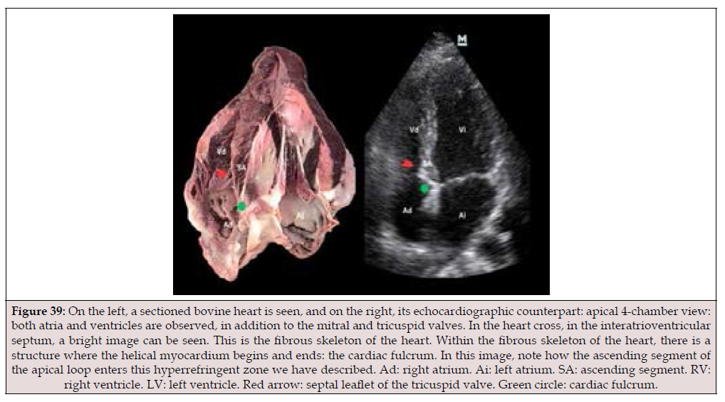
Figure 39:On the left, a sectioned bovine heart is seen, and on the right, its echocardiographic counterpart: apical 4-chamber view: both atria and ventricles are observed, in addition to the mitral and tricuspid valves. In the heart cross, in the interatrioventricular septum, a bright image can be seen. This is the fibrous skeleton of the heart. Within the fibrous skeleton of the heart, there is a structure where the helical myocardium begins and ends: the cardiac fulcrum. In this image, note how the ascending segment of the apical loop enters this hyperrefringent zone we have described. Ad: right atrium. Ai: left atrium. SA: ascending segment. RV: right ventricle. LV: left ventricle. Red arrow: septal leaflet of the tricuspid valve. Green circle: cardiac fulcrum.
Torrent Guasp believed that the myocardium lacks a fixed point of support like those found in the muscular system, which are essential for generating force. He analyzed that it would function similarly to the circular muscles of the arteries, relying on the contents of the cavity itself (the hemoskeleton). In our research, we consistently maintained that the myocardium must have a fixed support structure that allows it to rotate helically, enabling its movements with sufficient muscular power to perform the fundamental actions of shortening-torsion and lengtheningdetorsion. This investigation into a continuous support structure in the myocardium is related to the concept of an organic machine, such as the heart, which, without a solid attachment to a resistant core, would lack the mechanical capabilities necessary for its considerable power. Regarding the autochthonous muscle bundles that constitute the left ventricle, the ascending segment terminates at the base of the aorta. Recall that the descending segment lies continuously between the left and ascending segments, without any attachment to the fulcrum. Before attaching to the fulcrum, the ascending segment gathers into a bundle whose more external fibers curve to reach this attachment point. The more internal fibers enter directly without any slant. The cardiac fulcrum provides a solid point of attachment for this segment to the structure.
The musculature that conforms to the current ventricle corresponds to the start of the continuous myocardium (current segment) that originates in the cardiac fulcrum [52]. To diverge the segment afterward into the main groups of fibers, forming the paraepic and paraendocardial joints, a line was delimited between the so-called pulmotricuspídeo cord, ubicado between the pulmonary artery and the tricuspid valve.
a) This point of attachment, as in any muscle, serves as a support for the muscular lever and also acts as a cushion, preventing the ventricular rotational force, whether due to torque or torsional stress, from being transferred to the aorta. This dissipates the energy produced by the movement of the muscular helix and prevents strangulation of the artery or kinking during the systolic ejection period (40). In the human hearts studied, the findings are surprising from an interpretive standpoint, given that it is logical to consider its presence throughout the evolutionary chain of mammals. It should be noted that this structure, whenanalyzed in different specimens, shares the common function of supporting the helical myocardium to generate the power required by any muscle, which varies among different mammals. Therefore, its presence is constant in all the hearts studied, both bovine and human, but its structural characteristics differ. And this difference in the intimate analysis of the cardiac fulcrum is undoubtedly related to the resistance it must offer to the energetic action of the myocardium in hearts of different sizes.
b) It is important to consider that what makes the fulcrum (support) concept significant is the network between the myocardial fibers and the chondroma. It is the functional element that gives value to this structure, which supports and stabilizes myocardial movements.
c) The fulcrum should not be considered a nucleus with sharp, rigid edges. The insertion of the myocardium into such a structure would be undesirable, as it would generate a sudden tension at that point during movement, potentially causing tears at the insertion, given the force exerted by the myocardial band to eject the ventricular contents. Furthermore, its consistency decreases between cattle and humans because the force exerted differs due to body weight.
d) Both macroscopic and histological examinations of bovine and human hearts (from pregnant, lactating, and adult animals) have revealed a structure whose density increases toward a more solid center. In this gradual increase in density at the point of attachment, the fibers insert in a manner similar to a tendon matrix, equivalent to the insertion of skeletal muscle tendons into bone. This should be understood as a need to dissipate energy gradually with the least possible traction (support mechanism), avoiding a sudden and repetitive action-reaction pattern, and also preventing the aorta from being pulled by the helical movement of the band.
The heart would not only be subject to external fluctuations in connection with the central nervous system, but also to an internal organizing process that provides it with information management that does not deviate from the required homeostasis. We consider a structure to be valuable in its function. The fulcrum’s function of supporting the myocardium is important. Without this attachment, it is impossible to understand the movements and energy of the myocardial band required to sustain the necessary circulatory physiology.
Acknowledgments
None.
Conflict of Interest
None.
References
- Trainini J, Lowenstein J, Beraudo M, Mora Llabata V, Carreras Costa F, et al. (2022) El corazón helicoidal. Fulcro y Torsion. UNDAV Ed, Buenos Aires; Argentina.
- Trainini J, Lowenstein J, Beraudo M, Valle Cabezas J, Wernicke M, et al. (2023) Anatomy and organization of the helical heart. UNDAV Ed, Buenos Aires.
- Torrent Guasp F (1998) Estructura y función del corazó Rev Esp Cardiol 51: 91-102.
- Beaumont A, Grace F, Richards J, Hough J, Oxborough D, et al. (2017) Left Ventricular Speckle Tracking-Derived Cardiac Strain and Cardiac Twist Mechanics in Athletes: A Systematic Review and Meta-Analysis of Controlled Studies. Sports Med 47(6): 1145-1170.
- Helle Valle T, Crosby J, Edvardsen T, Lvseggen E, Amundsen BH, et al. (2005) New noninvasive method for assessment of left ventricular rotation: speckle tracking echocardiography. Circulation 112(20): 3149-3156.
- Mora Llabata V, Roldán Torresa I, Saurí Ortiza A, Fernández Galera R, Monteagudo Viana M, et al. (2016) Correspondence of myocardial strain with Torrent-Guasp’ s theory. Contributions of new echocardiographic parameters. Rev Arg de Cardiol 84: 541-549.
- Mora V, Roldán I Romero E, Saurí A, Romero D, Perez Gozabo J, et al. (2018) Myocardial contraction during the diastolic isovolumetric period: analysis of longitudinal strain by means of speckle tracking echocardiography. J Cardiovasc Dev Dis 5(3): 41.
- Nakatani S (2011) Left ventricular rotation and twist: why should we learn? J Cardiovasc Ultrasound 19(1): 1-6.
- Karamchandani JR, Nielsen TO, van de Rijn M, West RB (2012) Sox10 and S100 in the diagnosis of soft-tissue neoplasms. Appl Immunohistochem Mol Morphol 20(5): 445-450.
- Trainini J, Beraudo M, Wernicke M, Trainini A, Lowenstein J, et al. (2021) Myocardial Torsion and Cardiac Fulcrum. J Morphologie 105(348): 15-23.
- Trainini JC, Beraudo M, Wernicke M, Carreras Costa F, Trainini A, M, et al. (2022) Evidence that the myocardium is a continuous helical muscle with one insertion. REC: CardioClinics 57: 194-202.
- Trainini J, Beraudo M, Wernicke M, Herrero E, Trainini A, et al. (2025) Propiedades del Fulcro Cardí Rev Argent Cardiol 93: 43-49.
- Trainini J (2024) En busca del fulcro cardí El hallazgo del soporte del corazón. Ed Biblos, Buenos Aires, Argentina.
- Trainini JC, Elencwajg B, López Cabanillas N, Herreros J, Lago N, et al. (2015) Electrical Propagation in the Mechanisms of Torsion and Suction in a Three-phase. Rev Argent Cardiol 83: 416-28.
- Trainini JC, Elencwajg B, López Cabanillas N, Herreros J, Lago N, et al. (2015) Stimuli propagation, muscle torsión and cardiac suction effect through electrophysiological research. In Basis of the New Cardiac Mechanics. The Suction Pump, Trainini JC and col. Lumen, Bs: 39-61.
- Trainini JC, Elencwajg B, López Cabanillas N, Herreros J, Lago N, et al. (2015) Propagación eléctrica en los mecanismos de torsión y succión en un corazón de tres tiempos. Rev Argent Cardiol 83: 420-428.
- Trainini JC, Herreros J, Elencwajg B, López Cabanillas N, Lago N, et al. (2015) Fundamentos de la Nueva Mecánica Cardí La Bomba de Succión. Ed UCAM Murcia, España.
- Carreras F, Garcia Barnes J, Gil D, Pujadas S, Li CH, et al. (2012) Left ventricular torsion and longitudinal shortening: two fundamental components of myocardial mechanics assessed by tagged cine-MRI in normal subjects. Int J Cardiovasc Imaging 28(2): 273-284.
- Trainini J, Beraudo M, Wernicke M, Freire G, Trainini A, et al. (2025) Helical Heart Suction in Heart Failure with Preserved Ejection Fraction. Am J Biomed Sci Res 25(6): 808-812.
- Trainini J, Beraudo M, Wernicke M, Trainini A (2026) The Cardiac Fulcrum. E-book, Editorial Biblos, Buenos Aires.
- Trainini J, Beraudo M, Wernicke M, Trainini A (2026) The Cardiac Fulcrum. The Journal of Anatomy 15(1).
- Rushmer RF, Crystal DK, Wagner C (1953) The functional anatomy of ventricular contraction. Circ Res 1(2): 162-170.
- Rushmer RF (1964) Initial ventricular impulse. A potential key to cardiac evaluation. Circulation 29: 268-283.
- Rushmer RF (1972) Structure and Function of the Cardiovascular System. Philadelphia, Saunders Company.
- Sosa E, Scanavacca M, D Avila A, Pilleggi F (1996) A new technique to perform epicardial mapping in the electrophysiology laboratory. J Cardiovasc Electrophysiol 7: 531-536.
- Trainini J C, Herreros J (2019) El explorador del corazón: biografía de Francisco Torrent Guasp. Biblos, Buenos Aires.
- Karamchandani JR, Nielsen TO, van de Rijn M, West RB (2012) Sox10 and S100 in the diagnosis of soft-tissue neoplasms. Appl Immunohistochem Mol Morphol 20(5): 445-450.
- Trainini JC (2023) El Compromiso Médico con el factor Humano. Biblos Ed, Buenos Aires.
- Trainini J, Beraudo M, Wernicke M, Trainini A, Elencwajg B, et al. (2025) The Three Electromechanical Units of the Myocardium. Am J Biomed Sci Res 29(1): 61-75.
- Trainini J, Lopez Santoro R, Wernicke M, Beraudo M, Trainini A, et al. (2025) Hypothesis on the self-organizing process in the function of the helical myocardium. Am J Biomed Sci Res 29 (3): 476-489.
- Trainini J, Wernicke M, Beraudo M, Trainini A, Hernández López M P, et al. (2026) Clinical and Experimental Research on Negative Intraventricular Pressure. Contributions to the Pathophysiology of Heart Failure with Preserved Ejection Fraction. Am J Biomed Sci Res 30(1).
- Prigogine I, Stengers I (1991) Entre el tiempo y la eternidad. Buenos Aires, Alianza Ed.
- Ballester M, Ferreira A, Carreras F (2008) The myocardial band. Heart Fail Clin 4(3): 261-272.
- Trainini JC, Wernicke M, Beraudo M, Lowenstein J, Trainini A, et al. (2021) Hyaluronic acid in the heart. J Trends Biomedical Research 4: 1.
- Trainini J, Beraudo M, Wernicke M, Carreras Costa F, Trainini A, et al. (2022) The hyaluronic acid in intramyocardial sliding. REC: CardioClinics.
- Rodríguez Padilla J, Petras A, Magat J, Bayer J, Bihan Poudec Y, et al. (2022) Impact of intraventricular septal fiber orientation on cardiac electromechanical function. Am J Physiol Heart Circ Physiol 322(6): H936-H952.
- Trainini JC, Lowenstein J, Beraudo M, Trainini A, Mora Llabata V, et al. (2019) Myocardial Torsion. Ed Biblos, Buenos Aires, Argentina.
- Armour JA, Randall WC (1970) Structural basis for cardiac function. Am J Physiol 218(6): 1517-1523.
- Valle J, Herreros J, Trainini J, García Jimenez E, Talents M (2017) Three-dimensional definition of Torrent Guasps ventricular muscle band and its correlation with electric activation of the left ventricle (abstract). XXIII Congreso SEIQ, Madrid, España BJS, 105.
- Trainini J, Beraudo M, Wernicke M, Trainini A, Lowenstein J, et al. (2026) The Cardiac Fulcrum in the physiology of the Helical Heart. Editorial Biblos, Buenos Aires.
- Trainini J, Wernicke M, Beraudo M, Cohen M, Trainini A, et al. (2023) Cardiac Fulcrum, its Relationship with the Atrioventricular Node. Rev Argent Cardiol 91: 429-435.
- Trainini J, Beraudo M, Wernicke M, Trainini A, Lowenstein J, et al. (2024) Fundamental Conclusions on Research into the Anatomy and Organization of the Helical Heart. Am J Biomed Sci Res 23(5).
- Trainini J, Beraudo M, Wernicke M, Trainini A, Cohen M, et al. (2024) Relationship of the cardiac fulcrum and the AV node with ventricular torsion. Am J Biomed Sci Res 24(2).
- Jerison HJ (1973) Evolution of the Brain and Intelligence. Academic Press, Nueva York.
- Mayr E (1964) The Evolution of Living Systems. Proc Nat Acad Sci 51: 934-941.
- Ballester Rodés M, Carreras F, Narula J, Oschman JL (2016) A New Look at the T Wave. Fluid Mech Open Acc 3: 128.
- Trainini J, Trainini A (2025) La Circulación de la Sangre. Ed Biblos, Buenos Aires.
- Oschman JL, Oschman NH (2015) The Heart as a Bi-directional Scalar Field Antenna. J Vortex Sci Technol 2: 121.
- Henson RE, Song SK, Pastorek JS, Ackerman JH, Lorenz CH (2000) Left ventricular torsion is equal mice and humans. Am J Physiol Heart Circu Physiol 278(4): H1117-H1123.
- Trainini J, Beraudo M, Wernicke M, Trainini A, Cohen M, et al. (2024) Relationship of the cardiac fulcrum and the AV node with ventricular torsion. Am J Biomed Sci Res 24(2).
- Basilico F (2025) El corazón y el campo cuantico. Ed del autor, Buenos Aires.
- Herrero E, Trainini J, Hernández López MP, Lowenstein J (2026) Cardiac Fulcrum: Its Relationship with Two-dimensional Echocardiography. Rev Argent Cardiol 94(3): 195-202.
Video 1
Cardiac electrical activation. The left ventricular endo and epicardial electrical
activation sequence was studied using three-dimensional electroanatomical mapping
with a navigation system and Carto mapping, enabling three-dimensional anatomical
representation, with activation and electrical propagation maps. https://youtu.be/h1jpoPlEl8U.
Video 2
Ventricular torsion-detorsion. Video seen perpendicular to the apex in a human
heart. The torsion movement is observed and at the end of the systole, strictly at the
tip, an umbilication of the apex that corresponds to the protodiastolic suction phase
is observed. https://youtu.be/aIiZYfwKqX8




 We use cookies to ensure you get the best experience on our website.