
Research Article 
 Creative Commons, CC-BY
Do Embryo Morphokinetics Vary According to Female Infertility Indication?
*Corresponding author:Tulay Irez, Department of Histology and Embryology,Yeni Yuzyil University Faculty of Medicine, Istanbul 34010, Turkey.
Received:May 21, 2026; Published:May 28, 2026
DOI: 10.34297/AJBSR.2026.31.004026
Abstract
Objective: This study aimed to characterize embryo morphokinetic dynamics in patients with polycystic ovary syndrome (PCOS) and endometriosis, and to investigate their association with basal hormonal parameters
Materials and Methods: Patients treated at the Istanbul Memorial Hospital Assisted Reproductive Technologies and Reproductive Genetics Center between [year] and [year] were retrospectively evaluated. Embryo development in women aged 25–38 years diagnosed with polycystic ovary syndrome (PCOS) (n=50), endometriosis (n=50) or unexplained infertility (UEI) (n=50) was compared with that of a control group (n=1,043) using outcomes obtained from the Time-Lapse Embryo Monitoring System (TLEMS). A total of 150 egg retrieval cycles and the corresponding embryos were retrospectively analyzed. Clinical pregnancy was defined by elevated serum hCG levels and confirmation of a positive gestational sac. Embryo transfer involved a single embryo selected based on embryoscope assessment, while sperm selection was performed using IMSI. Statistical analyses were conducted using SPSS version 26.
Results: The study demonstrated that embryos derived from women with endometriosis exhibited significantly delayed developmental kinetics compared with those from the other study groups, particularly at the tPNf, t2, tSC, tM, tSB, tB, and tEB stages (p<0.05). In contrast, embryos from patients with PCOS showed accelerated developmental progression during the tSC stage compared with the EMS group (p<0.05). AMH levels were significantly lower in the endometriosis group and significantly higher in the PCOS group relative to the other groups (p<0.05). Basal LH concentrations were also significantly elevated in the PCOS group (p<0.05), whereas oocyte maturation rates were significantly reduced compared with the remaining groups (p<0.05). Despite these differences in morphokinetic and hormonal parameters, pregnancy outcomes were comparable among all groups.
Conclusion: Embryos from women with endometriosis exhibited slower development. Additionally, low AMH levels were noted in patients with endometriosis. The relationship between oocyte maturation, AMH, and LH levels with rapid embryo morphokinetics in patients with PCOS warrants further investigation using molecular markers and PCOS phenotypes.
Keywords:Endometriosis, PCOS, UEI, embryo morphokinetics, AMH, LH
Abbreviations: PCOS: Polycystic Ovarian Syndrome; IVF: In vitro fertilization; TLS: Time Lapse Embryo Screening; UEI: Unexplained Infertility; HCG: Human Chorionic Gonadotrophin; IMSI: Intracytoplasmic Morphologically Selected Sperm Injection; AMH: Antimullerian Hormone; LH: Luteinizing Hormone; DOR: Diminished Ovarian Response; FSH: Follicle Stimulating Hormone; ART: Assisted Reproductive Technology; TSH: Thyroid Stimulating Hormone; OPU: Ovum Pick Up; GnRH: Gonadotrophin Releasing Hormone; βHCG: Beta Human Chorionic Gonadotrophin; ICSI Intracytoplasmic Sperm Injection; HTF: Human Tubal Fluid Media; BMI: Body Mass Index
Introduction
Assisted Reproductive Technologies (ART) have undergone substantial advances, and embryo development has increasingly been investigated using time-lapse imaging systems [1]. Women seeking IVF treatment are frequently diagnosed with conditions such as PCOS, endometriosis, or UEI [2-6]. PCOS is a prevalent endocrine disorder that affects at least 10% of women of reproductive age and is associated with infertility. Endometriosis is characterized by the presence of functional endometrial glands and stroma outside the uterine cavity and is recognized as a major cause of female infertility [3,4]. Furthermore, in a subset of infertile couples, despite comprehensive diagnostic evaluations, the etiology of infertility remains unexplained (UEI) [6].
Previous studies have investigated the impact of these conditions on embryo morphokinetics and IVF outcomes [3,7]. Flores, et al. (2022) demonstrated prolonged developmental timings in embryos derived from women with endometriosis, particularly at the t2, t5, tC, tM, and tB stages, compared with embryos from unaffected women. In contrast, research evaluating morphokinetic parameters in PCOS has yielded inconsistent findings. Basheer, et al. (2020) reported accelerated embryo development in women with PCOS and observed that embryos from PCOS patients reached the morula stage more rapidly than those from non-PCOS patients. Additionally, they noted higher miscarriage rates among hyperandrogenic PCOS patients and suggested that these patients exhibited faster progression to the t5, t8, and morula stages. Conversely, several studies have reported no significant differences in embryo morphokinetics or IVF outcomes between women with and without PCOS.
Furthermore, altered ovarian physiology in PCOS, including disrupted in vitro maturation (IVM) and reduced oocyte quality, has been associated with impaired embryo developmental competence [8]. Some investigators have reported delayed blastocyst formation and prolonged timing to the eight-cell stage in embryos derived from PCOS patients compared with embryos from non-PCOS patients. Serum estradiol concentrations have been reported to be elevated in women with endometriosis and reduced in those with PCOS [9]. Several studies have also demonstrated increased anti-Müllerian hormone (AMH) levels in patients with PCOS [10-12]. However, the existing literature investigating embryo morphokinetics in both PCOS and endometriosis remains limited and yields conflicting results.
In the present study, in patients diagnosed with PCOS and endometriosis, embryo morphokinetic parameters, oocyte yield, maturation and fertilization rates, as well as basal hormonal profiles, including FSH, LH, AMH, estradiol, TSH, and PRL, were prospectively evaluated. In addition, demographic and clinical characteristics were analyzed in relation to pregnancy outcomes. The primary objective of this study was to investigate differences in embryo morphokinetics and IVF outcomes in patients with different infertility indication, and to identify potential predictive factors associated with clinical outcomes and pregnancy rates.
Material and Methods
The present study included patients diagnosed with PCOS, endometriosis, and UEI, whose embryos were monitored using a Time-Lapse System (TLS) incubator (EmbryoScope®). Participants underwent ART treatment at the Istanbul Memorial Hospital Assisted Reproductive Technologies and Reproductive Genetics Center between October 2011 and December 2021. A total of 150 patients were enrolled, with 50 individuals allocated to each study group. The study evaluated embryo morphokinetic parameters in addition to basal hormonal profiles, including FSH, LH, E2, PRL, AMH, and TSH levels measured on the third day of the menstrual cycle, as well as serum estradiol (E2) and LH concentrations on the day of trigger administration. Total and mature oocyte yield following oocyte pick-up (OPU), semen parameters, and pregnancy outcomes were also analyzed. Exclusion criteria included severe male factor infertility, maternal age >38 years, recurrent pregnancy loss, and endometrial thickness <7 mm on the day of embryo transfer.
Ethics Approval
This study received ethical approval on March 8, 2022, with approval number 2022/03-822, in accordance with the ethics committee guidelines established under Articles 14 and 42 of the Higher Education Law No. 2547.
Ovarian Stimulation
For ovarian stimulation, 150-225 IU of recombinant folliclestimulating hormone (rFSH, (Gonal-F) or a combination of rFSH and recombinant LH (rLH) (Pergoveris) Merck Serono, Turkey) or (Human Menopausal Gonadotropin (HMG) (Menopur, Ferring, Switzerland) were administered on the second day of menstruation, depending on the woman’s age and Body Mass Index (BMI). When the primary follicle reached a diameter of 12-13 mm, 0.25 mg gonadotropin-releasing hormone (GnRH) antagonist (Cetrotide; Merck Serono, Turkey) was administered daily. When two or more follicles reached at least 18 mm in diameter, final follicular maturation was triggered using 250 μg recombinant human chorionic gonadotropin (r-hCG; Ovitrelle®; Merck Serono, Switzerland) or GnRH analogue (Lucrin; Abbott Laboratories, USA). OPU was performed 36 h after the trigger.
Follicle Aspiration, Denudation and ICSI
Cumulus-oocyte complexes were retrieved from aspirated follicles, washed in human tubal fluid (HTF; Life Global®, Seattle, USA), and incubated at 6% CO2, 5% O2, and 37°C for 3.5 hours. Denudation was then performed using a hyaluronidase enzyme solution prepared at 40 IU/ml in HTF (Life Global, Seattle, USA). Microinjection was performed 4 h after OPU in HEPES-containing HTF medium (Life Global, Seattle, USA) using Olympus IX70 and IX71 inverted microscopes at ×400 magnification, with conditions of 6% CO2, 5% O2, and 37°C, following a minimum of 4 h of pre-conditioning. The complexes were placed in pre-incubated containers (EmbryoSlide®, Unisense Fertilitech, Aarhus, Denmark).
Embryo Culture
There were 12 wells in an EmbryoSlide® container. Each well was filled with 25 μL of one-step embryo culture fluid (Life Global, Seattle, USA) containing 10% Human Serum Albumin (HSA) protein supplement (Life Global) and 1.5 mL of paraffin oil (Life Global). After ICSI, embryos were left in these wells and loaded into SEIS (EmbryoScope, Unisense Fertilitech, Aarhus, Denmark). Embryos were cultured in a 6% CO2, 5% O2 incubator at 37°C for five days. On the third day of embryo development, culture media and oil were exchanged by loading them onto a new pre-incubated EmbryoSlide as described above.
Luteal Phase Support
In luteal phase support, Progestan vaginal capsule 200 mg (Koçak Farma, Turkey) was used at different doses in all (fresh and frozen) ET cycles in fresh embryo transfer cycles, starting the day after oocyte retrieval Progestan vaginal capsule three times a day with daily progesterone injection (Progestan dex, 25mg, Koçak Farma, Turkey) were administered. In modified natural (mNC) FET cycles Progestan vaginal capsule 200 mg (twice a day) was administered two days after rhCG administration. In artificial (AC) FET cycles, luteal phase support was initiated as early as the 15th day of estrogen administration, once the endometrial thickness reached at least 8 mm. Patients received Progestan vaginal capsules 200 mg (two capsules twice daily) in combination with a daily subcutaneous injection of Progestan Dex 25 mg.
Nine days after blastocyst transfer, serum β-hCG was measured. When pregnancy occurred, the same daily dose of progesterone was continued until the 10th week of gestation. At 7 weeks, a transvaginal ultrasound was performed to monitor early pregnancy. A viable pregnancy was defined as the presence of a fetal heartbeat at 7 weeks. A live birth was defined as a live-born fetus after 24 weeks of pregnancy. Biochemical pregnancy loss was defined as a spontaneous decrease in β-hCG level after an increase without a gestational sac by ultrasound. Clinical pregnancy loss was defined as pregnancy loss after the visualization of an intrauterine gestational sac by ultrasound.
Statistical Analysis
The morphokinetic parameters of the embryos in the automatically saved cases on an EmbryoViewer computer were transferred to Microsoft Excel for analysis. In this study, 365 embryos from the unexplained infertility (1) group, 266 from the endometriosis (2) group, and 412 from the PCOS (3) group were included in statistical analysis. Embryos that could not be visualized in the EmbryoScope® incubator and that were not fertilized were excluded from the study. All statistical analyses were performed using SPSS version 26 (SPSS Inc., Chicago, IL, USA) and Excel 2007 (Microsoft Inc., California, USA). The Shapiro-Wilk test was used to determine whether the cases fit the normal distribution. Those with p-values greater than 0.05 were considered to be within the normal distribution. The ANOVA test was used for cases with a normal distribution, and the Kruskal-Walli’s test was used for those without. The post-hoc Tukey test was applied to groups with a significant difference in the normally distributed groups. Bonferroni test was used for groups that did not fit the normal distribution and showed a substantial difference. The Chi-Square test was used to determine the pregnancy rates. When evaluating demographic and clinical parameters according to pregnancy outcomes, Student’s t-test was used for normally distributed data and the Mann-Whitney U test for non-normal data. Statistical significance was set at p<0.05.
Results
Comparison of Demographic and Clinical Characteristics of Study Groups
Demographic and clinical characteristics of the study groups are shown in Table 1. According to the statistical results, a significant difference was found between the clinical parameters BMI, female age, trigger day E2, and trigger day LH in the groups diagnosed with unexplained infertility and PCOS, as well as between the groups diagnosed with endometriosis and PCOS. AMH levels were found to be significant among all groups. Age was significantly different between the UEI and PCOS groups (p = 0.001) and between the endometriosis and PCOS groups (p <0.001), with younger patients in the PCOS group. The BMI was significantly higher in the UEI and PCOS groups (p < 0.001) and in the endometriosis and PCOS groups (p < 0.001), indicating a higher BMI in the PCOS group. Significant differences in AMH, UEI, and endometriosis (p = 0.050) were observed between the UEI and PCOS groups (p < 0.001), and between the endometriosis and PCOS groups (p < 0.001). Trigger day values were significantly different between the E2, UEI, and PCOS groups (p < 0.001) and between the endometriosis and PCOS groups (p < 0.001), with the highest values observed in the PCOS group. LH levels were significantly different between the UEI and PCOS groups (p = 0.008) and between the endometriosis and PCOS groups (p = 0.001). The highest LH level was observed in the PCOS group (Table 1).
Comparison of Oocyte Count, Maturation, Fertilization and Sperm Parameters in Study Groups
Metaphase II (MII) oocytes, fertilized oocytes, maturation (%), fertilization (%) rates, and sperm parameters in the study groups are presented in Table 2. In the groups diagnosed with Unexplained Infertility (UEI) and PCOS, there was a significant difference between the endometriosis diagnosed and PCOS groups in the groups diagnosed with UEI and PCOS. The maturation rate was significantly different between the UEI and PCOS groups. The number of MII oocytes was significantly different between the UEI and PCOS groups (p = 0.014) and between the endometriosis and PCOS groups (p < 0.001), with the highest MII oocytes observed in the PCOS group. The number of fertilized oocytes was also statistically significant between UEI and PCOS (p = 0.003) and between endometriosis and PCOS (p <0.001), with the highest number in the PCOS group. Similarly, the number of oocytes retrieved was significantly different between the UEI and PCOS groups and between the endometriosis and PCOS groups (both p < 0.001), with the highest number in the PCOS group. The maturation differences between the UEI and PCOS groups were statistically significant (p = 0.035), with lower values in the PCOS group (Table 2).
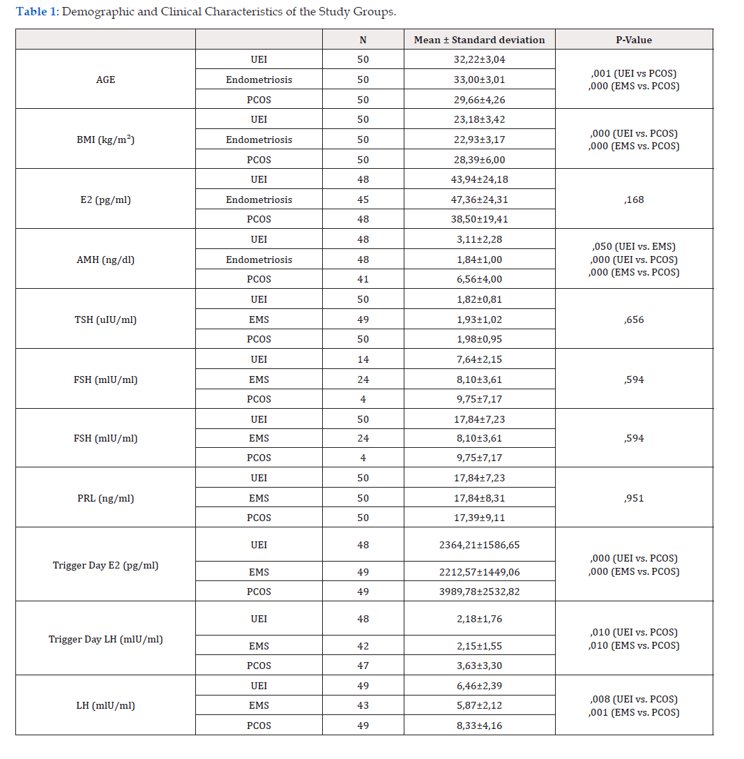
Table 1:Demographic and Clinical Characteristics of the Study Groups.
*Note: UEI, unexplained infertility; EMS, endometriosis; PCOS, polycystic ovary syndrome; BMI, body mass index; E2, estradiol; AMH, anti-Müllerian hormone; TSH, thyroid-stimulating hormone; FSH, follicle-stimulating hormone; PRL, prolactin; LH, luteinizing hormone; N, number of cases. Values are presented as mean±standard deviation. Comparisons between groups were made using Student’s t-test. Statistical significance (p<0.05) is indicated in bold.

Table 2:Comparison of Oocyte Count, Maturation, Fertilization and Sperm Parameters in Study Groups.
*Note: UEI: Unexplained Infertility; EMS: Endometriosis; PCOS: Polycystic Ovary Syndrome. N: Number of Cases. Values are presented as mean±standard deviation. Comparisons between groups were made using Student’s t-test. Statistical significance (p<0.05) is indicated in bold.
Comparison of Morphokinetic Characteristics of Embryos in Study Groups
A comparison of the morphokinetic characteristics of the embryos in the study groups is presented in Table 3. The tPNf stage was statistically significant (p = 0.001) between the endometriosis and PCOS groups, showing that embryos developed more slowly in the endometriosis group. The t2 stage was significantly different between the UEI and endometriosis groups (p = 0.005) and between the endometriosis and PCOS groups (p = 0.011), with embryos in the endometriosis group developing more slowly. The tSC stage (p=0.030) was significantly different between the UEI and endometriosis groups, as well as between the UEI and PCOS (p=0.018) and EMS and PCOS (p<0.001) groups. The results indicated the lowest embryo development rate in the endometriosis group and the highest rate in the PCOS group. The TM stage differed significantly between the UEI and endometriosis groups (p = 0.004) and between the endometriosis and PCOS groups (p < 0.001), with endometriosis showing the slowest progression to the morula. The tSB stage was statistically significant between UEI and endometriosis (p = 0.004) and between EMS and PCOS (p < 0.001), with endometriosis embryos developing more slowly. The tB stage was significantly different between UEI and endometriosis (p < 0.001) and between endometriosis and PCOS (p < 0.001), indicating the slowest development in the endometriosis group. The tEB stage showed significance between UEI and endometriosis (p = 0.001) and between endometriosis and PCOS (p = 0.001), with the endometriosis group developing more slowly. The transition from the compaction stage to the morula stage (TSC-tM) was significantly faster in the UEI group than in the endometriosis (p < 0.001) and PCOS (p < 0.001) groups, indicating that UEI embryos developed more rapidly during this phase.

Table 3:Morphokinetic Characteristics of Embryos in Study Groups.
*Note: UEI, Unexplained Infertility; PCOS: Polycystic Ovary Syndrome; N: Embryo Count. Values are presented as mean±standard deviation. Comparisons between groups were made using Student’s t-test. Statistical significance (p<0.05) is indicated in bold.
Comparison of Pregnancy Outcomes in Study Groups
In the UEI group, 33 women (n=42) achieved pregnancy; in the endometriosis group, 30 women (n=44) reached pregnancy; and in the PCOS group, 32 women (n=41) became pregnant. However, no significant differences were observed between the groups (p>0.05). Pregnancy outcomes were similar among the groups (UEI: 78%, PCOS: 68%, endometriosis: 69%).
Comparison of Demographic and Clinical Parameters According to Pregnancy Outcomes
All patient data showed that cases with high fertilization rates (p=0.001) and progressive motility (+4 progressive motile sperm) values in sperm parameters were statistically significant (p = 0.021) and were directly linked to positive pregnancy outcomes. Fertilization rate and sperm progressive motility were found to be essential factors in achieving pregnancy in all cases.
Comparison of Demographic and Clinical Parameters by Pregnancy in the Unexplained Infertility Group
A comparison of the demographic and clinical parameters according to pregnancy in the UEI group is shown in Table 4.
Comparison of Demographic and Clinical Parameters According to Pregnancy in the Endometriosis Group
A comparison of the demographic and clinical parameters according to pregnancy in the endometriosis group is shown in Table 5. The results indicated that cases with a high fertilization rate were statistically significant (p = 0.014) and directly proportional to positive pregnancy outcomes (Table 5).

Table 4:Comparison of demographic and clinical parameters by pregnancy in the unexplained infertility group.
*Note: N, number of cases; BMI, body mass index; AMH, anti-Müllerian hormone; E2, estradiol; TSH, thyroid-stimulating hormone; FSH, follicle-stimulating hormone; PRL, prolactin; LH, luteinizing hormone. The Mann-Whitney U test was performed, as shown in this table. Statistical significance was set at P<0.05.
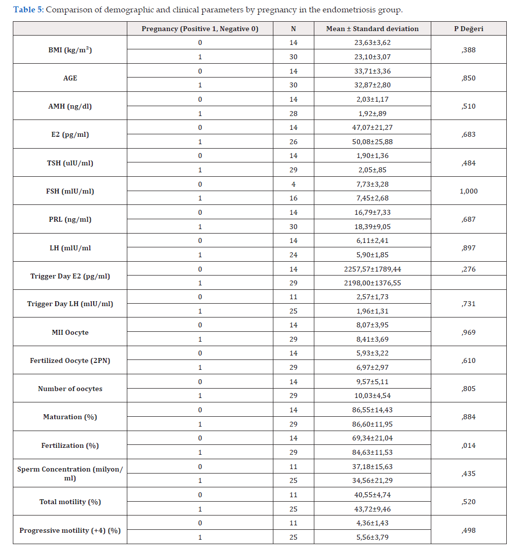
Table 5:Comparison of demographic and clinical parameters by pregnancy in the unexplained infertility group.
*Note: N, number of cases; BMI, body mass index; AMH, anti-Müllerian hormone; E2, estradiol; TSH, thyroid-stimulating hormone; FSH, follicle-stimulating hormone; PRL, prolactin; LH, luteinizing hormone. The Mann-Whitney U test was performed, as shown in this table. Statistical significance was set at P<0.05.
Comparison of Demographic and Clinical Parameters According to Pregnancy in the PCOS Group
A comparison of the demographic and clinical parameters according to pregnancy in the PCOS group is shown in Table 6. The results indicated that cases with a high fertilization rate were statistically significant (p < 0.001) and this percentage was directly related to positive pregnancy outcomes (Table 6).

Table 6:Table of Comparison of demographic and clinical parameters by pregnancy in the PCOS group.
*Note: N, number of cases; BMI, body mass index; AMH, anti-Müllerian hormone; E2, estradiol; TSH, thyroid-stimulating hormone; FSH, follicle-stimulating hormone; PRL, prolactin; LH, luteinizing hormone. The Mann-Whitney U test was performed, as shown in this table. Statistical significance was set at P<0.05.
Discussion
In the present study, embryos derived from women with endometriosis exhibited slower developmental kinetics compared with those from the PCOS and UEI groups. This finding may be attributed to oxidative stress–related impairment of embryo development in endometriosis. In contrast, embryos from women with PCOS demonstrated significantly accelerated developmental progression, potentially associated with elevated LH and AMH levels observed in this group. With respect to clinical parameters, serum AMH concentrations were significantly lower in the endometriosis group compared with both the PCOS and control groups? This observation may reflect oxidative stress–induced ovarian damage in patients with endometriosis, resulting in diminished ovarian reserve and reduced AMH production. Conversely, the highest AMH levels were detected in the PCOS group, likely secondary to increased follicular number and elevated AMH secretion. In addition, serum E2 levels, trigger-day LH concentrations, and day-2 LH levels were significantly higher in women with PCOS than in the other study groups.
In the present study, embryos derived from women with endometriosis exhibited slower developmental kinetics compared with those from the PCOS and UEI groups. This finding may be attributed to oxidative stress–related impairment of embryo development in endometriosis. In contrast, embryos from women with PCOS demonstrated significantly accelerated developmental progression, potentially associated with elevated LH and AMH levels observed in this group. With respect to clinical parameters, serum AMH concentrations were significantly lower in the endometriosis group compared with both the PCOS and control groups? This observation may reflect oxidative stress–induced ovarian damage in patients with endometriosis, resulting in diminished ovarian reserve and reduced AMH production. Conversely, the highest AMH levels were detected in the PCOS group, likely secondary to increased follicular number and elevated AMH secretion. In addition, serum E2 levels, trigger-day LH concentrations, and day-2 LH levels were significantly higher in women with PCOS than in the other study groups.
Freis, et al. (2017) conducted a study with patients in a group with Unexplained Infertility (UEI) and women with endometriosis [13]. According to the TLS results in this study, a significant difference was found between the two groups in CS (2-8) and CS (4-8) times. Based on this difference, the embryos of women with endometriosis develop slower than those of patients with unexplained infertility. We found that only the tPNf and t2 stages developed significantly slower in the EMS group until t8 stage. However, the embryos of women with endometriosis generally developed markedly slower than those of other groups. Additionally, Freis et al. reported that patients with endometriosis have lower oocyte and embryo quality. They stated that the results were similar between the groups with unexplained infertility and endometriosis in terms of AMH, age, and BMI. The clinical parameter results obtained from our study showed that the AMH level was significantly lower in the EMS group than in the UEI group. Age and BMI did not differ considerably between the EMS and UEI groups.
Ramezani Tehrani, et al. [14] found that AMH levels were low in women with endometriosis [15]. We also found that the AMH levels were low in patients with endometriosis. This finding could be related to poor oocyte quality and slow embryo morphokinetics. This warrants further investigation. According to the study by Ramezani Tehrani F, et al., in contrast to the age-dependent threshold observed in the control group, AMH levels in women with endometriosis do not decline more sharply before the age of 27. These findings suggest that endometriosis can modify the typical pattern of AMH, and therefore, clinicians should interpret AMH levels carefully in this population [14].
Schenk, et al. [16] found no statistically significant differences in morphokinetic parameters between the control and endometriosis groups in their study, which included 1148 embryos (control: n = 596, endometriosis: n = 552) [16]. Additionally, no significant relationship was observed between fetal heartbeats in groups with and without endometriosis [16]. The morphokinetic parameter results showed that embryos developed more slowly in the EMS group than in the UEI group (tPNf, t2, tSC, tM, tSB, tEB, tB, and tSCtM were significantly slower in the EMS group).
Irez, et al. [17] showed that oocyte fertilization and pregnancy rates in PCOS cases at different ICSI timings were different compared to those in cases with a normal response, and prolonging the duration caused a decrease in fertilization and pregnancy outcomes [17]. Thus, it was inferred that oocyte aging is shorter and faster in PCOS cases related to oocyte sufficiency. In this study, embryo selection methods used today, especially Preimplantation Genetic Diagnosis (PGD), were not applied. The similarity in pregnancy outcomes between the groups in our study can be attributed to embryo selection via PGD. Similarly, our study showed the rate at which PCOS embryos developed, and we believe that molecular studies support this finding.
Chappel, et al. [18] formed two groups of women of similar age and body mass index (BMI), including women with PCOS (n=64) and women without PCOS (n=64) (control group) [18]. They studied 990 and 628 embryos in the PCOS and control groups, respectively. In this retrospective study, when embryos were examined morphokinetically, the rates of reaching stages t7, t8, and t9 (i.e., reaching the morula) were significantly higher in embryos from women with PCOS than in those from the control group. They showed that the other morphokinetic parameters were similar. In this retrospective study, we demonstrated that the embryos of women with PCOS at the morula stage (tSC-tM) were slower than those in the UEI group. We also showed that, during the compaction stage (tSC), embryos from women with PCOS were faster than those from the UEI group. Additionally, Chappel et al. [18] showed that women with PCOS had a significantly higher abortion rate (PCOS group,38.1%; control group, 18.8%). However, we found no significant differences in pregnancy outcomes [18].
Wissing, et al. [19] showed in their study that women with hyperandrogenic PCOS exhibited delayed morphokinetic progression from the tPNf to t8 stages compared to women without PCOS [19]. However, they did not demonstrate a significant difference in morphokinetic parameters between women with normoandrogenic PCOS and those without PCOS. They also found no significant differences in the implantation and clinical pregnancy rates between the groups. Similarly, Tabibnejad et al. [20] showed that the tPNf-t8 interval in embryo development parameters of women with PCOS was slower than that in women without PCOS [20]. They found no significant differences between groups in terms of implantation and clinical pregnancy rates. Based on our findings, there were no significant differences in implantation and pregnancy rates.
Llarena, et al. [21] compared embryo morphokinetics between two groups with and without endometriosis [21]. The continuous embryo monitoring system examined 3,471 embryos from the EMS (n = 1078) and non-EMS (n = 2393) groups until day 6. Morphokinetic results showed that the EMS group was significantly slower to reach the t2 and t8 stages, as well as the compaction (tSC), morula (tM), and blastocyst stages (tB), compared to the non- EMS group [21]. However, there were no significant differences in IVF outcomes between these groups. We found a similar result for t2, although the difference was not evident until the tSC stage: tSC was significantly slower between tM and tB. Kitajima, et al. [11] compared serum AMH levels between patients diagnosed with endometriosis (n = 90) and those without endometriosis (n = 30) [11]. They showed that serum AMH levels are inversely proportional to age. However, the authors stated that there was no significant difference between the groups. Our study revealed that AMH levels were substantially lower in women with endometriosis than in those without endometriosis.
Bungum, et al. [12] examined serum AMH, FSH, LH, progesterone, testosterone, and estradiol levels in 18 patients n=10 in the control group and n=8 in the PCOS group [12]. According to their results, the serum AMH, LH, and testosterone levels were significantly higher in the PCOS group. FSH levels were considerably lower in the PCOS group. They did not find a significant difference in progesterone or estradiol levels [12].
Blood glucose, TSH, testosterone, FSH, and LH levels of women diagnosed with PCOS (n=33) and a control group of healthy women (n=32). Their results showed that luteinizing hormone (LH), testosterone, and blood glucose levels were significantly higher in women with PCOS than in the control group. However, they did not find a statistically significant difference in the TSH and FSH levels [12].
Conclusion
In patients with endometriosis, embryo development was delayed, with fewer MII oocytes and slower progression through various fertilization stages (tPN, t2, tSC, tM, tSB, tEB, and tSCtM). Despite the differences in embryo morphokinetics, clinical pregnancy rates were similar between the endometriosis and control groups, regardless of disease severity. This study highlights the potential usefulness of embryo morphokinetics in selecting high-quality embryos in patients with endometriosis. The association between low AMH levels and embryo quality in endometriosis warrants further investigation. In PCOS patients, higher BMI and trigger-day E2 and LH levels were associated with more M2 oocytes, fertilized oocytes, and a lower rate of mature oocytes. Additionally, tSC was faster, while delays were noted in the tSB-tM processes. Future prospective studies should focus on selecting embryos with the best potential for implantation, pregnancy, and live birth in patients with PCOS and endometriosis, considering established parameters.
Declarations
Disclosure Statement /Competing Interests
The authors declare that they have no affiliations with or involvement in any organization or entity with any financial interest in the subject matter or materials discussed in this manuscript.
Ethics Approval
This study received ethical approval on March 8, 2022, with approval number 2022/03-822, in accordance with the ethics committee guidelines established under Articles 14 and 42 of the Higher Education Law No. 2547.
Consent for Publication
Not applicable.
Data Availability
The data analyzed during the current study are available from the corresponding author on reasonable request.
Funding Statement
This study was supported by data from Memorial IVF and the Genetics Center. This study was conducted as a thesis within the scope of the Clinical Embryology Master’s program at Istanbul Yeni Yuzyil University.
Author Contributions
TI and SK: Supervision. TI, GO, AO, T.M.A, YKC: Conceptualization, investigation, methodology, and formal analysis. AO, TI: Writing— original draft. TI, AO: Writing—review and editing.
Acknowledgements
We thank the Memorial Sisli Hospital IVF Center team, all clinicians who made this study possible, and all the women who participated in the study.
References
- Kalra Kansal S, Barnhart K T (2011) In Vitro Fertilization and Adverse Childhood Outcomes: What We Know, Where We Are Going, and How Will We Get There? A Glimpse into What Lies Behind and Beckons Ahead. Fertil Steril 95(6): 1887-1889.
- Countinho E A, Kaufmann A S (2019) The Role of the Brain in the Pathogenesis and Phsiology of Polycystic Ovary Syndrome (PCOS). Med Sci (Basel) 7(8): 84.
- Sanchez A M, Pagliardini L, Cermisoni G C, Privitera L, Makieva S, et al. (2020) Does Endometriosis Influence the Embryo Quality and/or Development? Insights from a Large Retrospective Matched Cohort Study. Diagnostics 10(2): 83.
- Rolla E (2019) Endometriosis: Advances and Controversies in Classification, Pathogenesis, Diagnosis and Treatment. F1000Res (p: 8-11).
- Koç O, Gündüz B, Topçuoğlu A, Buğdaycı G, Yılmaz F, et al. (2010) Effects of Pinealectomy and Melatonin Supplementation on Endometrial Explants in a Rat Model. Eur J Obstet Gynecol Reprod Biol 153(1): 72-76.
- Farquhar C (2020) What should the first-line treatment for couples with unexplained infertility be: intrauterine insemination or in vitro fertilization? Fertile Battle 114(6): 1140.
- Minh Tam Le, Nguyen TV, Nguyen TT, Thai Thanh Thi Nguyen, Nguyen TT (2019) Does polycystic ovary syndrome affect morphokinetics or abnormalities in early embryonic development?. Eur J Obstet Gynecol Reprod Biol X 17:3:100045.
- Hart RJ, Walls M (2022) The Role of In Vitro Maturation in Polycystic Ovary Syndrome. In: Kovacs GT, Fauser B, Legro RS, eds. Polycystic Ovary Syndrome. Cambridge University Press 142-149.
- RajkovicA, Pangas SA, Matzuk MM (2006) Follicular development: Mouse, sheep, and human models. In Neill J. D. (Ed.), Knobil & Neill's physiology of reproduction 383-424.
- Pigny P, Jonard S, Robert J, Dewailly D (2005) Serum anti-Mullerian hormone as a surrogate for antral follicle count for definition of the polycystic ovary syndrome. J Clin Endocrinol Metab 91(3):941-945.
- Kitajima M, Matsumoto K, Murakami N, Kajimura I, Harada A, et.al (2020) AMH Concentrations in Peritoneal Fluids of Women with and Without Endometriosis. Front Surg 2-7: 11; 7:600202.
- Bungum L, Franssohn F, Bungum M, Humaidan P, Giwercman A (2013) The Circadian Variation in Anti-Müllerian Hormone in Patients with Polycystic Ovary Syndrome Differs Significantly from Normally Ovulating Women. PLoS One 4;8(9): e68223.
- Wiweko B, Indra I, Susanto C, Natadisastra M, Hestiantoro A (2018) The correlation between serum AMH and HOMA-IR among PCOS phenotypes. BMC Res Notes 9;11(1):114.
- Ramezani Tehrani F, Mousavi M, Noori Ardebili S, Saei Ghare Naz M, Azizi F, et.al (2025) Behboudi-Gandevani S. Association between anti-Mullerian hormone levels and age in women with endometriosis: insights from a population-based study. BMJ Open 5;15(7): e102774.
- Freis A, Dietrich JE, Binder M, Holschbach V, Strowitzki T, et.al (2018) Relative Morphokinetics Assessed by Time-Lapse Imaging Are Altered in Embryos from Patients with Endometriosis. Reprod Sci. 25(8):1279-1285.
- Schenk M, Kröpfl J M, Hörmann-Kröpfl M, Weiss G (2019). Endometriosis Accelerates Synchronization of Early Cell Divisions but Does Not Change Morphokinetic Dynamics in Endometriosis Patients. PLos One 14(8): e0220529.
- Irez T, Erel CT, Senturk LM, Kaleli S, Senol H, et.al (2005). Is ICSI timing predicting the fertilization and pregnancy rate in women with PCOS. 13th World congress on in vitro fertilization assisted reproduction & genetics, İstanbul Turkey, Conference Book 26-29.
- Chappel N R, Barsky M, Shah J, Peavey M, Yang L Sangi-Haghpeykar H, et al. (2020) Embryos from polycystic ovary syndrome patients with hyperandrogenemia reach morula stage faster than controls. F S Rep 1(2): 125–132.
- Wissing M L, Bjerge M R, Olesen A I, Hoest T, Mikkelsen A L (2014) Impact of PCOS on early embryo cleavage kinetics. Reprod Biomed Online 28(4): 508-514.
- Tabibnejad N, Sheikhha MH, Ghasemi N, Fesahat F, Soleimani M, Aflatoonian A (2019) Association between early embryo morphokinetics plus cumulus cell gene expression and assisted reproduction outcomes in polycystic ovary syndrome women. Reprod Biomed Online 38(2):139-151.
- Llarena N C, Hur C E, Yao M, Schwartz K, Falcone T, et al. (2022) The impact of endometriosis on embryo morphokinetics: embryos from endometriosis patients exhibit delayed cell cycle milestones and decreased blastulation rates. J Assist Reprod Genet 39(3): 619-628.




 We use cookies to ensure you get the best experience on our website.